1918
Assessment of Daily Variations of GABA Levels within the Parietal Lobe and Anterior Cingulate Gyrus Regions of Healthy Young Adults Based on MRS.1Department of Radiology, The First Affiliated Hospital of Soochow University, SuZhou, China, 2Center for Circadian Clocks, Soochow University, Suzhou, China, 3Urinary surgery, The First Affiliated Hospital of Soochow University, SuZhou, China, 4Suzhou Medical College of Soochow University, Soochow University, SuZhou, China, 5Philips Healthcare, Shanghai, China, 6Center for Circadian Clocks, Soochow University, SuZhou, China
Synopsis
Keywords: Neurotransmission, Brain
Motivation: The assessment of GABA and Glx levels within the brain with MEscher-Garwood Point RESolved Spectroscopy (MEGA-PRESS) has an important role in the diagnosis and treatment of neuropsychiatric disorders.
Goal(s): We evaluate the daily fluctuations of GABA levels within the parietal lobe (PL) and anterior cingulate gyrus (ACC) regions.
Approach: The GABA+, GABA+/Cr , Glx/Cr ,Glx were measured at six different time points throughout the day using MEGA-PRESS.
Results: Significant variations in GABA+/Cr levels within the PL region, with the lowest point occurring at 9:00 and the highest peak occurring at 21:00 . The melatonin levels were positively correlated with GABA+/Cr within the ACC region.
Impact: GABA changes in localized brain regions are strongly associated with many psychiatric disorders. The outcomes of this study could be used to guide the diagnosis and treatment of neuropsychiatric disorders.
Introduction
Alterations in the GABA levels within the brain have also been associated with multiple psychiatric and neurological disorders, including insomnia, Parkinson's disease (PD), Alzheimer's disease (AD), and multiple sclerosis (MS) [1]. GABA interacts with melatonin and is involved in the regulation of blood pressure and heart rate [2,3]. Therefore, there is a need to understand the changes that occur in the GABA levels within specific brain regions throughout the day and their impact on physiological parameters such as blood pressure and heart rate. MEscher-Garwood Point RESolved Spectroscopy (MEGA-PRESS) is commonly used to measure the GABA levels within the brain of healthy individuals and in patients suffering from various neurological disorders, including insomnia, Parkinson's disease, Alzheimer's disease[4,5].To address this issue, in this study, we aimed to make use of MRS to measure the daily variations in GABA levels and assess the impact of these changes on melatonin levels, blood pressure, and heart rate in healthy individuals.Methods
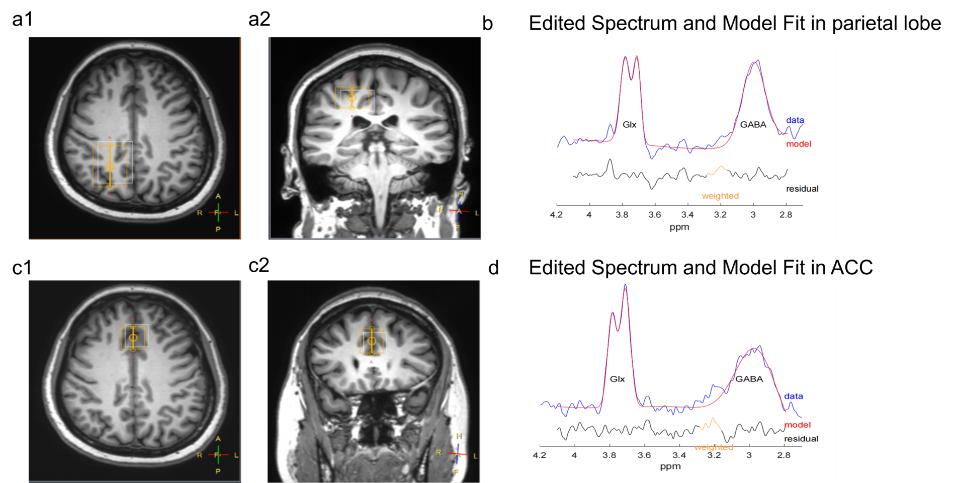
Twenty-six healthy subjects from Soochow University aged between 22 and 30 years who met the eligibility criteria were invited to participate in this study. All images were acquired on a 3.0 T Philips MRI (Ingenia I, Best, Netherlands). The acquired GABA signal contained the overlapping signals of macromolecules and homocarnosine, hence expressed as GABA+. The creatine (Cr) signal was applied as an endogenous reference. The GABA+, GABA+/Cr, ratio of glutamine/glutamate complex to creatine (Glx/Cr), Glx were measured at six different time points (1:00 h, 5:00 h, 9:00 h, 13:00 h, 17:00 h, and 21:00 h) throughout the day using magnetic resonance spectroscopy. To measure the MRS spectrum, a region of interest (ROI) measuring 2×2×2 cm3 and 2×3×4 cm³ was placed on the PL and ACC regions, respectively, as stated by the MEGA-PRESS guidelines [6,7] (Figure 1). The acquisition parameters for MAGE-PRESS were a TR of 2000 ms, TE of 68 ms, and a scan bandwidth (acquisition bandwidth) of 2000 Hz. The Gannet software version 3.0 (GitHub - richardedden/Gannet3.0)[8,9] was used to apply a Gaussian curve to fit the GABA+, Glx, GABA+/Cr, and Glx/Cr. The one-way repeated-measures analysis of variance (ANOVA) was used to evaluate the GABA, Glx, blood pressure, heart rate, and melatonin levels throughout the day.Results
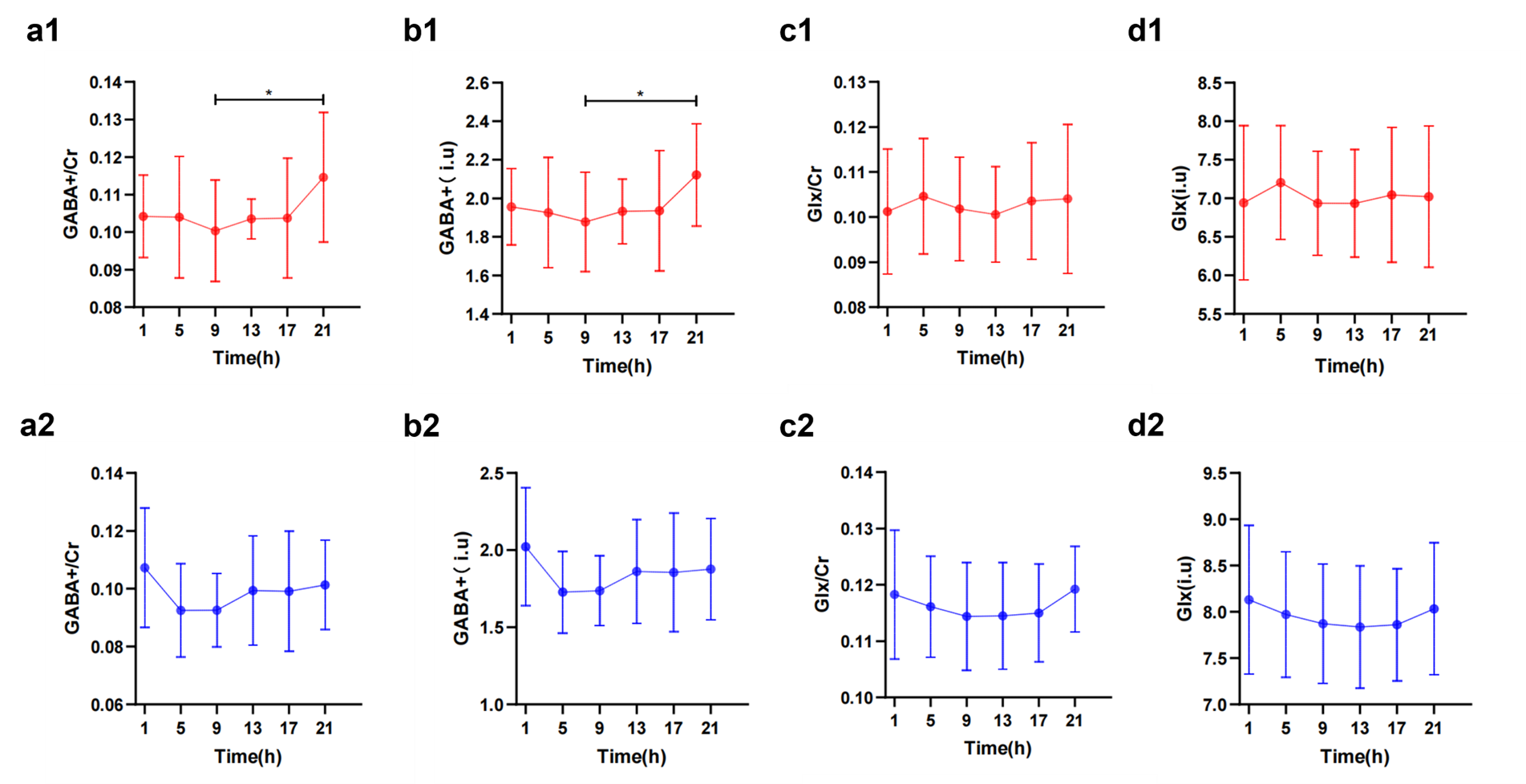
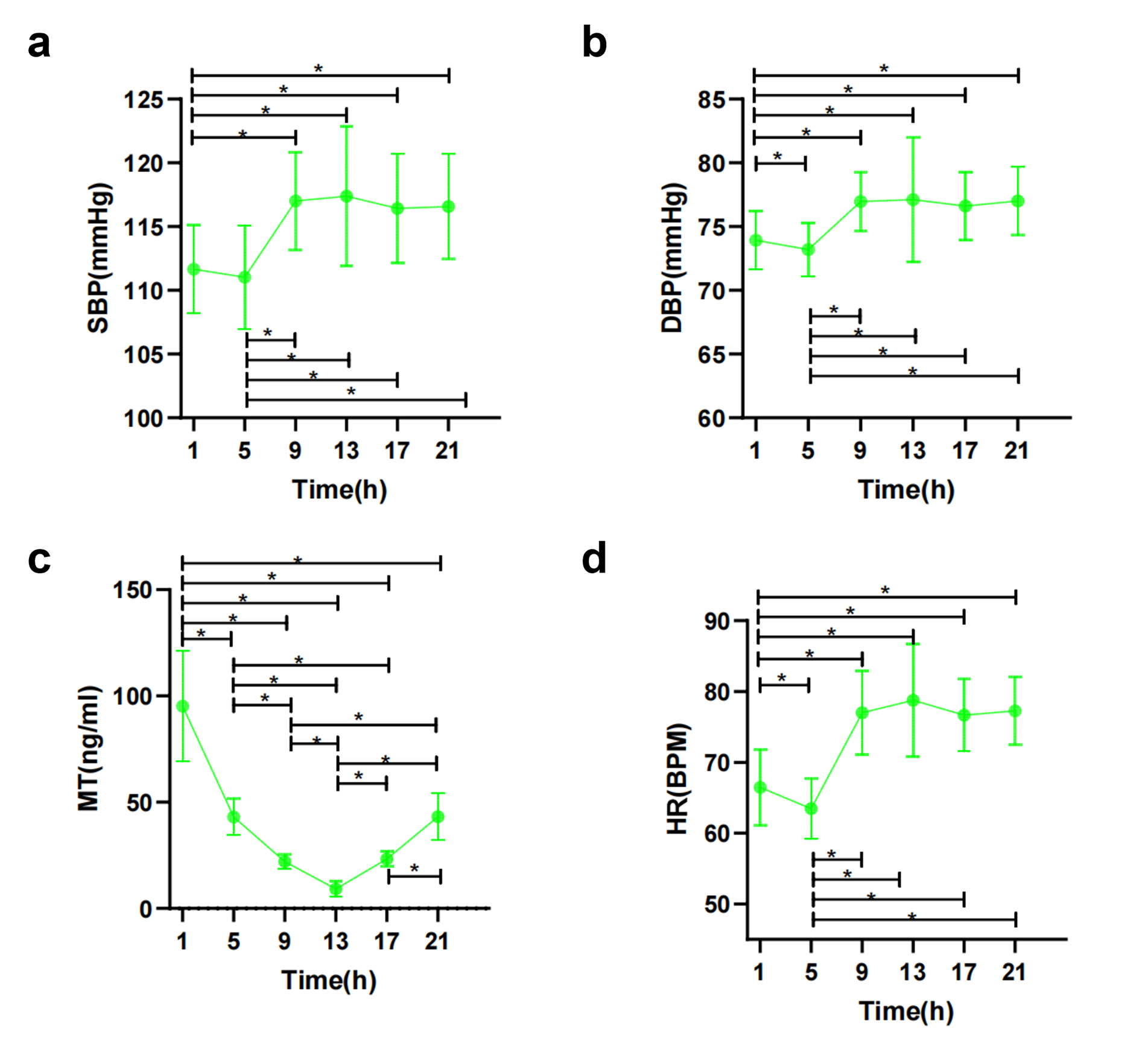
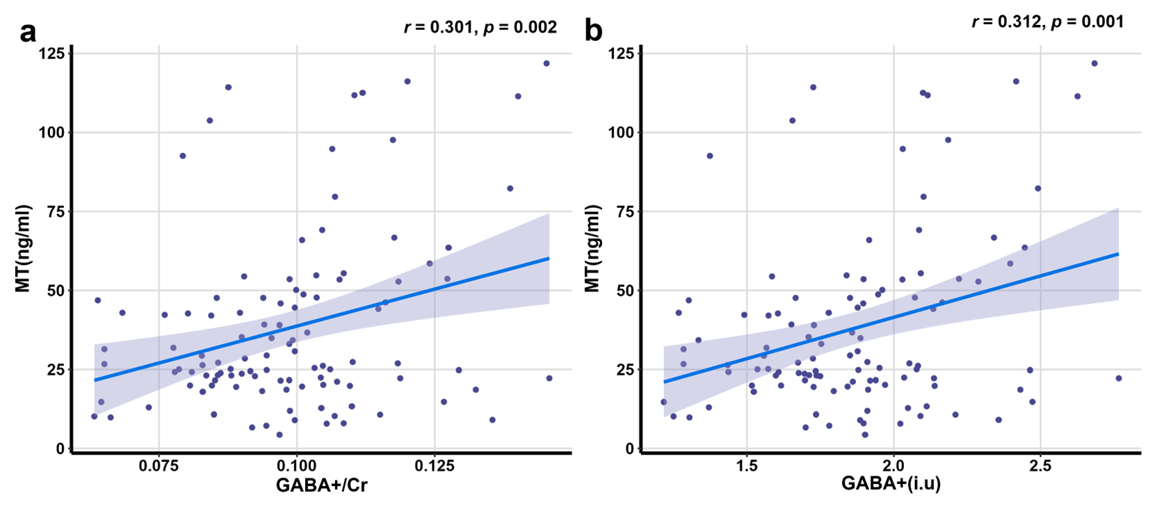
1. The daily variations of GABA+/Cr, GABA+, Glx/Cr, and Glx within the PL and ACC regions are summarized in Figure 2. 2. The mean GABA+/Cr and GABA+ in the PL region were significantly higher than those found in the ACC region. Conversely, the mean Glx/Cr and Glx levels in the PL region were significantly lower than those found in the ACC region. 3. The changes of systolic blood pressure (SBP), diastolic blood pressure (DBP), heart rate (HR) and melatonin (MT) at six time points are shown in Figure 3. 4. Within the ACC region, the mean GABA+/Cr (r=0.301, P=0.002) and GABA+ (r=0.312, P =0.001) were positively correlated with the melatonin levels(Figure 4).Discussion
Statistically significant differences were noted in the GABA levels in the PL region between 9:00 h and 21:00 h. Glx/Cr, and Glx levels did not differ significantly between the ACC and PL regions at any time point. Our results, while intriguing, emphasize the importance of considering time effects when investigating function-related GABA changes within the brain, especially when studying the PL region. We recommend selecting consistent time points when scanning the same patient to minimize potential errors in future studies. Research has demonstrated the influence of melatonin levels on GABA levels. Melatonin enhances GABAergic inhibition of neuronal activity [10] and exerts its effects on GABA receptors, augmenting the GABA binding within the body [11]. In our study, we noted a positive correlation between the GABA levels within the ACC region and the melatonin levels, suggesting a potential interaction between GABA receptors in the ACC and melatonin. Considering that both GABA and melatonin are commonly targeted in the treatment of insomnia, these findings provide a solid theoretical basis for exploring pharmacological interventions for insomnia.Conclusion
In healthy individuals, the GABA levels within the PL vary throughout the day as part of the circadian rhythm. These variations should be taken into account in GABA-related studies within this region, particularly in the evaluation of neuropsychiatric disorders. Furthermore, we also found a positive correlation between the GABA levels in the ACC region and melatonin levels. These findings indicate a possible interaction between the GABA receptors in the ACC region and melatonin levels and thus offer some interesting theoretical basis for exploring pharmacological interventions in the treatment of insomnia.Acknowledgements
The authors would like to acknowledge healthy volunteers taking part in this study and the support of the funding sources. We would also like to thank TopEdit (www.topeditsci.com) for English language editing of this manuscript.References
[1] Zhang W, Xiong BR, Zhang LQ, et al. The Role of the GABAergic System in Diseases of the Central Nervous System. Neuroscience. 2021 Aug 21; 470:88-99. [2] Bangsumruaj J, Kijtawornrat A, Kalandakanond-Thongsong S. Effects of chronic mild stress on GABAergic system in the paraventricular nucleus of hypothalamus associated with cardiac autonomic activity[J]. Behavioural Brain Research, 2022, 432: 113985. [3] Albers H E, Walton J C, Gamble K L, et al. The dynamics of GABA signaling: Revelations from the circadian pacemaker in the suprachiasmatic nucleus[J]. Frontiers in Neuroendocrinology, 2017, 44: 35-82. [4] Bai X, Edden R A E, Gao F, et al. Decreased γ-aminobutyric acid levels in the parietal region of patients with Alzheimer's disease[J]. Journal of magnetic resonance imaging: JMRI, 2015, 41(5): 1326-1331. [5] Chiang G C, Mao X, Kang G, et al. Relationships among Cortical Glutathione Levels, Brain Amyloidosis, and Memory in Healthy Older Adults Investigated In Vivo with 1H-MRS and Pittsburgh Compound-B PET[J]. AJNR. American journal of neuroradiology, 2017, 38(6): 1130-1137. [6] Park S, Kang I, Edden R A E, et al. Shorter sleep duration is associated with lower GABA levels in the anterior cingulate cortex[J]. Sleep Medicine, 2020, 71: 1-7. [7] Volk C, Jaramillo V, Merki R, et al. Diurnal changes in glutamate + glutamine levels of healthy young adults assessed by proton magnetic resonance spectroscopy[J]. Human Brain Mapping, 2018, 39(10): 3984-3992. [8] Myers J F M, Evans C J, Kalk N J, et al. Measurement of GABA using J-difference edited 1H-MRS following modulation of synaptic GABA concentration with tiagabine[J]. Synapse (New York, N.Y.), 2014, 68(8): 355-362. [9] Edden RA, Puts NA, Harris AD, et al. Gannet: A batch-processing tool for the quantitative analysis of gamma-aminobutyric acid–edited MR spectroscopy spectra. J Magn Reson Imaging. 2014 Dec;40(6):1445-52. [10] Stankov B, Biella G, Panara C, et al. Melatonin signal transduction and mechanism of action in the central nervous system: using the rabbit cortex as a model[J]. Endocrinology, 1992, 130(4): 2152-2159. [11] Niles L P, Peace C H. Allosteric modulation of t-[35S] butylbicyclophosphorothionate binding in rat brain by melatonin[J]. Brain Research Bulletin, 1990, 24(4): 635-638.Figures