1915
Coil Sensitivity Estimation and Complex Image Combination for 96-Channel Receive Array at 7T1Electrical Engineering and Computer Sciences, University of California, Berkeley, Berkeley, CA, United States, 2Bernard and Irene Schwartz Center for Biomedical Imaging, Department of Radiology, New York University Grossman School of Medicine, New York, NY, United States, 3Center for Advanced Imaging Innovation and Research (CAI2R), Department of Radiology, New York University Grossman School of Medicine, New York, NY, United States, 4Helen Wills Neuroscience Institute, University of California, Berkeley, CA, United States
Synopsis
Keywords: Image Reconstruction, Data Processing
Motivation: High density receive arrays can improve SNR and parallel imaging capability; however, they also introduce significant image reconstruction challenges.
Goal(s): We aim to find a reconstruction method that will produce consistent and high-quality complex images for high-channel-count receive arrays at 7T.
Approach: Several existing sensitivity map estimation methods and coil combination methods were tested for 8-channel and 32-channel datasets, and an ultrahigh resolution 96-channel dataset acquired at 7T.
Results: Existing reconstruction methods did not produce consistent results for the 96-channel dataset. Compression of high-quality sensitivity maps reduced data size by a factor of 100 while maintaining image quality.
Impact: This work explores the unique reconstruction challenges in high-channel-count receive arrays by assessing performance of existing reconstruction techniques on an ultrahigh resolution dataset acquired with a 96-channel receive array, establishing a need for more research in effective reconstruction methods.
Introduction
High density receive arrays have many potential benefits including improved g-factor and better SNR. However, massive receive arrays also introduce significant image reconstruction challenges such as data handling, coil sensitivity map estimation and coil combination. Most existing coil combination algorithms have been developed based on data using receive arrays with up to 32 channels; furthermore, most of them report performance based on the combined magnitude images, while rarely investigating recovered phase maps. This work explores the unique reconstruction challenges in high-channel-count receive arrays by assessing the performance of existing coil combination techniques on an ultrahigh resolution dataset of a postmortem brain acquired with a 96-channel receive array at Berkeley's NexGen 7T scanner1,2.Methods
A postmortem brain specimen was scanned on the NexGen 7T MRI scanner at UC Berkeley equipped with a 16Tx96Rx head RF coil and high-performance gradient coils. A 3D gradient echo (GRE) sequence was used with the parameters: voxel size=0.18x0.18x0.18 mm3, matrix size=1000x930x512, TR=36 ms, TE=20 ms, FA=20°, PAT=off, raw data size=575.5 GB. Multiple existing coil sensitivity map estimation and coil combination techniques were tested to assess reconstruction quality in both magnitude and phase images. For comparison, the same techniques were repeated for data of the same specimen acquired with a 32-channel array on a 7T scanner and in vivo 3T data from a different specimen acquired with an 8-channel array. Five common methods for sensitivity map estimation (Walsh adaptive coil estimation3,4, ESPIRiT5, SOFTSENSE6, direct calibration7, and nonlinear inversion8,9) were paired with four coil combination techniques (SENSE10, L1-regularized reconstruction with a radial basis, L2-regularized reconstruction11, and simple combination—defined as complex summation of coil images multiplied by the complex conjugate of the coil sensitivity). In total, 20 unique combinations of reconstruction methods were tested on all three datasets. Coil compression, specifically low-rank approximation of adaptive estimation and ESPIRiT sensitivity maps, was explored for the 96-channel dataset. Most methods were implemented with Berkeley’s Advanced Reconstruction Toolbox (BART)12-14. The recovered phase images were processed with STI Suite15: unwrapping was done using a Laplacian-based approach16 and background removal was done using VSHARP17 for the phase contrast evaluation.Results
The methods resulted in large variations in sensitivity estimation for the 32-channel and 96-channel arrays, with higher variation observed for the 96-channel array data (Fig. 1b). In general, adaptive estimation and ESPIRiT produced maps with less apparent artifacts. Direct calibration and nonlinear inversion (NLINV) yielded many obvious artifacts in sensitivity maps, and NLINV generally produced maps with low SNR or no substantial signal. Direct calibration failed to remove anatomical information for some coil sensitivity maps.Figure 2 compares the coil-combined magnitude and phase images of the 96-channel array. While most sensitivity map estimation methods paired with at least one combination technique produced a high-quality magnitude image, phase image quality was almost solely dependent on sensitivity map estimation. Adaptive estimation with all combination methods yielded high quality magnitude images, but noisy phase images (Fig. 2a). ESPIRiT estimated coil sensitivity maps with all combination techniques produced high-quality magnitude and phase images (Fig. 2b). SOFTSENSE produced the highest-quality phase images in this particular 96-channel dataset, despite inhomogeneous magnitude images for all but one combination technique (Fig. 2c). Direct calibration (Fig. 2d) and NLINV (Fig. 2e) methods introduced significant artifacts in magnitude and phase. Similar results were found in the 32-channel array (Fig. 3). In contrast, each coil sensitivity map estimation technique for the 8-channel dataset at 3T produced generally consistent maps per channel (Fig. 4).
Low rank approximation for 96-channel coil sensitivity maps was highly effective. Magnitude images were comparable to full-rank images with only 8 components (Fig. 5a), and phase images were recoverable with only 2 components (Fig. 5b). This corresponds to a data reduction by over a factor of 100 and 400 in the 96-channel slice, respectively.
Discussion & Conclusion
These results establish a need for more research in effective reconstruction techniques with massive receive arrays. Inconsistencies in sensitivity map estimation results for the 96-channel array dataset suggest that, while existing methods are highly reliable for low-channel-count arrays, they are not reliable for massive arrays. Furthermore, low-rank approximation of coil sensitivity maps may preserve image quality while substantially reducing data size and computational demand. While some techniques produced high quality magnitude images for the data acquired with the 96-channel array, no method produced artifact-free phase images. As such, more development in techniques catered to high-channel coil datasets is necessary and should be quantitatively evaluated with more datasets.Acknowledgements
Research reported in this publication was supported by the National Institutes of Health through grants U01EB025162, R01AG070826, and the Weill NeuroHub. The content is solely the responsibility of the authors and does not necessarily represent the official views of the National Institutes of Health.
References
1. Feinberg DA, Dietz P, Liu C, Setsompop K, Mukherjee P, Wald LL, Vu AT, Beckett A, Insua IG, Schröder M, Stocker S. Design and development of a next-generation 7T human brain scanner with high-performance gradient coil and dense RF arrays. In Proceedings of the 29th Annual Meeting of ISMRM, Online, 2021. Abstract 0562.
2. Gunamony S, et al. A 16-channel transmit 96-channel receive head coil for NexGen 7T scanner. In Proceedings of the 29th Annual Meeting of the ISMRM, Online, 2021. Abstract 0182.
3. Walsh DO, Gmitro AF, Marcellin MW. Adaptive reconstruction of phased array MR imagery. Magn Reson Med. 2000 May;43(5):682–90.
4. Griswold M, Walsh D, Heidemann R, Haase A, Jakob A. The Use of an Adaptive Reconstruction for Array Coil Sensitivity Mapping and Intensity Normalization. In Proceedings of the 10th Annual Meeting of ISMRM, Honolulu, HI, United States, 2002. Abstract 2410.
5. Uecker M, Lai P, Murphy MJ, Virtue P, Elad M, Pauly JM, Vasanawala SS, Lustig M. ESPIRiT - An Eigenvalue Approach to Autocalibrating Parallel MRI: Where SENSE meets GRAPPA. Magn Reson Med. 2014; 71:990-1001.
6. Uecker M, Virtue P, Vasanawala S, Lustig M. ESPIRiT Reconstruction Using Soft SENSE. In Proceedings of the 21st Annual Meeting of ISMRM, Salt Lake City, UT, United States, 2013. Abstract 0127.
7. McKenzie CA, Yeh EN, Ohliger MA, Price MD, Sodickson DK. Self-calibrating parallel imaging with automatic coil sensitivity extraction. Magn Reson Med. 2002; 47:529-538.
8. Uecker M, Virtue P, Vasanawala SS, Lustig M. Image reconstruction by regularized nonlinear inversion-joint estimation of coil sensitivities and image content. Magn Reson Med. 2008; 60:674-682.
9. Holme HCM, Rosenzweig S, Ong F, Wilke RN, Lustig M, Uecker M. ENLIVE: An Efficient Nonlinear Method for Calibrationless and Robust Parallel Imaging. Sci Rep. 2019; 9:3034.
10. Pruessmann KP, Weiger M, Scheidegger MB, Boesiger P. SENSE: Sensitivity encoding for fast MRI. Magn Reson Med. 1999; 42:952-962.
11. Pruessmann KP, Weiger M, Boernert P, Boesiger P. Advances in sensitivity encoding with arbitrary k-space trajectories. Magn Reson Med. 2001; 46:638-651.
12. Uecker M, Ong F, Tamir JI, Bahri D, Virtue P, Cheng JY, Zhang T, Lustig M. Berkeley Advanced Reconstruction Toolbox. In Proceedings of the 23rd Annual Meeting of ISMRM, Toronto, ON, Canada, 2015. Abstract 2486.
13. Uecker M, Virtue P, Ong F, Murphy MJ, Alley MT, Vasanawala SS, Lustig M. Software Toolbox and Programming Library for Compressed Sensing and Parallel Imaging. In ISMRM Workshop on Data Sampling and Image Reconstruction, Sedona, AZ, United States, 2013.
14. Tamir JI, Ong F, Cheng JY, Uecker M, Lustig M. Generalized Magnetic Resonance Image Reconstruction using The Berkeley Advanced Reconstruction Toolbox. In ISMRM Workshop on Data Sampling and Image Reconstruction, Sedona, AZ, United States, 2016.
15. Li W, Wu B, Liu C. STI Suite: A Software Package for Quantitative Susceptibility Imaging. In Proceedings of the 22nd Annual Meeting of ISMRM, Milan, Italy, Canada, 2014. Abstract 3265.
16. Li W, Avram AV, Wu B, Xiao X, Liu C. Integrated Laplacian-based phase unwrapping and background phase removal for quantitative susceptibility mapping. NMR Biomed. 2014;27(2):219-27.
17. Wei H, Zhang Y, Gibbs E, Chen N-K, Wang N, Liu C. Joint 2D and 3D phase processing for quantitative susceptibility mapping: Application to 2D echo-planar imaging. NMR Biomed. 2017 Apr;30(4):e3501. DOI: 10.1002/nbm.3501.
Figures

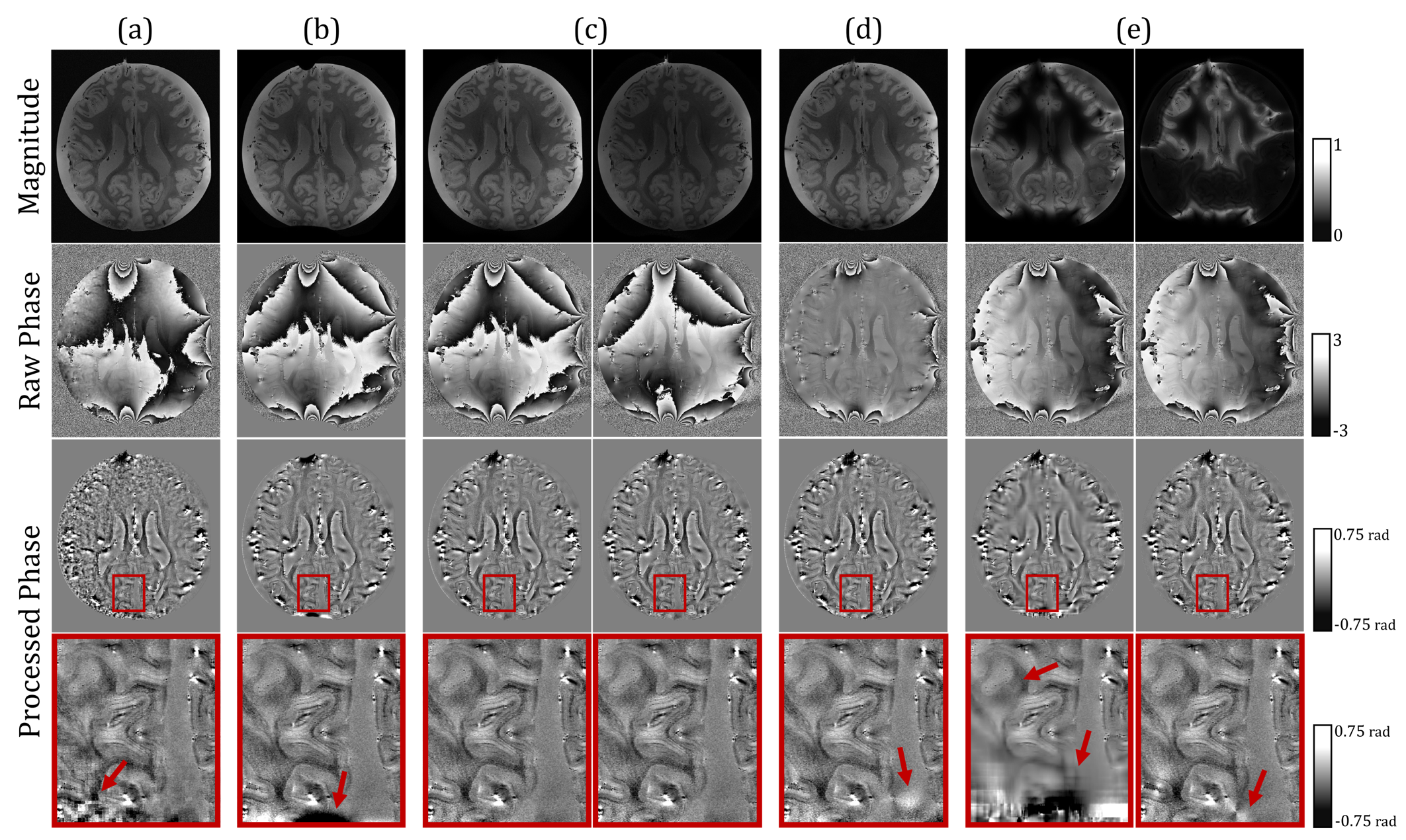
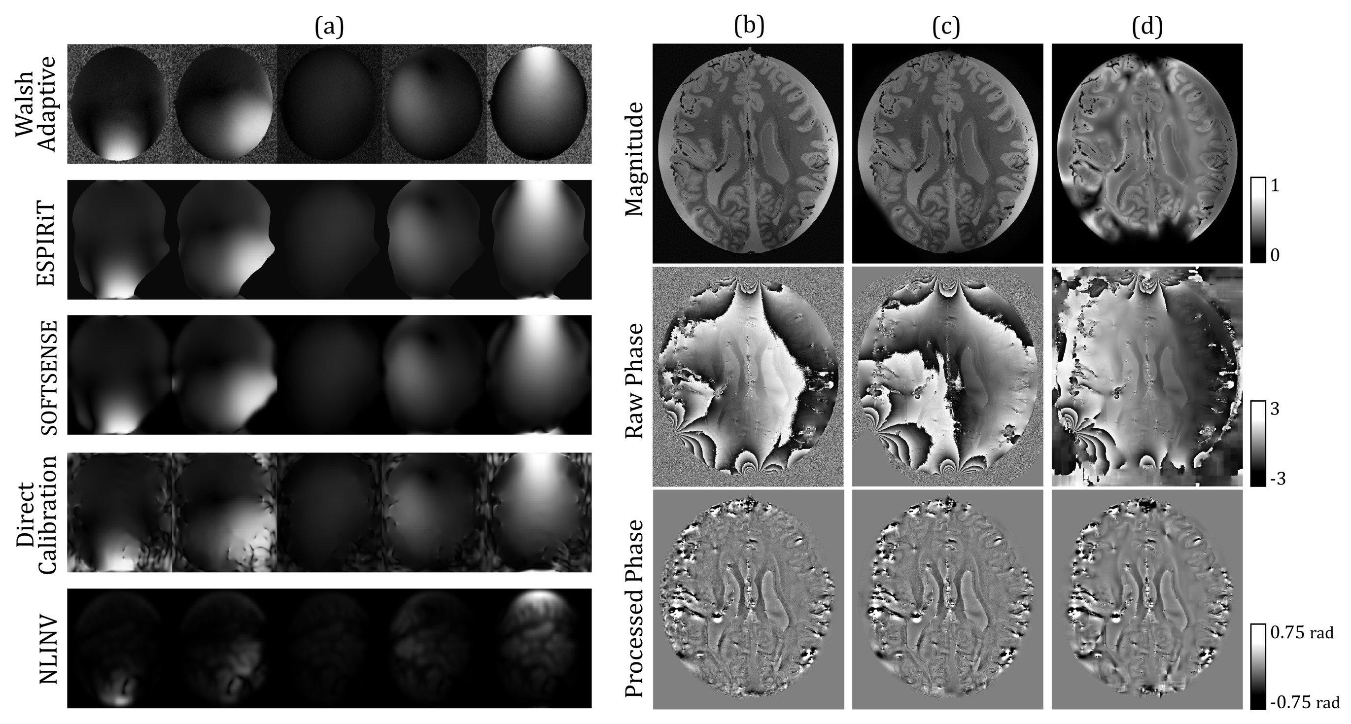
Figure 3. Estimated Sensitivity Maps and Reconstructed Images for the 32-Channel Array. (a) Sensitivity maps of select channels in 32-channel array for each coil sensitivity estimation technique. (b-d) 32-channel coil-combined magnitude (top row), raw phase (second row) and processed phase (third row) images for select methods. Methods include: (b) Walsh adaptive estimation with simple combination, (c) ESPIRiT with simple combination, and (d) NLINV with L1-regularized combination.
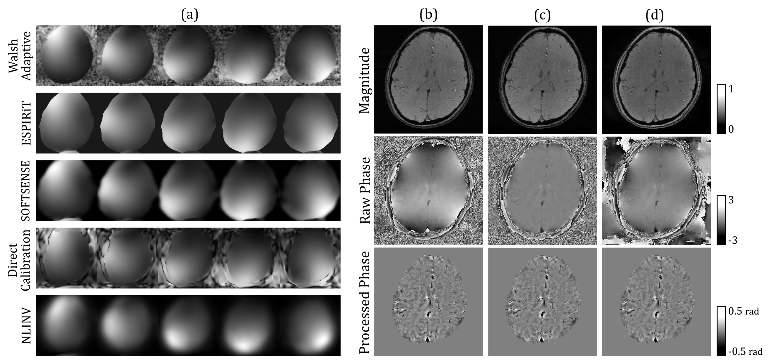
Figure 4. Estimated Sensitivity Maps and Reconstructed Images for the 8-Channel Array. (a) Sensitivity maps of select channels in 8-channel array. (b-d) 8-channel coil-combined magnitude (top row), raw phase (second row) and processed phase (third row) images for select methods. Methods include: (b) Walsh adaptive estimation with simple combination, (c) direct calibration with SENSE combination, and (d) NLINV with L1-regularized combination.
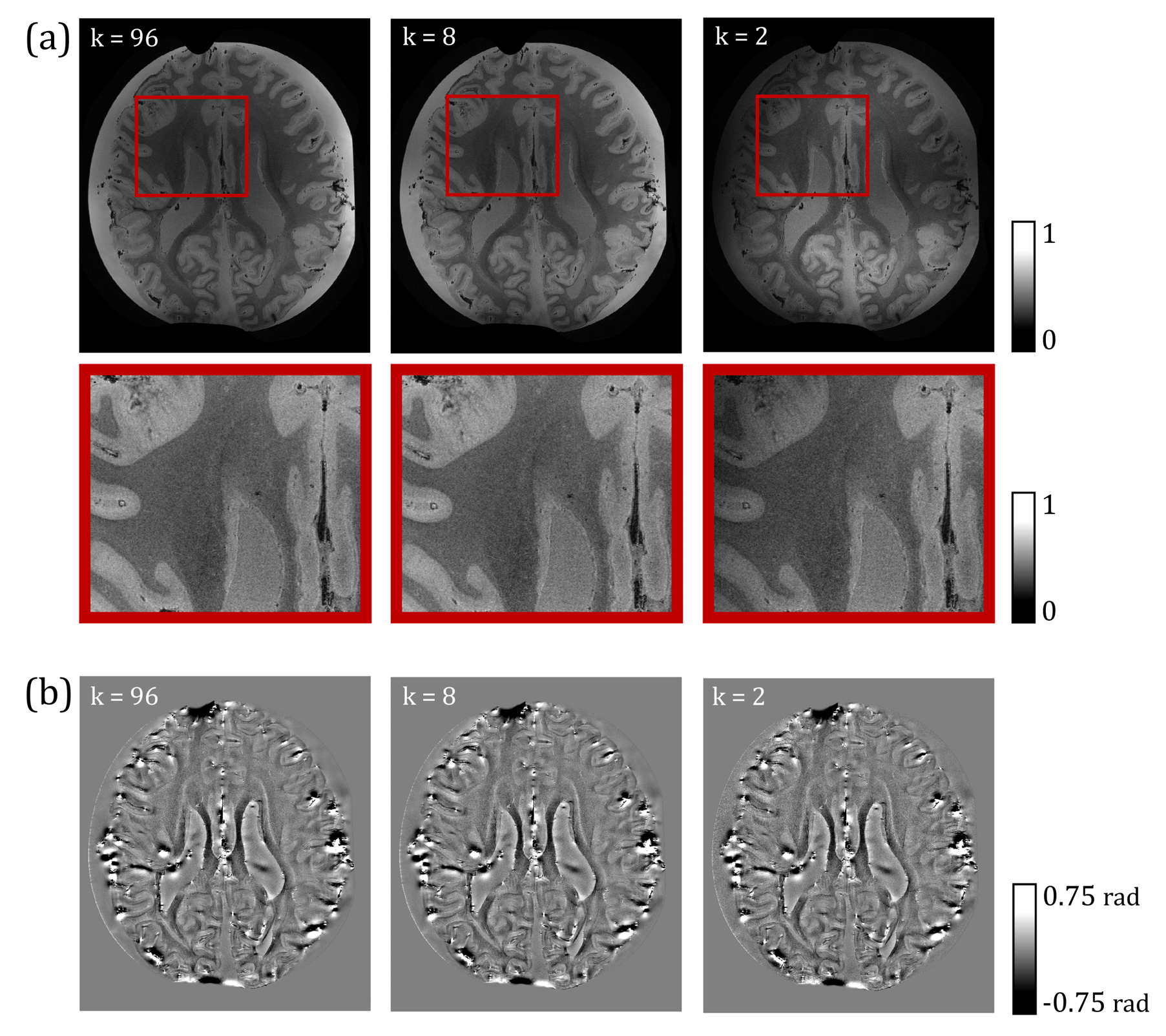
Figure 5. Compressed ESPIRiT Reconstructed Images for the 96-Channel Array. 96-channel array reconstructions with compressed ESPIRiT sensitivity maps followed by simple combination; (a) magnitude images, (b) zoom-in of magnitude images, and (c) processed phase images are displayed for full-rank (left column), rank-8 (center column), and rank-2 (right column) approximations.