1914
Optimizing the T1-mapping GOAL-SNAP MRA with Histogram-Matching-Based Multi-Frame Combination1Center for Biomedical Imaging Research, Tsinghua University, Beijing, China, 2Department of Radiology, Union Hospital, Tongji Medical College, Huazhong University of Science and Technology, Beijing, China, 3Hubei Province Key Laboratory of Molecular Imaging�, Wuhan, China, 4Philips Healthcare, Beijing, China
Synopsis
Keywords: Image Reconstruction, Vessels
Motivation: GOAL-SNAP, a sequence designed for T1 value measurement of vessel walls, shows promise in generating MRA images for intracranial vessel visualization, albeit with opportunities for further improvement.
Goal(s): To introduce a novel approach to optimize GOAL-SNAP MRA.
Approach: A histogram-matching-based approach was developed to optimize GOAL-SNAP MRA. This technique combined multiple GOAL-SNAP frames and utilizes histogram matching to suppress background signals and improve the contrast between vessels and background.
Results: Contrast-to-noise (CNR) and visualization evaluation demonstrated the effectiveness of the optimized GOAL-SNAP MRA, exhibiting comparable performance to TOF and superior visualization of distal vessels.
Impact: A novel histogram-matching-based multi-frame combination approach improved GOAL-SNAP MRA for intracranial vessel visualization and vascular stenosis assessment, with comparable performance to TOF and superior visualization of distal vessels.
Introduction
Non-contrast magnetic resonance angiography (MRA) techniques, such as Time of Flight (TOF)1, 2 and Simultaneous Non-Contrast Angiography and Intraplaque Hemorrhage (SNAP)3, are widely used for the assessment of vascular stenosis. GOAL-SNAP4, an extension of SNAP, which was designed for measuring T1 values of vessel walls, incorporates a 3D golden-angle radial readout, allowing for the reconstruction of multiple 3D image volumes along the inversion recovery (IR) curve. This study introduced a histogram-matching-based approach for optimizing GOAL-SNAP MRA by combining multiple GOAL-SNAP frames.Methods
Study Population and Data AcquisitionThis study was approved by the local institutional review board with all patients having provided written informed consent. Patients with middle cerebral artery (MCA) atherosclerotic plaques were included in this study. Intracranial vessels of all subjects were imaged on a 3T scanner (Ingenia CX, Philips Healthcare, Best, The Netherlands) with a 32-channel head coil, using GOAL-SNAP sequence, with the following scan parameters: FOV = 100x100x100mm3, spatial resolution = 0.7x0.7x0.7mm3, IRTR = 2000 ms, TFE factor = 155. Axial 3D time-of-flight (TOF) MR angiography with FOV = 200x200x84mm3 and spatial resolution = 0.4x0.4x1.2mm3 was acquired for comparison.
GOAL-SNAP Sequence and Multi-Frame MRA
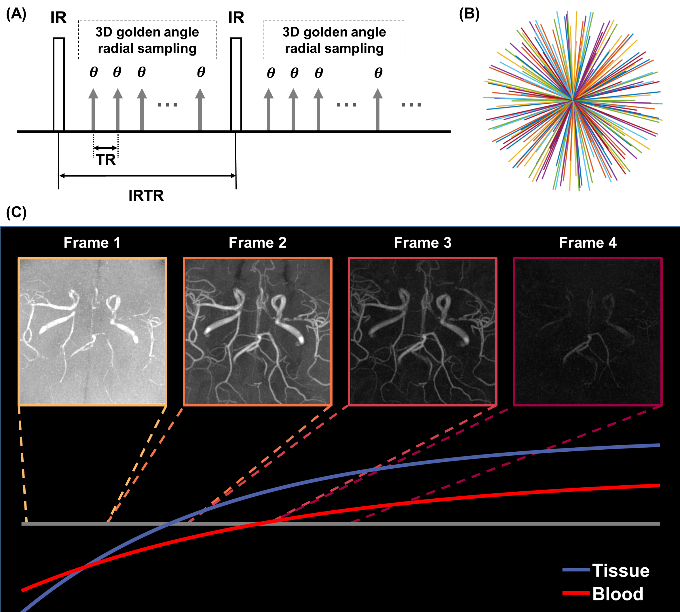
Figure 1 illustrates the GOAL-SNAP sequence diagram, golden-angle sampling trajectory, and simulated signal evolutions of tissue and blood. Following reference4, a TFE factor of 155 and a reconstruction window width of 19 were employed, yielding 8 frames along the IR curve. Real-value signal representation and computation of absolute values for negative signals were implemented, thus rendering blood as positive. The first four frames are shown in Figure 1C. With increasing inversion time, both background and blood signals gradually weaken. Frame 2 exhibited the best vascular visualization, while Frame 3 demonstrated the highest vessel-to-background contrast (Figure 1C).
Optimization of GOAL-SNAP MRA Based on Histogram Matching
Frames 2 and 3 of GOAL-SNAP were utilized for optimizing GOAL-SNAP MRA. Before combining the two frames, full histogram matching5 was utilized to align the input probability density function (PDF) with the reference PDF. This process involved the calculation of the mapping from the input cumulative distribution function (CDF) to the reference CDF and the application of the mapping function to the input distribution. Frame 2 served as the input, while Frame 3 acted as the reference. Then, the optimized GOAL-SNAP MRA was derived by summing the adjusted Frame 2 and Frame 3.
Qualitative and Quantitative Evaluation of the Optimized GOAL-SNAP MRA
For qualitative evaluation, the optimized GOAL-SNAP MRA was compared to TOF for vessel visualization and stenosis detection. For quantitative evaluation, vessel segmentation was performed using threshold segmentation and then the signal-to-noise (SNR)5 and contrast-to-noise (CNR)6 were calculated.
Results
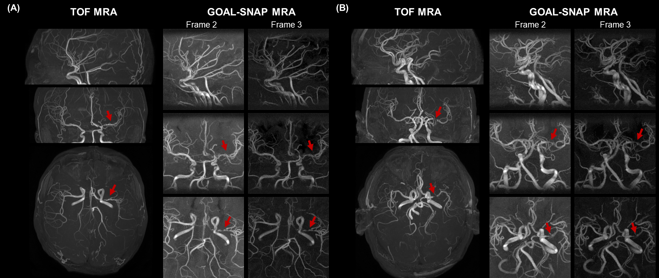
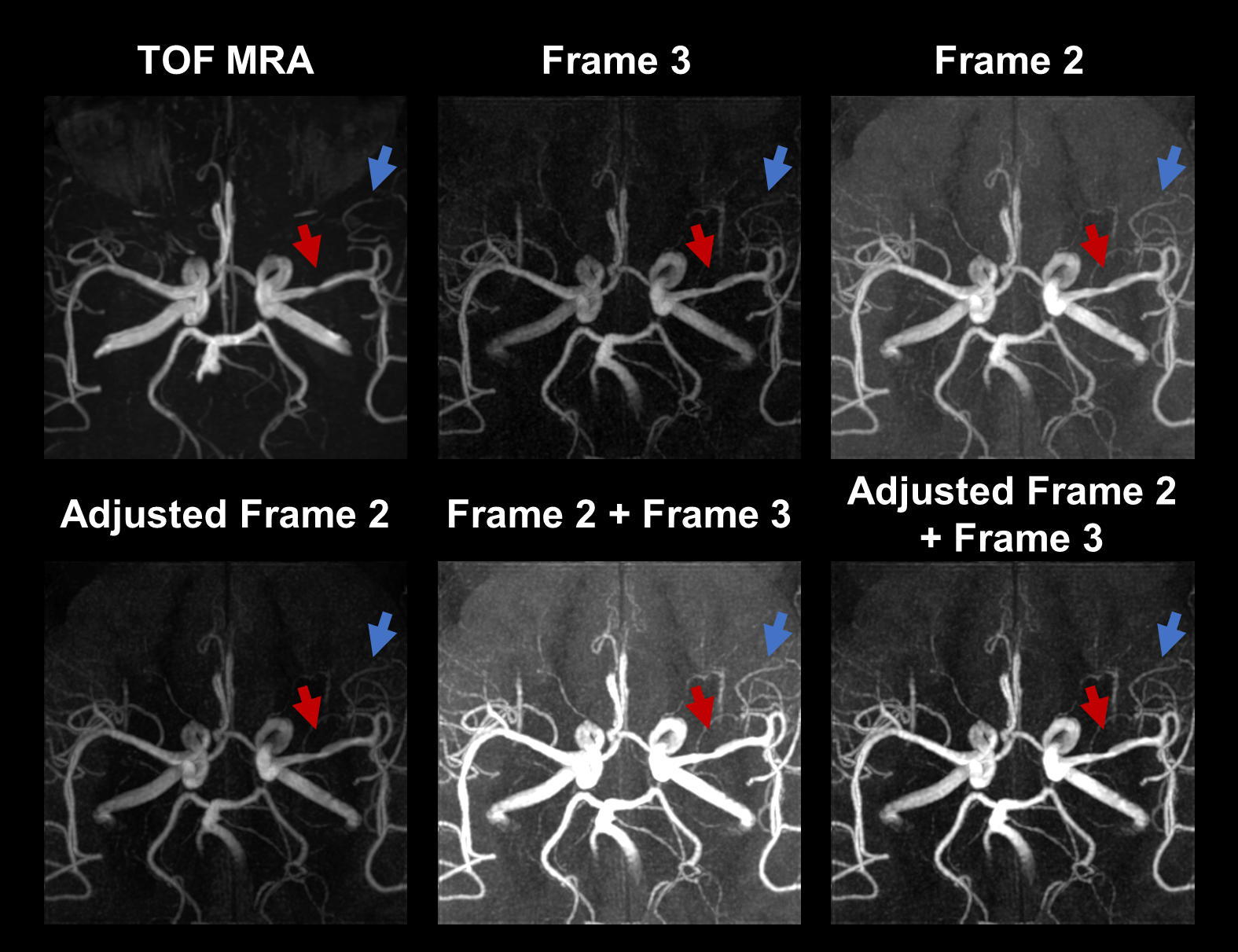
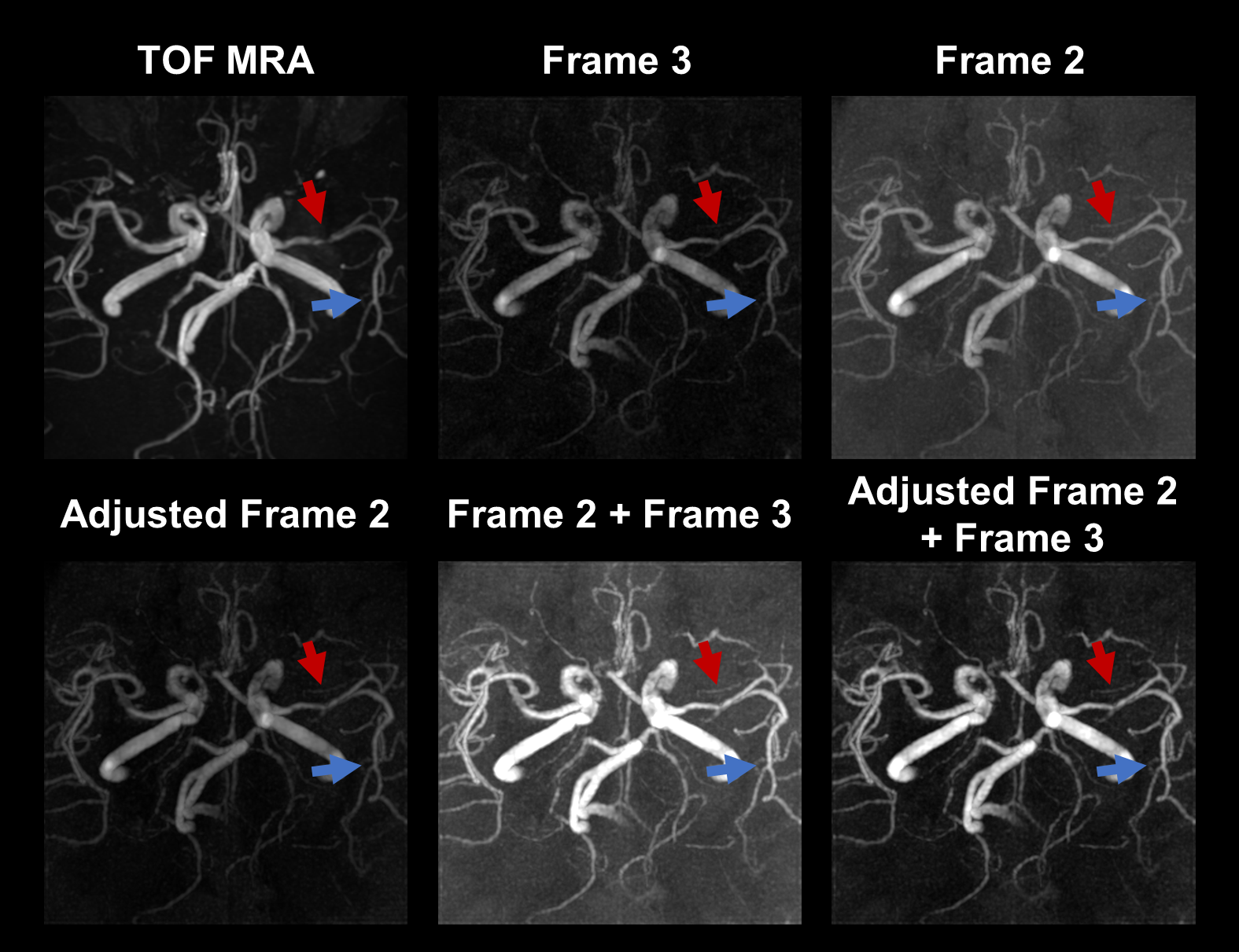
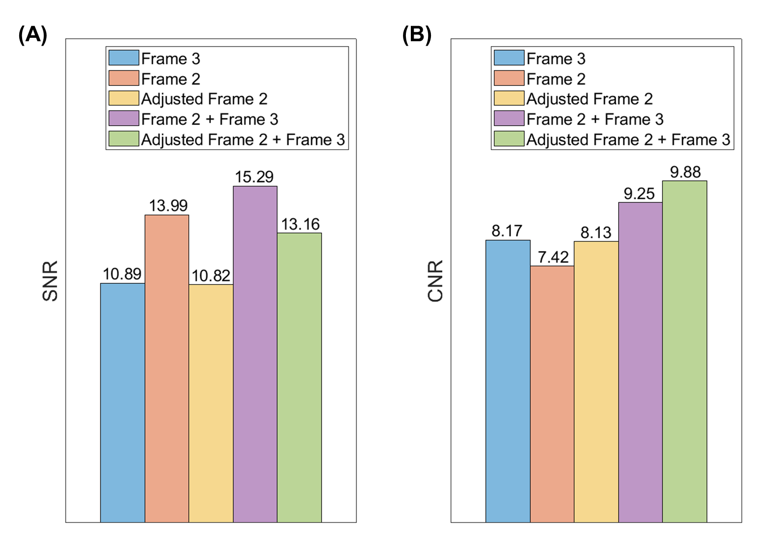
Eighteen patients (44.67 ± 17.61 years; 15 males) were recruited in this study. Figure 2 illustrates three orthogonal directions of GOAL-SNAP multi-frame MRA and TOF. GOAL-SNAP MRA effectively depicted intracranial vessels. Figure 3 presents the comparison of TOF MRA and GOAL-SNAP MRA variants in vessel visualization and stenosis detection. Notably, utilizing histogram matching with Frame 3 as a reference effectively suppressed the background of Frame 2. Furthermore, when compared to simply summing Frame 2 and Frame 3, adjusted_Frame 2 + Frame 3 yielded superior contrast between the vessels and background. The optimized GOAL-SNAP MRA (adjusted_Frame 2 + Frame 3) exhibited comparable capabilities to TOF in detecting stenosis (red arrows). Additionally, GOAL-SNAP MRA outperformed TOF in visualizing distal vessels (blue arrows). Figure 4 illustrates a patient with near-complete occlusion of the left MCA on TOF exhibited good continuity of stenosis in all GOAL-SNAP MRA variants, particularly in the optimized GOAL-SNAP MRA, indicating superior accuracy in stenosis depiction by GOAL-SNAP MRA. Figure 5 presents the SNR and CNR results of all GOAL-SNAP MRA variants. Frame 2 and Frame 2 + Frame 3 outperform adjusted_Frame 2 + Frame 3 in SNR, primarily due to the higher vascular signal in Frame 2 compared to Frame 3 and adjusted_Frame 2. However, in terms of CNR, adjusted_Frame 2 + Frame 3 exhibited the highest values, signifying its advantage in depicting intracranial vessels.Conclusion and Discussion
This study introduced a histogram-matching approach for optimizing GOAL-SNAP MRA, significantly enhancing its capabilities for intracranial vessel visualization and stenosis assessment. The optimized GOAL-SNAP MRA demonstrated superior contrast between vessels and background, as evidenced by visualization and CNR evaluation. Quantitative comparison of the optimized GOAL-SNAP MRA with TOF will be conducted in the future. This MRA technique holds promise for clinical and research applications, offering a valuable tool for diagnosing vascular conditions and potentially improving patient outcomes. Further performance advancements of GOAL-SNAP MRA will particularly focus on expanding the FOV and improving the resolution.Acknowledgements
None.References
1. Heiserman JE, Drayer BP, Keller P, et al. Intracranial vascular stenosis and occlusion: evaluation with three-dimensional time-of-flight MR angiography. Radiology. 1992;185(3):667-73.
2. Sawada M, Yano H, Shinoda J, et al. Symptomatic middle cerebral artery stenosis and occlusion: comparison of three-dimensional time-of-flight magnetic resonance angiography with conventional angiography. Neurologia medico-chirurgica. 1994;34(10):682-5.
3. Wang J, Börnert P, Zhao H, et al. Simultaneous noncontrast angiography and intraplaque hemorrhage (SNAP) imaging for carotid atherosclerotic disease evaluation. Magnetic resonance in medicine. 2013;69(2):337-45.
4. Qi H, Sun J, Qiao H, et al. Carotid intraplaque hemorrhage imaging with quantitative vessel wall T1 mapping: technical development and initial experience. Radiology. 2018;287(1):276-84.
5. Bottenus N, Byram BC, Hyun D. Histogram matching for visual ultrasound image comparison. IEEE transactions on ultrasonics, ferroelectrics, and frequency control. 2020;68(5):1487-95.
6. Shi Z, Zhao X, Zhu S, et al. Time-of-flight intracranial MRA at 3 T versus 5 T versus 7 T: Visualization of distal small cerebral arteries. Radiology. 2023;306(1):207-17.
Figures