1908
The software system of an MR compatible dedicated brain PET1Lauterbur Research Center for Biomedical Imaging, Shenzhen Institute of Advanced Technology, Chinese Academy of Sciences, Shenzhen, China, Shenzhen, China, 2Key Laboratory of Biomedical Imaging Science and System, Chinese Academy of Sciences., Shenzhen, China
Synopsis
Keywords: Image Reconstruction, Image Reconstruction, Simultaneous PET/MRI Imaging, dedicated scanner
Motivation: Dedicated brain PET devices can acquire high-quality images while also allowing for simultaneous imaging with MRI equipment.
Goal(s): The implemention of software system of a MR compatible brain PET including data acquisition, sinogram generation, imaging reconstructionis presented.
Approach: We designed a virtual crystal-based sinogram generation method and implemented OSEM image reconstruction software with various acceleration strategies.
Results: The functionality of the software system and the imaging capability of the PET scanner were demonstrated by simultaneous PET and MRI imaging of the human brain.
Impact: The sinogram generation method and image reconstruction acceleration strategies developed in this work can also be used for other PET scanners using high DOI resolution depth encoding detectors.
Introduction
Positron emission tomography (PET) is a molecular imaging technology that has gained widespread use in both clinical and research settings due to its ability to visualize the metabolic processes of the human body for the early diagnosis of various diseases [1-4]. Dedicated brain PET scanners achieve higher spatial resolution, increased sensitivity, and lower cost by reducing detector ring diameters compared to the most popular whole-body PET scanners, and they can also be seamlessly integrated into standard whole-body MR scanners for simultaneous dual-modality brain imaging if the detectors and electronics are carefully designed to be MR-compatible [5-7]. An MR-compatible brain PET scanner named SIAT bPET (Shenzhen Institute of Advanced Technology brain PET) was recently developed in our laboratory to simultaneously achieve high spatial resolution and sensitivity [8].The SIAT bPET scanner
SIAT bPET has 224 depth-encoding detectors with dual-ended readoutreadouts of segmented crystal arrays. The detectors are arranged in eight circular rings, each with 28 detectors, providing SIAT bPET with an axial field of view (FOV) of 329 mm. Each LYSO array has 26×26 LYSO crystals with a size of 1.4×1.4×20 mm3 and is read out by two 10×10 Hamamatsu silicon photomultiplier (SiPM) arrays placed at the opposite ends. The active area of the SiPM pixel is 3×3 mm2, and the pitch of the SiPM array is 4 mm. In total, SIAT bPET consists of 151,424 crystal elements arranged in 208 crystal rings with 728 crystals per ring and 44800 SiPM pixels. 16 Depth of Interaction (DOI) bins are used during PET scans based on a DOI resolution of ~2 mm of the detectors [9]. A photo of the SIAT bPET scanner is shown in Fig. 1.Sinogram generation
SIAT bPET has 151,424 crystals, a higher number than many whole-body PET scanners. If each DOI bin is regarded as a crystal, the size of the sinogram would be 16×16 times larger than that of a PET scanner without DOI measurement. To compress the size of the sinogram, a novel virtual crystal-based sinogram generation method was implemented. A virtual crystal ring with a diameter the same as the distance between the front of the two opposite crystal arrays (376.8 mm) of the SIAT bPET scanner and consisting of 800 virtual crystals of the same width as the crystal pitch of SIAT bPET detectors (1.48 mm) and 0 mm length was defined as shown in Fig 5. In the axial direction, two virtual crystals were inserted into the gaps between the detectors, leading to 222 virtual crystal rings. In each plane, a sinogram of 358 radial bins with a bin size of half of the virtual crystal width (0.74 mm) and 400 angle bins in 180° was generated using the virtual crystals, leading to a transaxial FOV of 265 mm. Thus, the final sinogram dimension is 358×400×222×222.Image reconstruction
As Fig 4 shown, to implement the OSEM reconstruction, the projection data are divided into 20 subsets according to the angle in the sinogram. During each forward-projection and back-projection operation, one subset of data is input to the GPU. Algorithm acceleration strategies, including system geometric symmetry on both axial and transaxial directions, precomputation of LOR-driven ray-tracing and the use of texture memory, are applied in the reconstruction [10]. In addition, the CUDA thread allocation parameters such as the block and thread sizes are optimized based on the performance of the GPU parallel calculation.Result
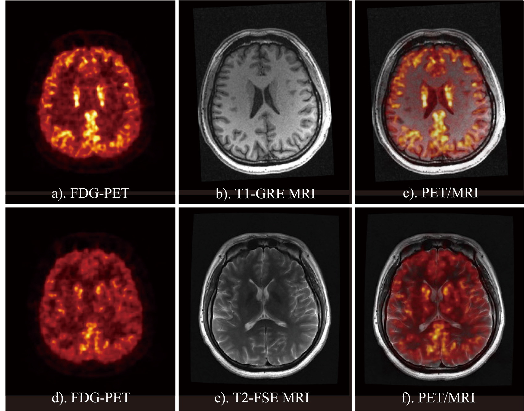
Fig 4 shows 6 transverse slices of the 3D Hoffman brain phantom image. Detailed structures of the phantom were clearly observed without major artifacts. Fig 5 shows simultaneous PET/MRI imaging of the human brain.Discussion and conclusion
In this work, the SIAT bPET software system was introduced. A virtual crystal-based sinogram generation method was developed to reduce the sinogram size to reduce the computational complexity of image reconstruction. Acceleration strategies based on GPU parallel computation were developed to accelerate OSEM image reconstruction. Quantitative evaluation of the spatial resolution loss can be performed in the future by developing a list mode-based imaging reconstruction algorithm that uses accurately measured LORs. The sinogram generation method and image reconstruction acceleration strategies developed in this work can also be used for other PET scanners using high DOI resolution depth encoding detectors.Acknowledgements
This work was supported by the National Natural Science Foundation of China (82372038), the Shenzhen Excellent Technological Innovation Talent Training Project of China (RCJC20200714114436080), the Key Laboratory for Magnetic Resonance and Multimodality Imaging of Guangdong Province (2023B1212060052) and the Shenzhen Science and Technology Program (JCYJ20220818101804009).References
[1] Cherry, S.R., The 2006 Henry N.Wagner lecture: Of mice and men (and positrons) - Advances in PET imaging technology. Journal of Nuclear Medicine, 1735-1745, 2006. [2] Cherry, S.R., Total-body imaging: Transforming the role of positron emission tomography. Science Translational Medicine, 9:381, 2017. [3] Vandenberghe S, Moskal P, Karp JS. State of the art in total body pet. EJNMMI Physics. 7(1),2020. [4] Jones, T. and D. Townsend, History and future technical innovation in positron emission tomography. Journal of Medical Imaging, 4(1), 2017. [5] Catana C. Development of dedicated brain pet imaging devices: Recent advances and future perspectives. Journal of Nuclear Medicine, 60(8):1044-1052, 2019. [6] Jones, T. and E.A. Rabiner, The development, past achievements, and future directions of brain PET. Journal of Cerebral Blood Flow and Metabolism, 32(7):1426-1454, 2012. [7] Kolb A, Wehrl HF, Hofmann M, et al. Technical performance evaluation of a human brain PET/MRI system. European Radiology 22(8):1776-1788, 2012. [8] Kuang Z, Sang Z, Ren N, et al. Development and performance of SIAT BPET: A high-resolution and high-sensitivity MR-compatible brain pet scanner using dual-ended readout detectors. European Journal of Nuclear Medicine and Molecular Imaging. Published online 2023. [9] Yang Q, Kuang Z, Sang Z, Yang Y, Du J. Performance comparison of two signal multiplexing readouts for SIPM-based pet detector. Physics in Medicine Biology, 64(23), 2019. [10] Meng F, Wang J, Zhu S, Cheng J, Liang J, Tian J. Comparison of GPU reconstruction based on different symmetries for dual-head pet. Medical Physics, 46(6):2696-2708, 2019.Figures