1906
Improvement in Fat-Water Separation Using Modeled Gradient Impulse Response with Two-Point Dixon Radial Imaging1Medical Physics, UW Madison, Madison, WI, United States, 2Radiology, UW Madison, Madison, WI, United States
Synopsis
Keywords: Image Reconstruction, Image Reconstruction, GIRF, Radial, PDFF, 2-Point Dixon
Motivation: Radial imaging is desirable for whole body imaging due to its motion robustness. However, two-point Dixon with bipolar readouts can have reduced quality due to gradient errors.
Goal(s): To develop improved imaging capabilities by leveraging gradient impulse response function (GIRF) measurements to correct for gradient errors and yield better image quality.
Approach: A radial two-point Dixon acquisition was compared using GIRF-corrected and uncorrected reconstructions in phantoms and a human volunteer.
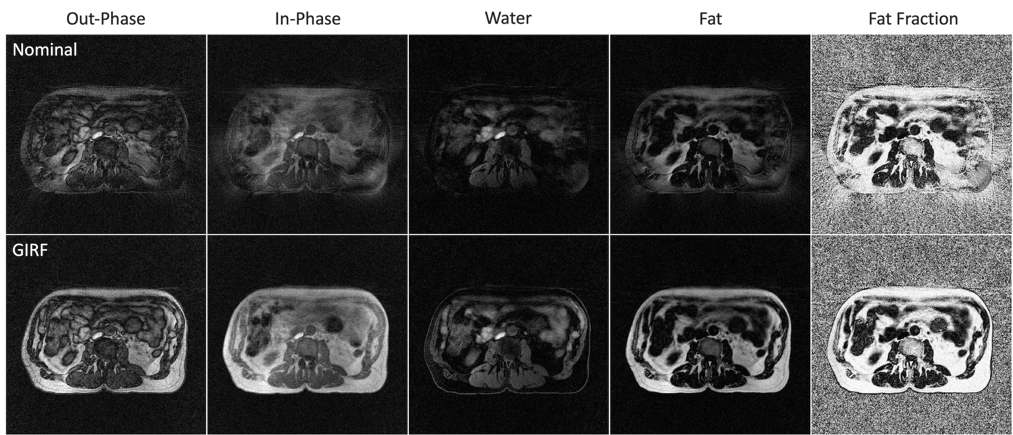
Results: The use of GIRF in the reconstruction of two-point Dixon radial imaging provides improved image quality and better fat-water separation and provided similar quality to a Cartesian acquisition.
Impact: This work demonstrates improved image quality and improved fat-water separation by incorporating gradient impulse response function compensation in two-point Dixon radial imaging for an intended application of motion-robust whole body MR imaging.
Introduction
Radial imaging provides important advantages for body MR imaging due to its inherent robustness to motion1. Likewise, chemical shift encoding (CSE) methods like two-point Dixon offer robust separation and suppression of water and fat signal compared to alternative approaches2. However, radial imaging is sensitive to gradient system imperfections and can lead reduced image quality. This is particularly true for CSE methods where water-fat separation capability is reduced due to image phase errors resulting from gradient eddy currents and other imperfections. The purpose of this work was to evaluate gradient impulse response function (GIRF) modeling to demonstrate robust water-fat separation in body MRI using radial acquisitions.Methods
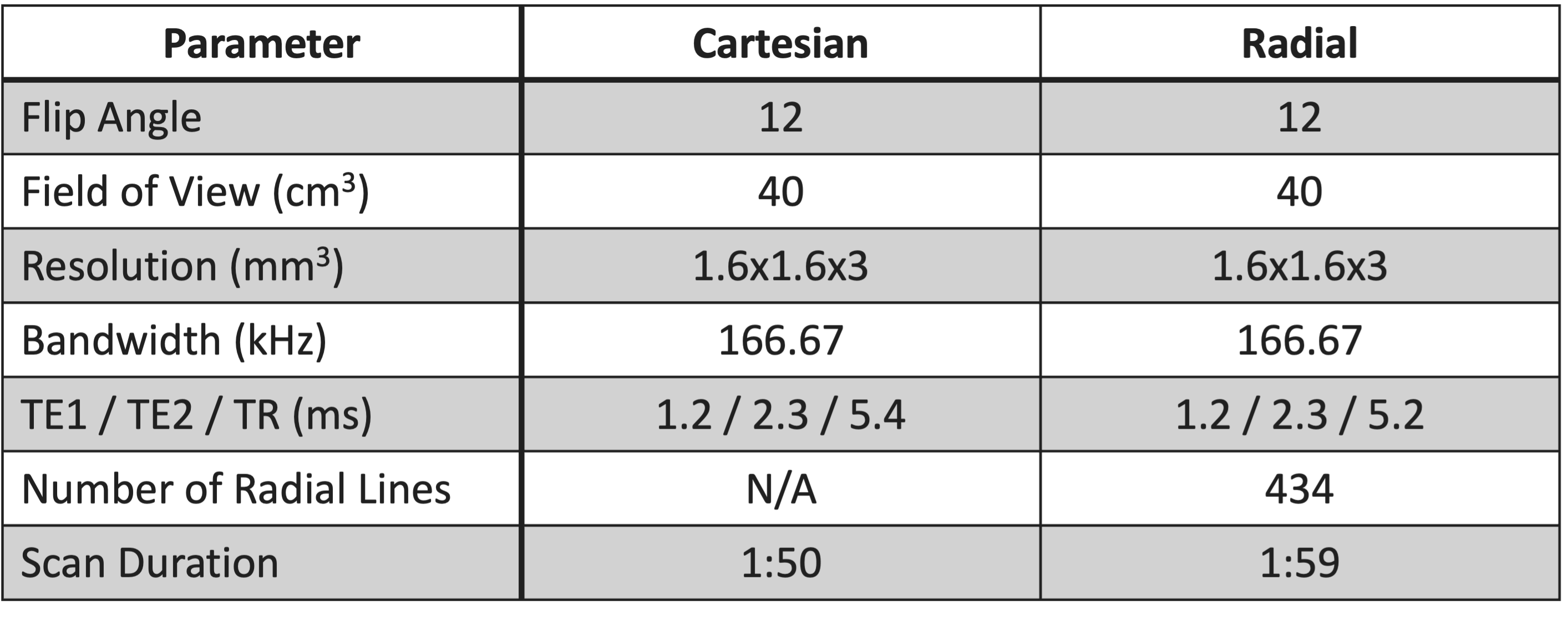
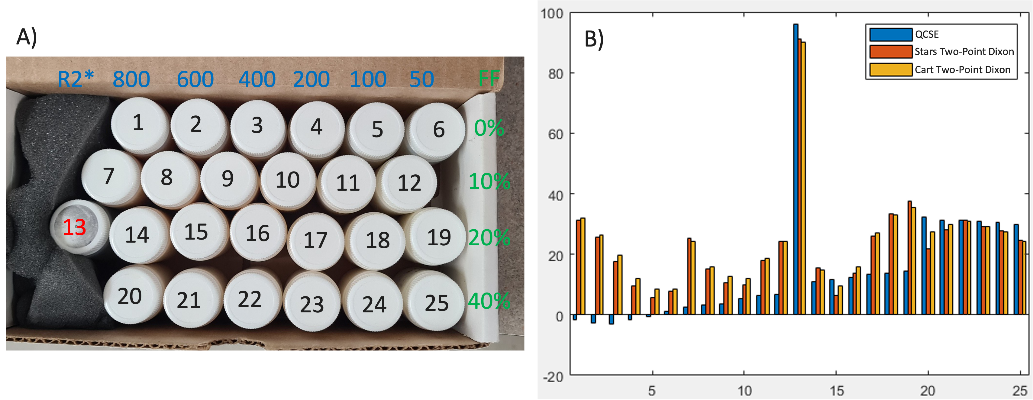
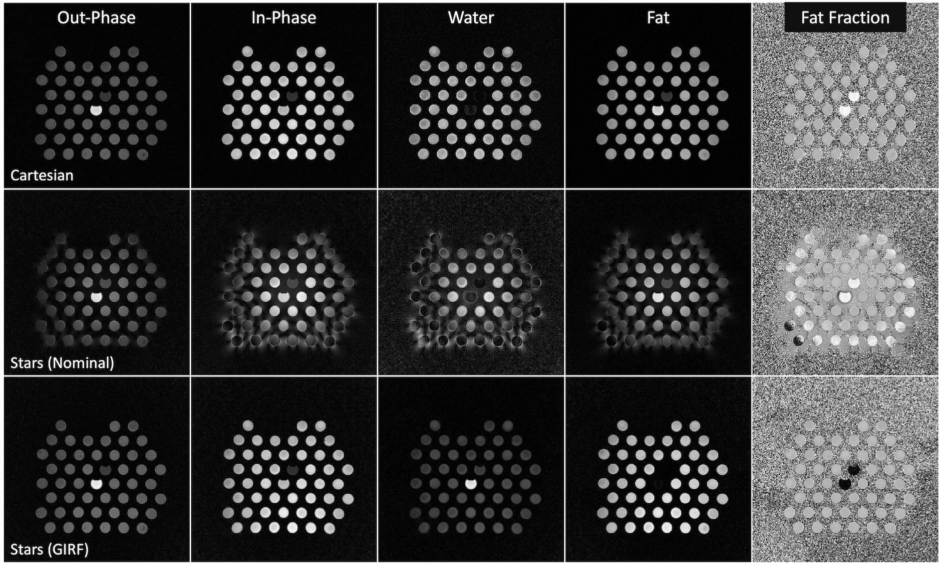
All experiments were conducted on a 3T PET/MR scanner (GE Signa PET/MR). GIRF was measured using a previously reported image-based approach3. Scans using Cartesian and radial (Stars) two-point Dixon were obtained in two types of phantoms and a research volunteer. Scan parameters are provided in Figure 1. To evaluate fat fraction quantification accuracy, fat-water phantom vials were used with varied fat-water fractions and R2* values. Reference values were obtained using the vendor’s quantitative CSE (QCSE) acquisition (IDEAL-IQ). The configuration of these vials along with their fat fractions and R2* values can be seen in Figure 1. To determine the consistency of the GIRF in a larger FOV, 40% fat fraction vials were evenly spaced to take up a larger field of view. In vivo scanning was performed on the abdomen of a research volunteer under a locally-approved IRB. All radial images were reconstructed with both the nominal and GIRF-corrected k-space trajectory. Cartesian, Radial-Nominal, and Radial-GIRF images were compared for image quality and fat fraction equivalence.Results
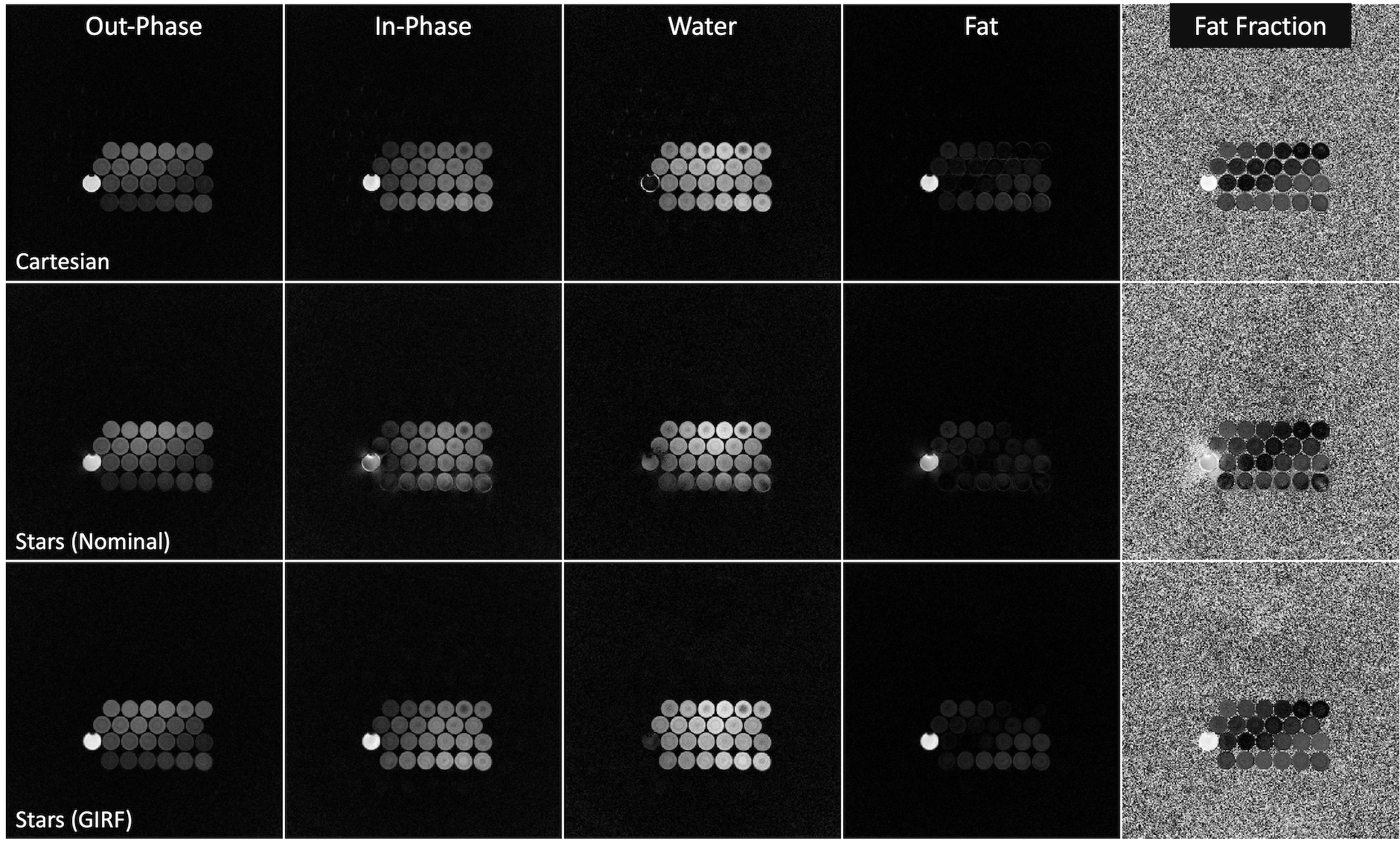
In the analysis of the first set of phantoms depicted in Figure 2, it was observed that uncorrected radial images exhibited prominent blurring artifacts. Without the application of the GIRF correction, these artifacts led to notable deviations in fat fraction values beyond the vial boundaries. Upon implementing GIRF correction on the radial images, the fat fraction differences were found to be around 4.47%. Region of interests (ROIs) were generated for each vial and their fat fractions are illustrated in Figure 3B. The comparison of the Cartesian and radial fat fraction images reveal that they produce similar fat fraction values. These values however are different from the quantitative CSE images. To assess the consistency of the GIRF correction near the edges of the field of view, another phantom was used. As shown in Figure 4, fat fraction differences between Cartesian and radial images remained consistent with differences found to be around 4.14%. An in-vivo image was acquired to evaluate the practicality of this method in human subjects. Notably, the uncorrected radial images show severe blurriness as seen in Figure 5. The application of the GIRF correction made a significant improvement in image quality. Visual inspection of the fat fraction maps demonstrates a marked improvement with many of the blurring artifacts mitigated.Discussion and Conclusion
In this work, we utilized GIRF to improve the quality of reconstruction in radial chemical shift encoded two-point Dixon imaging. With the use of GIRF, radial images were equivalent in fat-water separation to a Cartesian acquisition while benefitting from the motion robust characteristics of radial. While the fat quantification was similar between the Cartesian and radial acquisitions, it did not match well with the QCSE acquisition. This is likely a limitation of 2-point Dixon not being able to account for the R2* effects. In conclusion, GIRF correction can improve the quality of chemical shift encoded radial imaging. By compensating for gradient-induced phase errors, Radial-GIRF allows for robust water-fat separation, even in the presence of abdominal motion. This improvement opens the door for more reliable and motion-resistant imaging in the abdomen and other body regions where patient movement is prevalent, which is valuable for whole body PET/MR imaging.Acknowledgements
No acknowledgement found.References
1. Glover GH, Pauly JM. Projection reconstruction techniques for reduction of motion effects in MRI. Magn Reson Med. 1992 Dec;28(2):275-89. doi: 10.1002/mrm.1910280209. PMID: 1461126.
2. Ma J. Dixon techniques for water and fat imaging. J Magn Reson Imaging. 2008 Sep;28(3):543-58. doi: 10.1002/jmri.21492. PMID: 18777528.
3. Jang H, McMillan AB. A rapid and robust gradient measurement technique using dynamic single-point imaging. Magn Reson Med. 2017 Sep;78(3):950-962. doi: 10.1002/mrm.26481. Epub 2016 Oct 3. PMID: 27699867; PMCID: PMC5378680.
Figures