1904
Enhancing Self-Navigated Interleaved Spiral with ESPIRiT (eSNAILS)1Key Laboratory for Biomedical Engineering of Ministry of Education, Department of Biomedical Engineering, College of Biomedical Engineering & Instrument Science, Zhejiang University, Hangzhou, China, 2Athinoula A. Martinos Center for Biomedical Imaging, Massachusetts General Hospital, Charlestown, MA, United States, 3Department of Radiology, Harvard Medical School, Boston, MA, United States, 4Department of Radiology, Juntendo University, Tokyo, Japan, 5Department of Radiology, The University of Tokyo, Tokyo, Japan, 6Brigham and Women's Hospital, Harvard Medical School, Boston, MA, United States, 7Harvard/MIT Health Sciences and Technology, Cambridge, MA, United States
Synopsis
Keywords: Image Reconstruction, Data Acquisition, multi-shot; self-navigation
Motivation: Current methods for estimation of shot-to-shot phase variations in multi-shot DWI may not fully exploit the correlations in data.
Goal(s): To propose a method which efficiently uses correlations between shots and coils to calculate composite sensitivities and then improve multi-shot DWI reconstruction.
Approach: A multi-shot, dual density spiral sequence was designed, with each shot having fully sampled k-space center and undersampled periphery. The center of all shots and all coils are concatenated and fed into ESPIRiT to estimate sensitivities.
Results: The proposed method successfully estimated shot-to-shot phase variations and yielded comparable results to the reference locally low-rank regularized reconstruction which requires parameter tuning.
Impact: The proposed eSNAILS demonstrated the ability of estimating composite sensitivities that incorporate shot-to-shot phase variations. Compared to low-rank modeling methods that assume phase smoothness, eSNAILS can handle cases where there are abrupt phase changes and does not require parameter tuning.
Introduction
In diffusion MRI (dMRI), multi-shot encoding is used for segmenting the readout period into multiple portions to mitigate relaxation-related blurring, B0 inhomogeneity artifacts. In echo planar imaging (EPI), this also helps reduce the echo time (TE) of the acquisition. Despite these advantages, subject's motion during diffusion encoding gradients leads to shot-to-shot phase variations. When combining multi-shot data, significant artifacts arise if these phase variations are not properly handled. A straightforward way to address phase variations is to first estimate the phase contribution of each shot then remove them, and finally combine the phase-corrected shots[1]. Navigators are usually used to estimate the phase variations. Navigators can either be in the form of an extra readout immediately before/after imaging data or directly derived from the imaging data itself (self-navigation)[2,3]. After getting phase variation, it can be removed by subtracting it from imaging data[3]. However, this direct subtraction may have residual artifacts for undersampled trajectories. Alternatively, the phase variation can be deemed as an encoding function and combined with coil sensitivity map to form composite sensitivity profiles[4]. Local/Hankel low-rank approaches have also been developed to perform navigation-free multi-shot reconstruction via regularization[5,6]. An important application of self-navigation in multi-shot spiral imaging is SNAILS[4]. Here, phase variations and coil sensitivities are directly estimated from a low-resolution image obtained from k-space center, which may fail to fully harness the phase variations for image encoding. We propose eSNAILS where we use ESPIRiT[7] to estimate the composite sensitivities in multi-shot spiral data and combine this with a dual density trajectory design. This enables high fidelity diffusion MRI reconstruction while obviating the need for regularization parameter tuning.Theory
Motion-induced phase variation between shots can be incorporated into coil sensitivity, forming composite sensitivities. Thus, each shot has its own sensitivity profile. The encoding model can be written as $$s=Am$$$${\bf{A}}=\left({\begin{array}{*{20}{c}}{{e^{-i{k_{11}}{r_1}}}{S_{11}}({r_1})}&\ldots&{{e^{-i{k_{11}}{r_{{N^2}}}}}{S_{11}}({r_{{N^2}}})}\\\vdots&{{e^{-i{k_{ln}}{r_\rho}}}{S_{nj}}({r_\rho})}&\vdots\\{{e^{-i{k_{{N_L}{N_s}}}{r_1}}}{S_{{N_s}{N_c}}}({r_1})}&\cdots&{{e^{-i{k_{{N_L}{N_s}}}{r_{{N^2}}}}}{S_{{N_s}{N_c}}}({r_{{N^2}}})}\end{array}}\right)$$where N is the matrix size, l, n, j is the k-space sample, shot, coil index, respectively. While NL, NS, and NC represent the #samples per shot, #shots, and #coils respectively. Snj is the composite sensitivity of the j-th coil at the n-th shot. The acquired signal s is of size NL×NS×NC-by-1.
Methods
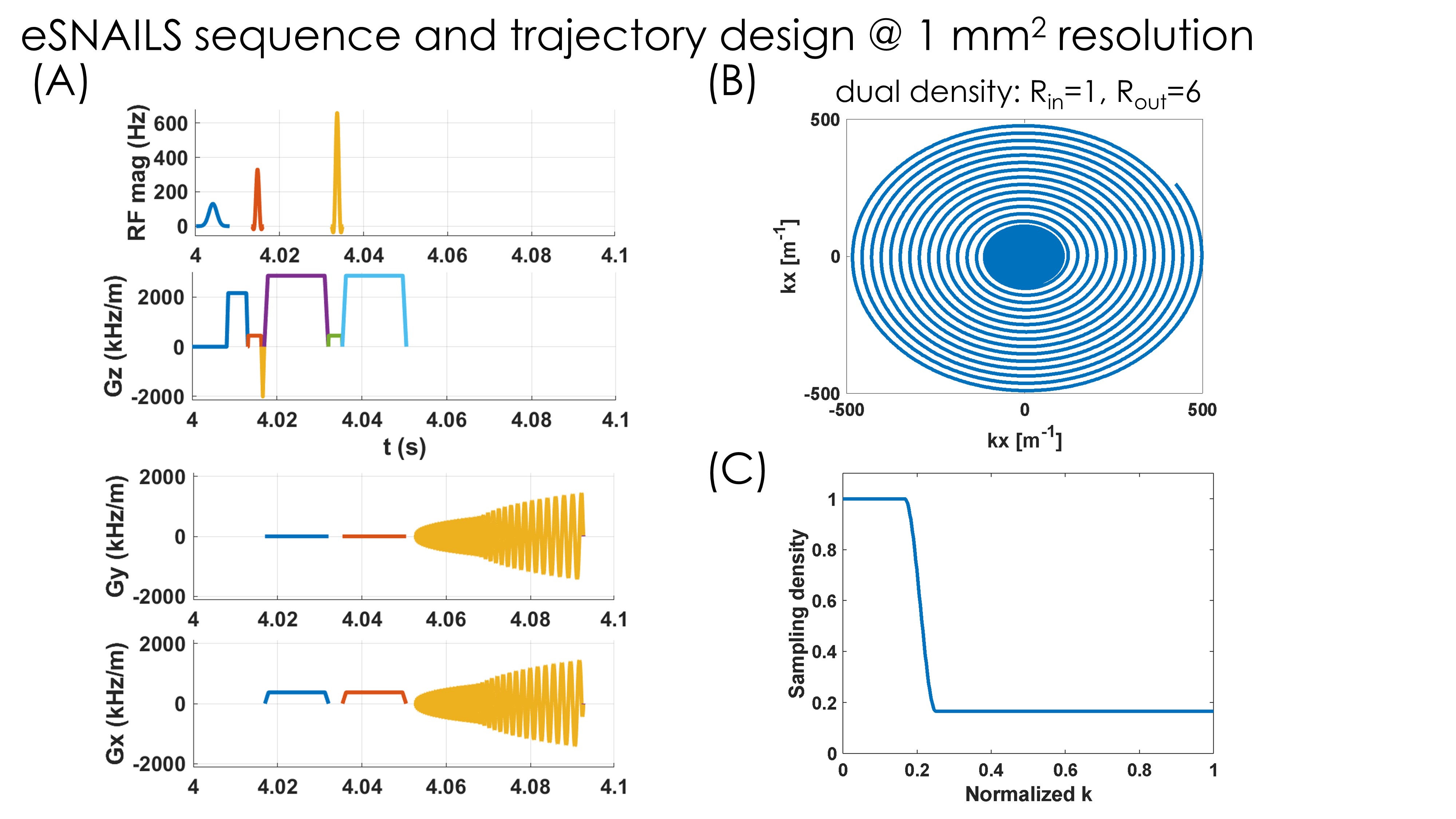
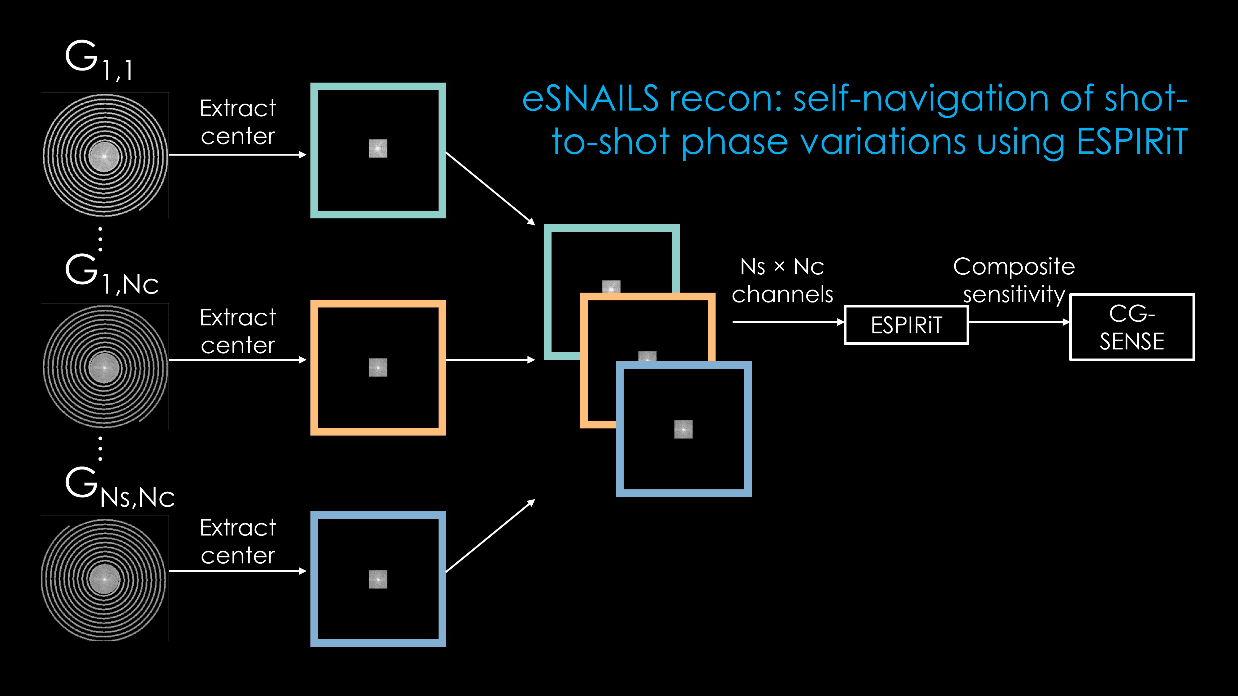
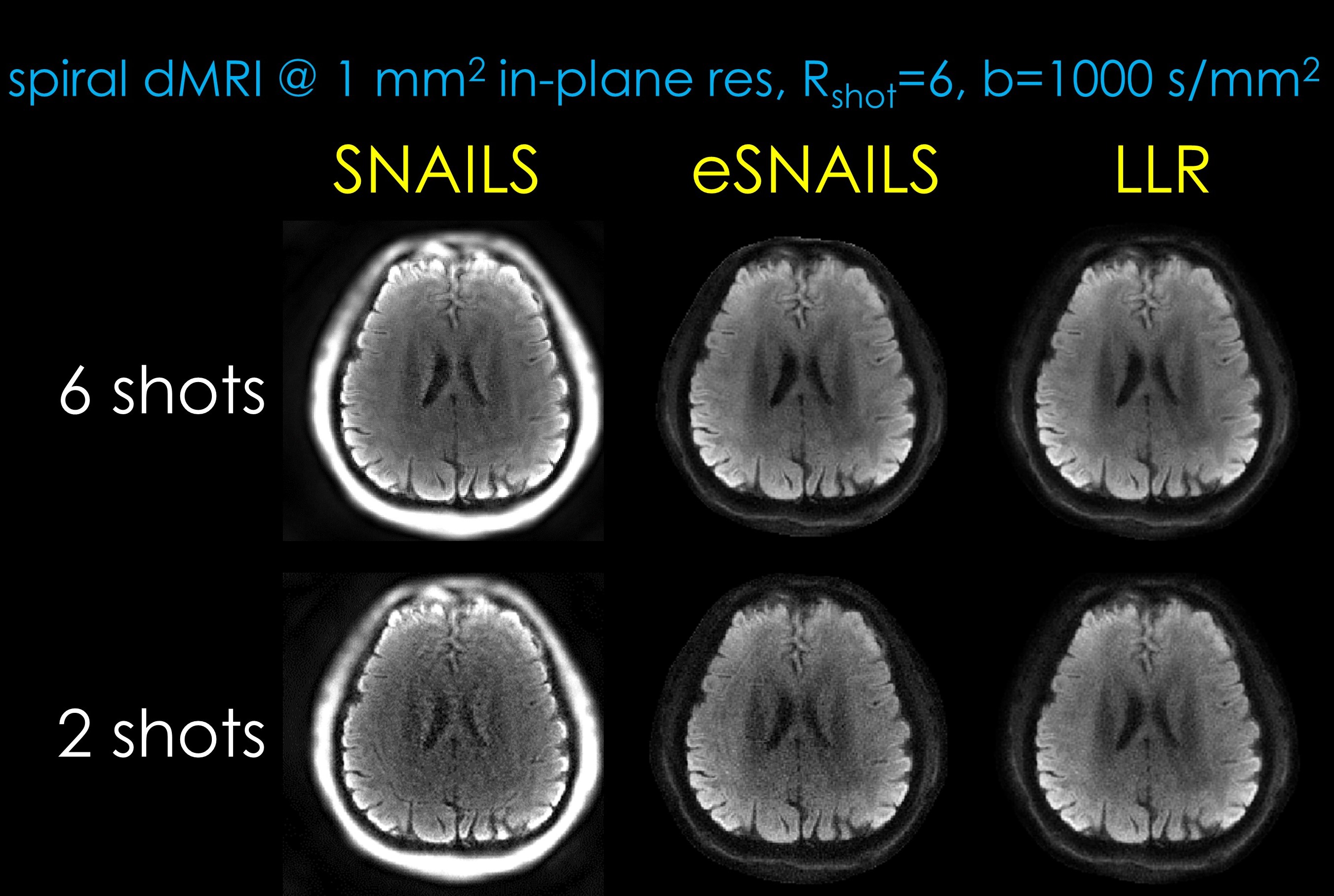
A multi-shot DWI sequence with dual density spiral readout was implemented with the open-source framework Pulseq[8]. Fig.1A shows the 2nd shot. The 6-interleave spiral was generated using time-optimal gradient design[9]. As shown in Fig.1B and Fig.1C, for k-space center, each interleave satisfies Nyquist criterion, while for k-space periphery, all interleaves combined satisfies that. The sequence, reconstruction code, and raw data can be accessed from https://anonymous.4open.science/r/esnails-2577. Healthy volunteers were imaged on a 3T Siemens Prisma scanner. The acquisition parameters for the DWI were TR=4000ms, TE=38ms, resolution=1x1mm2, slice thickness=3mm, #shots=6, spiral readout duration=39.6ms, b-value=1000s/mm². GRE images were acquired to estimate conventional coil sensitivity maps. Image reconstruction was conducted in Matlab with BART[10], using 3 methods, proposed eSNAILS, original SNAILS[4], and locally low rank (LLR)[6]. For eSNAILS, composite sensitivities were estimated from 32-by-32 k-space center using ESPIRiT[7]. Specifically, as shown in Fig.2, the data from n-th interleave and j-th coil Gn,j was first gridded onto Cartesian grid then masked to keep only k-space center, at last all interleaves from all coils are stacked in the channel dimension with NS×NC channels and fed into ESPIRiT. The original SNAILS used the low-resolution image reconstructed from the k-space center as composite sensitivities. Both eSNAILS and original SNAILS used CG-SENSE[11] for image reconstruction. The LLR used sensitivity maps from reference GRE.Results
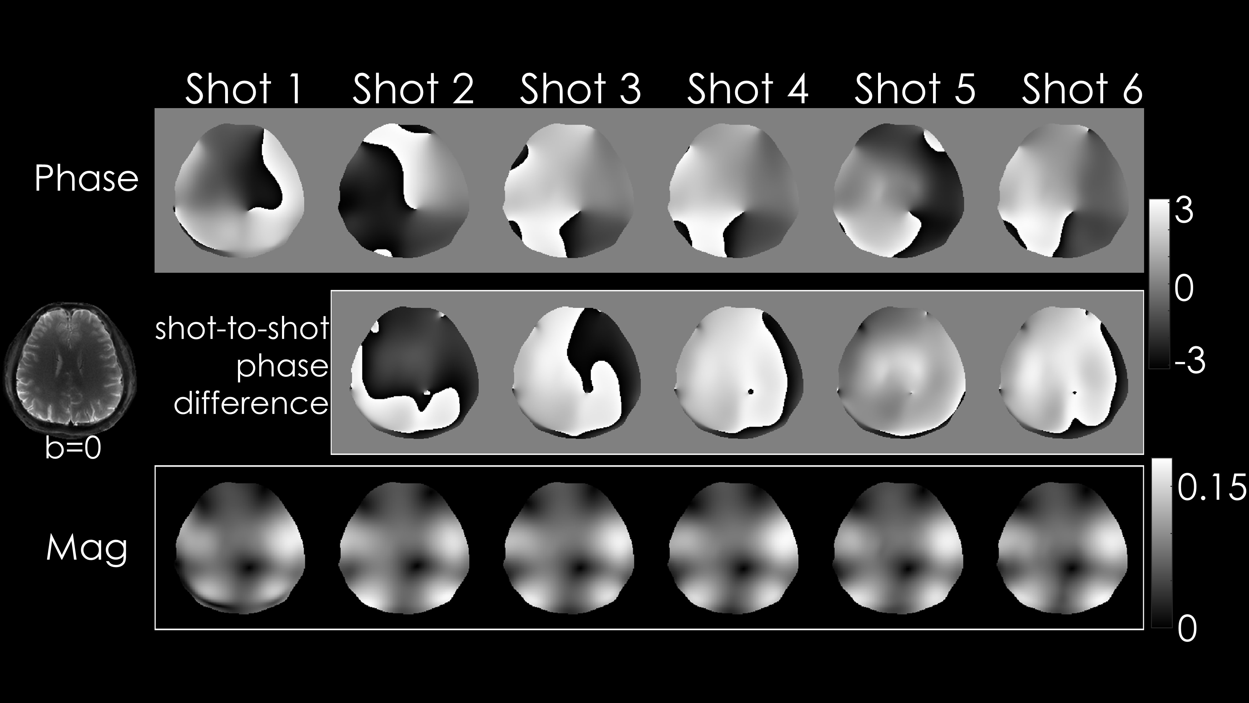
Fig.3 shows phase and magnitude of the estimated composite sensitivities using ESPIRiT, along with phase difference. The phase images clearly depicted the shot-to-shot phase variations, and as expected the magnitude of the sensitivity profiles were similar between shots. Fig.4 illustrates reconstructed multi-shot data using different methods. The SNAILS method yielded bright signals around the scalp due to mis-estimation of sensitivity profile around these regions. While eSNAILS and LLR yielded similar results, eSNAILS did not make use of any regularization.Discussion and conclusion
We proposed to use ESPIRiT to improve the composite sensitivity map estimation in multi-shot spiral dMRI. The resulting sensitivity maps captured the shot-to-shot phase variations without the need for any additional low-pass filtering (as in SNAILS) or parameter tuning. By incorporating this composite sensitivity into encoding model, high-quality distortion-free diffusion weighted images can be obtained by conventional CG-SENSE. The dual density spiral design provided a convenient and efficient way to extract the phase variation information. Possible applications of the proposed method include diffusion relaxometry with spiral, where there might be dead time between equidistant echoes. This dead time can be allocated to acquire the redundant k-space center.Acknowledgements
This work was supported by research grants NIH R01 EB028797, U01 EB025162, P41 EB030006, U01 EB026996, R03 EB031175, R01 EB032378, UG3 EB034875, and NVidia Corporation for computing support. National Natural Science Foundation of China: 81971605. Key R&D Program of Zhejiang Province: 2022C04031.References
[1] Miller KL, Pauly JM. Nonlinear phase correction for navigated diffusion imaging. Magn Reson Med 2003;50(2):343-353.
[2] Pipe JG, Farthing VG, Forbes KP. Multishot diffusion-weighted FSE using PROPELLER MRI. Magn Reson Med 2002;47(1):42-52.
[3] Liu C, Bammer R, Kim DH, Moseley ME. Self-navigated interleaved spiral (SNAILS): application to high-resolution diffusion tensor imaging. Magn Reson Med 2004;52(6):1388-1396.
[4] Liu C, Moseley ME, Bammer R. Simultaneous phase correction and SENSE reconstruction for navigated multi-shot DWI with non-cartesian k-space sampling. Magn Reson Med 2005;54(6):1412-1422.
[5] Mani M, Jacob M, Kelley D, Magnotta V. Multi-shot sensitivity-encoded diffusion data recovery using structured low-rank matrix completion (MUSSELS). Magn Reson Med 2017;78(2):494-507.
[6] Hu Y, Levine EG, Tian Q, Moran CJ, Wang X, Taviani V, Vasanawala SS, McNab JA, Daniel BA, Hargreaves BL. Motion-robust reconstruction of multishot diffusion-weighted images without phase estimation through locally low-rank regularization. Magn Reson Med 2019;81(2):1181-1190.
[7] Uecker M, Lai P, Murphy MJ, Virtue P, Elad M, Pauly JM, Vasanawala SS, Lustig M. ESPIRiT--an eigenvalue approach to autocalibrating parallel MRI: where SENSE meets GRAPPA. Magn Reson Med 2014;71(3):990-1001.
[8] Layton KJ, Kroboth S, Jia F, Littin S, Yu H, Leupold J, Nielsen JF, Stocker T, Zaitsev M. Pulseq: A rapid and hardware-independent pulse sequence prototyping framework. Magn Reson Med 2017;77(4):1544-1552.
[9] Lustig M, Kim SJ, Pauly JM. A fast method for designing time-optimal gradient waveforms for arbitrary k-space trajectories. IEEE Transactions on Medical Imaging 2008;27(6):866-873.
[10] Uecker M, Ong F, Tamir JI, Bahri D, Virtue P, Cheng JY, Zhang T, Lustig M. Berkeley advanced reconstruction toolbox. 2015.
[11] Pruessmann KP, Weiger M, Bornert P, Boesiger P. Advances in sensitivity encoding with arbitrary k-space trajectories. Magn Reson Med 2001;46(4):638-651.
Figures