1902
Spatiotemporally Encoded Single-Sided MRI with a Numerical Point-Spread Function Reconstruction1Promaxo, Oakland, CA, United States, 2Case Western Reserve University, Cleveland, OH, United States
Synopsis
Keywords: Image Reconstruction, Prostate
Motivation: Motivation: Single-sided MRI scanners offer excellent interventional access but require novel approaches to image acquisition.
Goal(s): To generate distortion-free spatiotemporally encoded images with a single-sided low-field MRI scanner and validate the acquisitions in vivo.
Approach: Spatiotemporally encoded images were collected in phantom and volunteer and reconstructed using a numerical PSF-based reconstruction.
Results: Images of male and female subjects showed anatomic structures. Reconstruction with a numerical modeled PSF increased visibility of these structures and improved signal across the field of view in both phantom and human images relative to using a sinc-based PSF.
Impact: This methodology can be used for translation of an xSPEN encoded sequence into clinical practice and aid other researchers facing similar reconstruction challenges.
Introduction
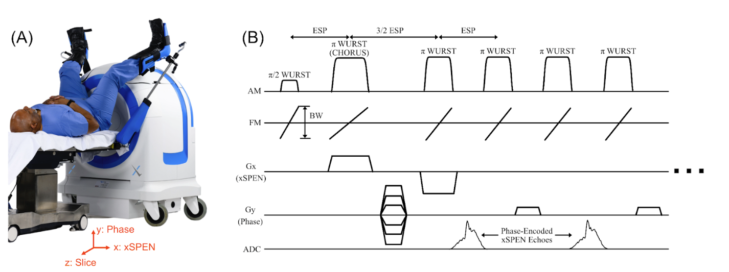
Single-sided, low-field MRI scanners provide image guidance during interventions such as prostate biopsies without restricting surgical access. For example, the Promaxo scanner in Fig. 1A (Promaxo, Oakland, CA, USA) has a built-in 58-73 mT z-gradient. Imaging is performed using a multi-slab, 3D sequence in which the large matrix dimensions (100s of voxels; x and y) are phase-encoded by the pulsed x and y gradients, while the small matrix dimension (10s of voxels; z) is frequency encoded by the static z gradient. This leads to anatomic scan times less than 10 minutes. However, faster scans are desirable for localizers and intraoperative imaging but are fundamentally limited by the fact that the fastest spatial encoding mechanism (frequency encoding) is used to encode the smallest matrix dimension. We recently demonstrated how spatiotemporal encoding using the xSPEN technique can be leveraged to swap phase and frequency encoding dimensions [1] on this scanner. However, those scans suffered geometric distortions due to the lack of an xSPEN reconstruction that accurately incorporates gradient non-linearity. Here we describe a numerical point-spread function (PSF)-based reconstruction method for new CPMG-RARE xSPEN acquisitions that resolves these distortions and demonstrate it in phantoms and in vivo female and male pelvic scans.Methods
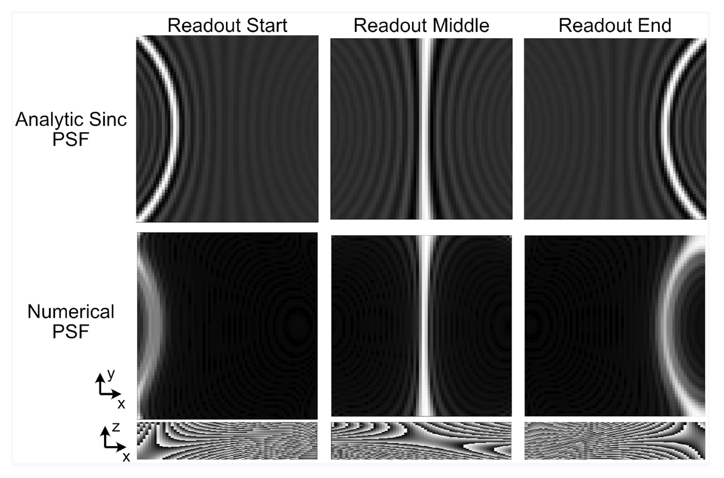
Figure 1b illustrates the CPMG-RARE xSPEN sequence, which uses opposite-polarity Gx pulses played during the first two refocusing pulses and in the presence of the static Gz gradient to apply a bilinear xSPEN spatial encoding phase function. To reconstruct images from this acquisition, Figure 2 illustrates how the bilinear xSPEN phase is calculated over each slice’s volume based on sequence parameters and measured gradient field maps. This phase is shifted to each readout time point to account for Gz-induced phase evolution over the readout which moves the sensitive point in x, and the shifted phase is summed through the slice to obtain a PSF for each time point in the xSPEN readout. The PSFs are collected into a matrix and cascaded with a Type-III NUFFT operator to apply phase encoding in y, and image reconstruction is performed using the combined operator in a CGLS algorithm.Imaging experiments were performed to compare the numerical PSF-based reconstruction with conventional inverse FFT- [4] and analytic sinc- [5] based reconstructions, to evaluate how finely the slice must be sampled when calculating the PSFs, and to compare analytic sinc and PSF-based reconstructions in vivo in pelvic scans of healthy adult female and male subjects scanned in accordance with a local IRB. The scans used the following parameters: TR=1.5s, spacing between spin echoes=10ms, TF=8, Number phase encodes=56, reconstructed matrix size 72x72 in plane, 36 slices, reconstructed FOV 18 cm x 18cm x 10cm, pixel spacing 2.5 mm x 2.5 mm, slice thickness 2.8 mm. All RF pulses were broadband WURST-40 pulses with a 40 kHz bandwidth.
Results
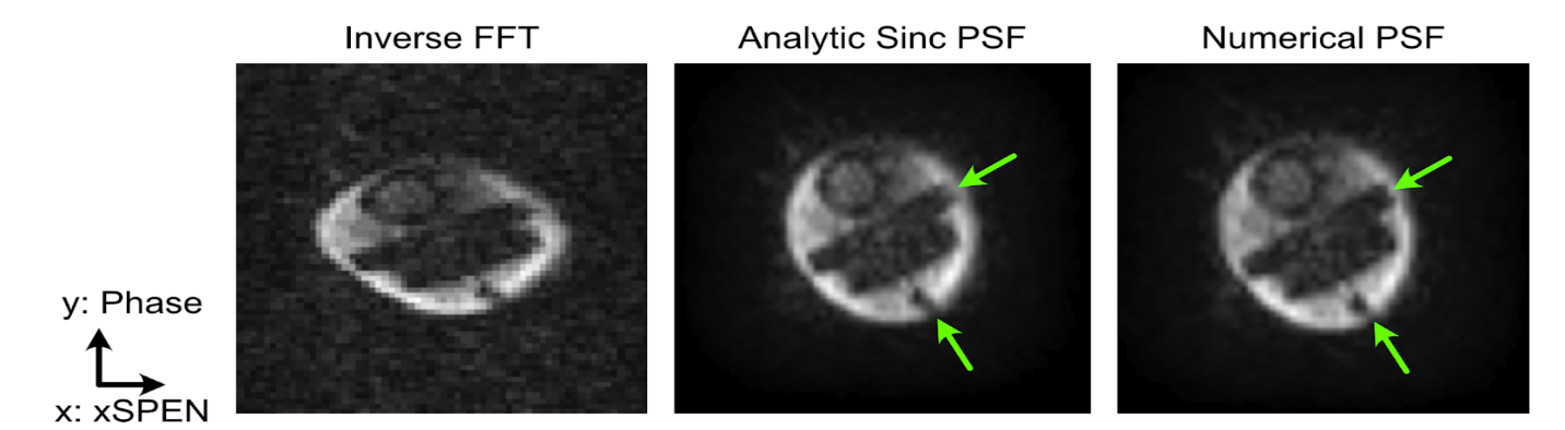
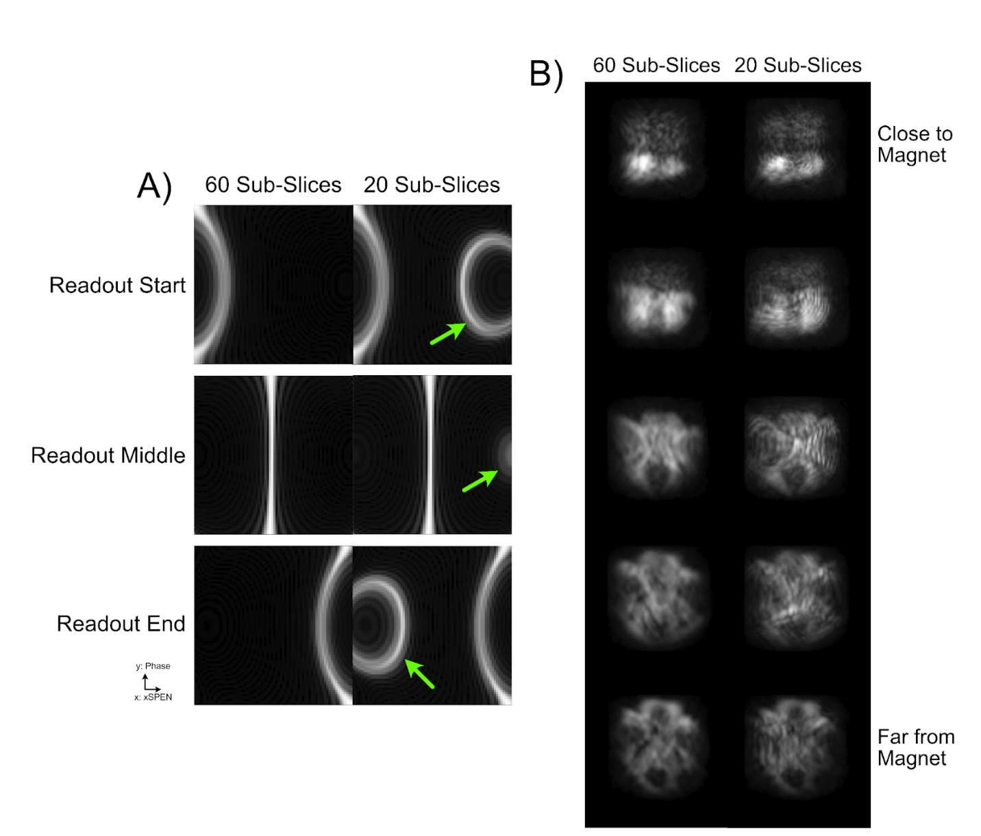
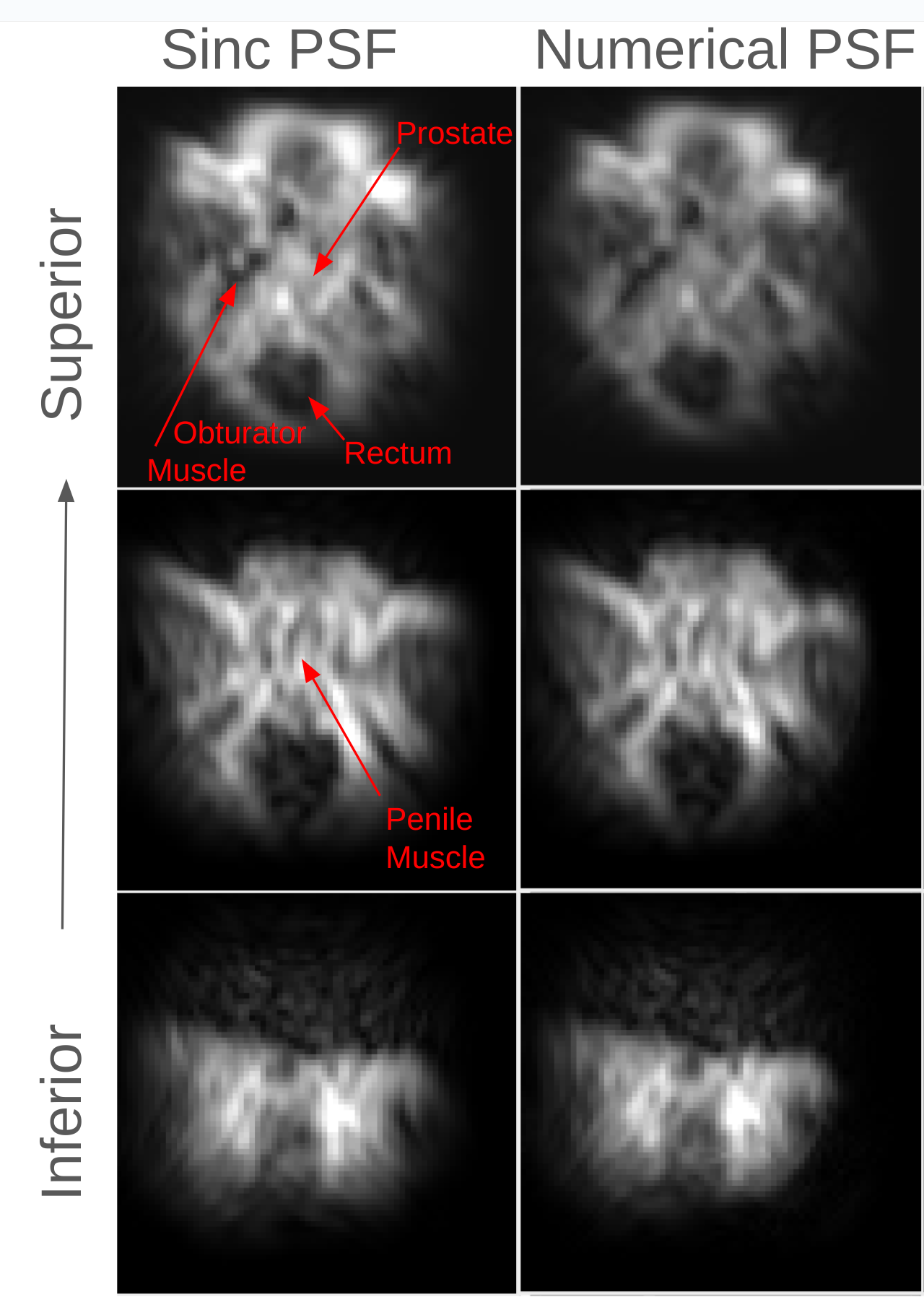
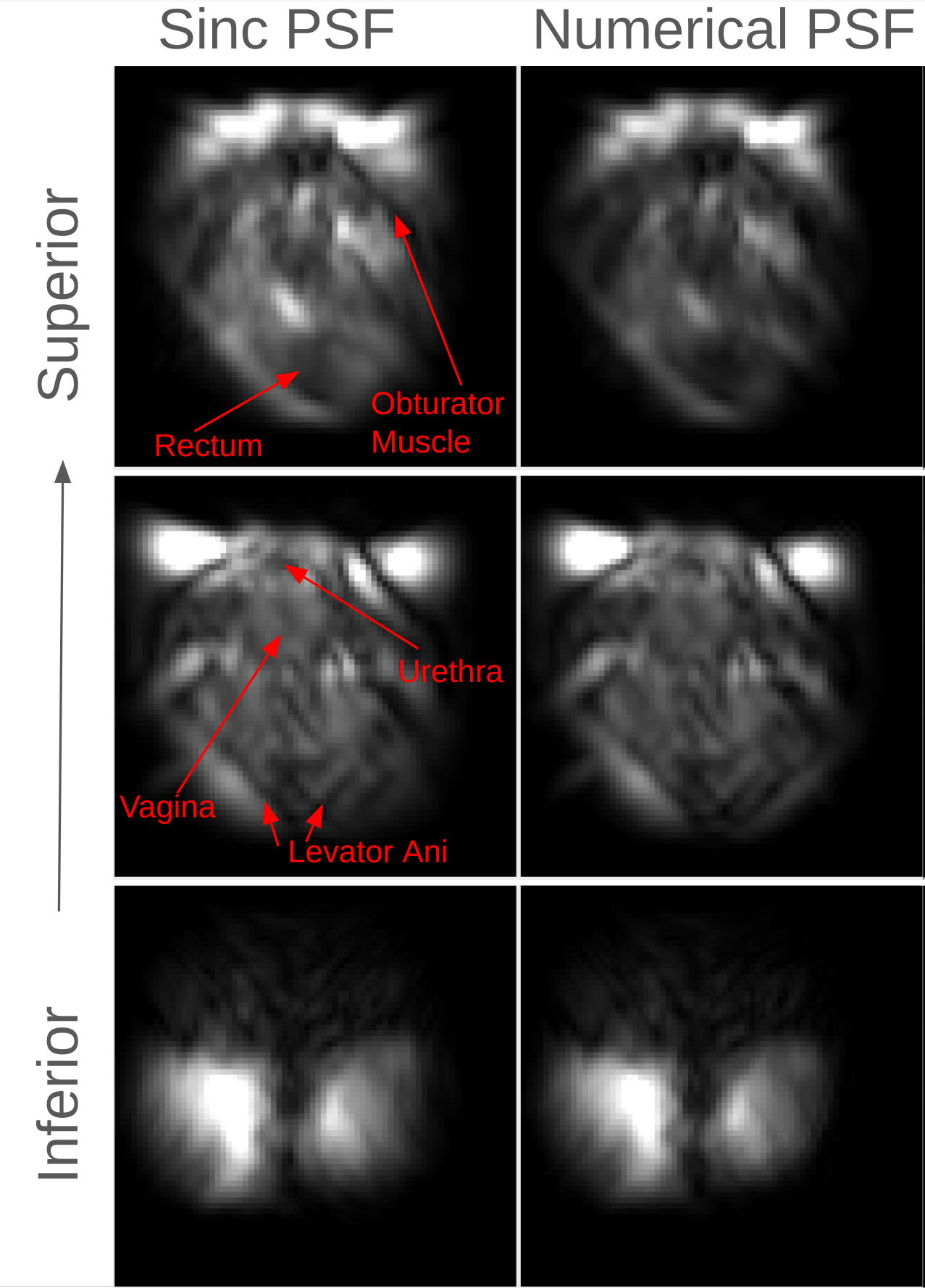
Figure 3 shows a comparison of inverse FFT (in y), analytic sinc, and numerical PSF reconstructions of an ACR phantom. The inverse FFT reconstruction is highly distorted because it does not account for gradient non-linearity or the xSPEN PSF. The analytic sinc reconstruction accounts for Gx non-linearity but neglects Gz non-linearity, resulting in blurring in the x-direction. The numerical PSF reconstruction yields a sharper, undistorted image. Figure 4 shows the effect of using too-few subslices when calculating the numerical xSPEN PSF, when reconstructing the male pelvic images. Using only 20 subslices causes aliasing of the calculated sensitive points, in particular at the beginning and end of the xSPEN readout, which then causes rippling artifacts in the xSPEN-encoded (x) dimension of the images which obscure such structures as the prostate and penile muscle. Using a sufficient number of subslices (60) resolves these artifacts. Figures 5 and 6 show a comparison of analytic sinc and numerical PSF reconstructions of the female and male pelvic data. The analytic sinc reconstructions contain vertical dark bands which obscure anatomy, while the numerical PSF reconstructions more clearly depict the pelvic anatomy, as labeled in the figures.Conclusion
We reported a numerical PSF-based reconstruction for xSPEN imaging on a single-sided low-field MRI scanner, which simultaneously resolves gradient non-linearity and ringing effects suffered by previous inverse-FFT and analytic sinc-based reconstructions. The method enables clear imaging of human pelvic anatomy with a single-sided scanner. Future work will focus on removing residual noise and ringing artifacts in the images.Acknowledgements
No acknowledgement found.References
1. Gomes et al, ISMRM Low-Field Workshop, 2022.
2. Power et al, Chem Commun, 52: 2916-2919, 2016.
3. https://github.com/mikgroup/sigpy/.
4. Z Zhang et al. MRM, 80:1492–1506, 2018.
5. Gomes et al, ISMRM Data Sampling Workshop, 2023.
Figures