1900
The peripheral branches of the trigeminal nerve: An illustrated article of neurogenic tumors imaging and related anatomy and pathway1third hospital of beijing university, beijing, China
Synopsis
Keywords: Head & Neck/ENT, Brain Connectivity
Motivation: Trigeminal neurogenic tumors involving the peripheral branches are relatively rare, and valueable.Luckly,I collect many kind of neurogenic tumor including rare case.
Goal(s): CT and MR imaging can illustrate related anatomy
and pathway of three peripheral branches of the trigeminal neurogenic tumors.
Approach: We obtained MR and CT images of all the adult Chinese patients during regular clinical trial with their informed consent.
Results: Imaging features include irregular thickening and enhancement of the nerve, furthermore to clearly display supra-orbital, infra-orbital, and inferior alveolar nerves respectively.Surprisely,it also widened the neural foramen those neurogenic nerves pass through.
Impact: A thorough understanding of its anatomy and pathwayis very important, both in diagnosis and treatment of tumors.It really provided a important clinical guiding for proper practice.
Materials and Methods:This study retrospectively analyzed 34 cases of peripheral trigeminal neurogenic tumors confirmed by surgery and pathology at Peking University Third Hospital from 2008 to 2022, including 21 cases of trigeminal schwannoma, 12 cases of trigeminal neurofibroma, and 1 case of trigeminal Intraneural perineuriomas,.All patients underwent enhanced brain MRI and CT reconstruction examinations.
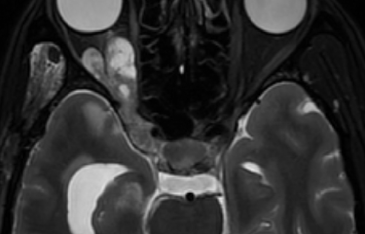
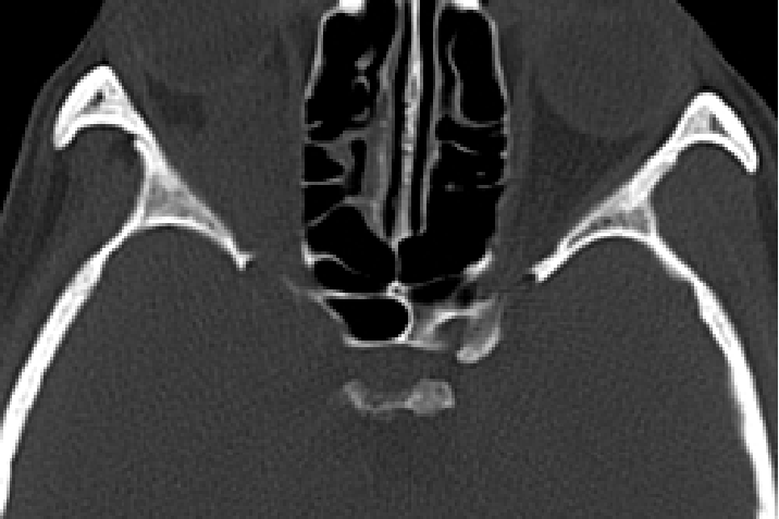
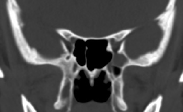
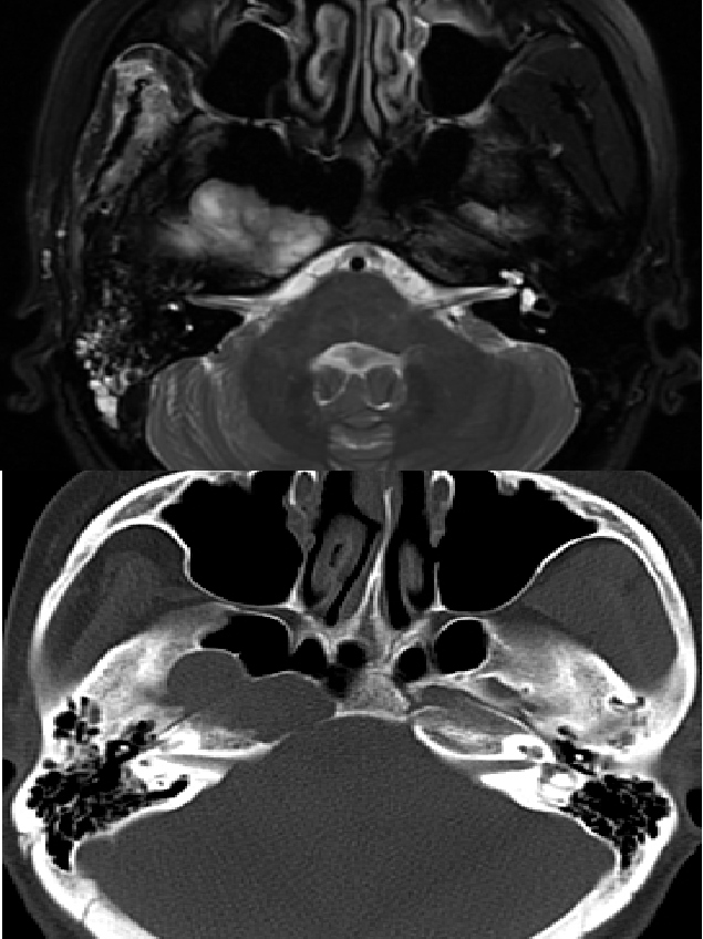
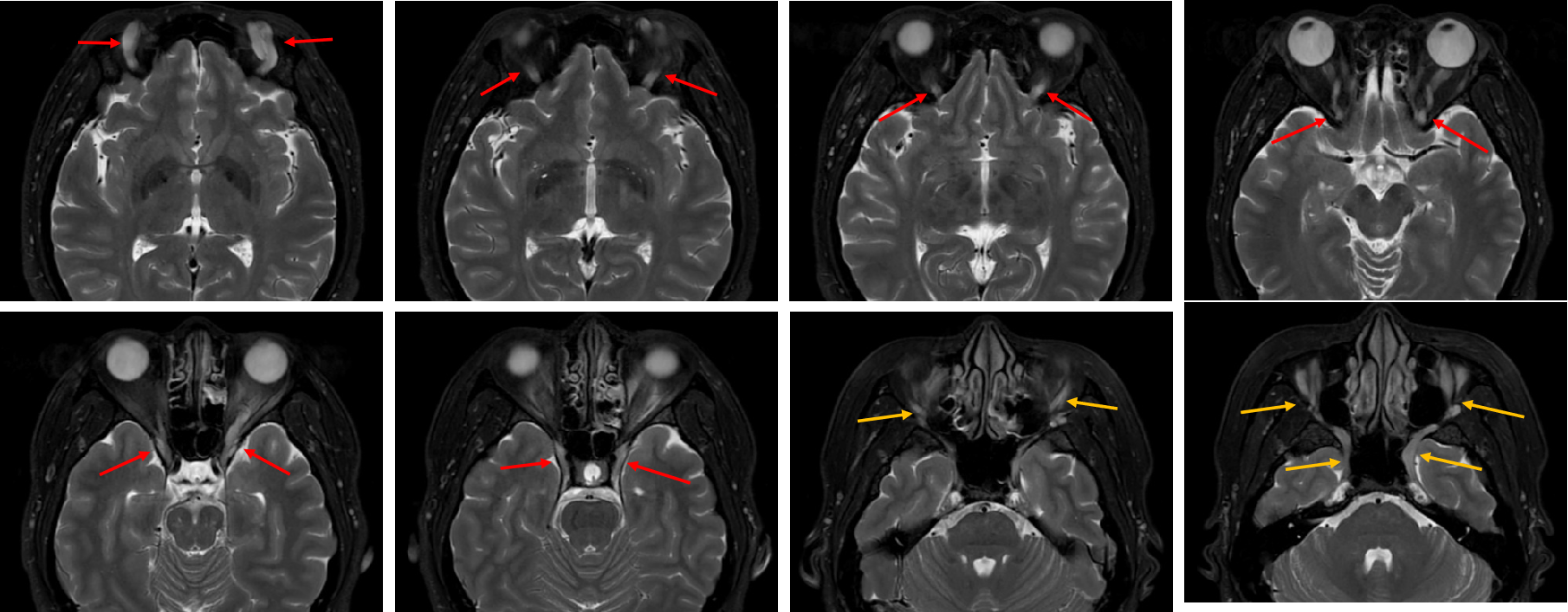
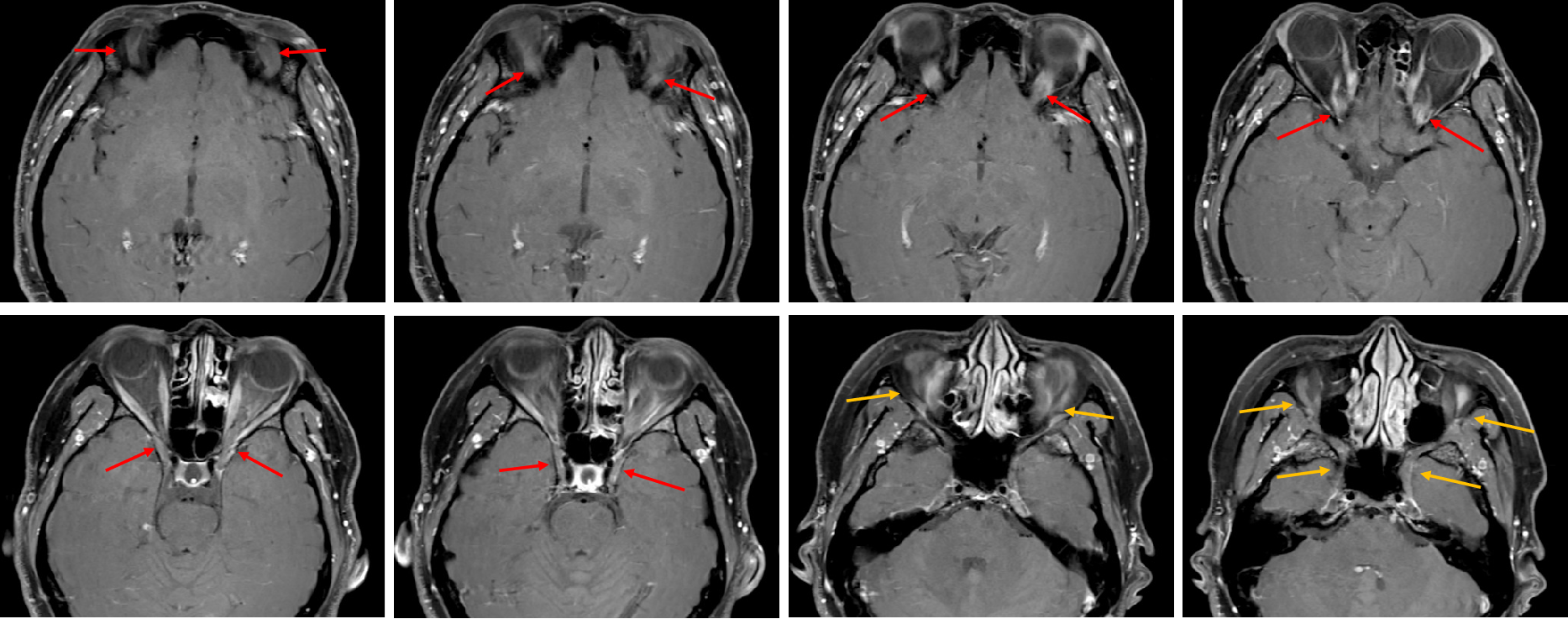
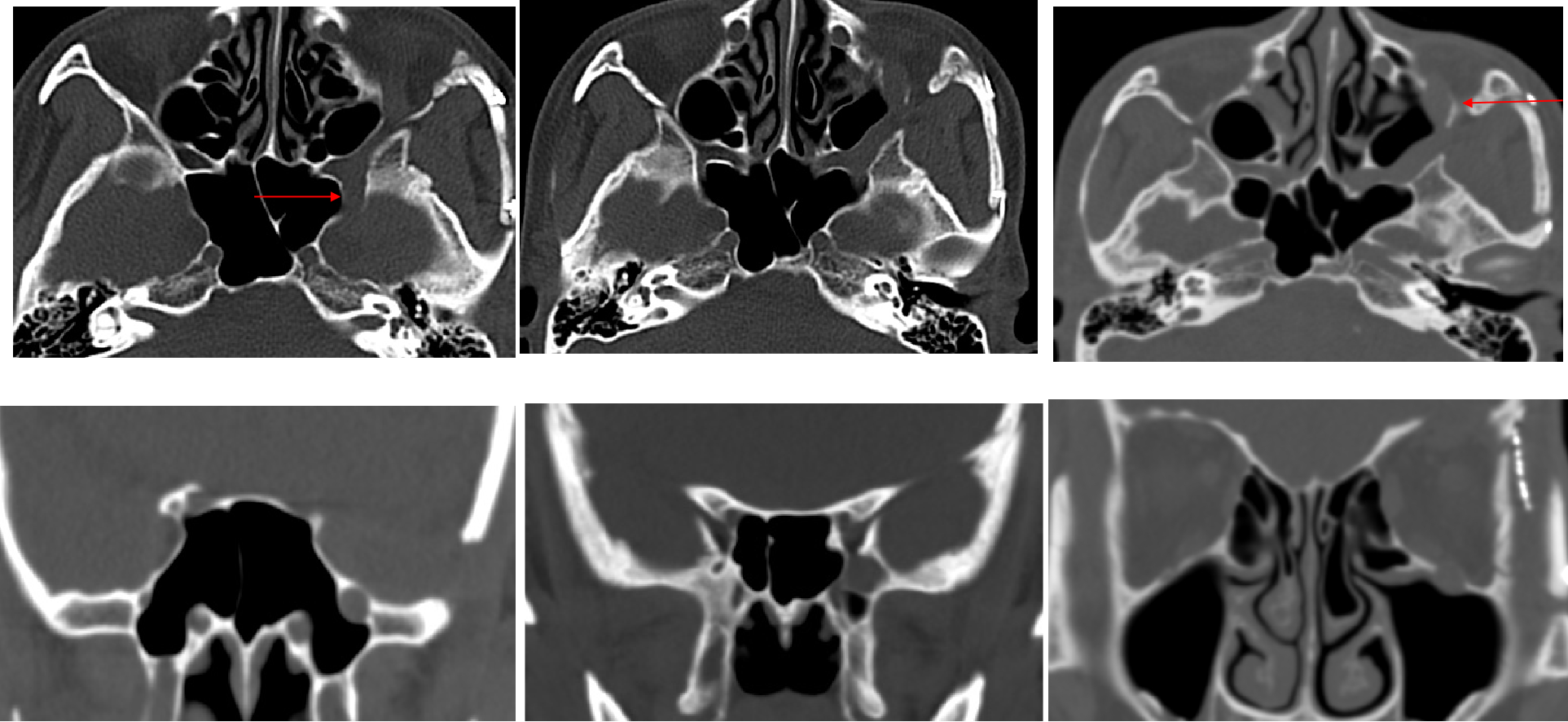
Result:There were 11 cases involving the ophthalmic branch, 8 cases involving the maxillary branch, 7 cases involving the mandibular branch, 6 cases involving both branches, and 2 cases involving all three branches.The schwannoma involving the ophthalmic branch shows cystic necrosis within the tumor, uneven enhancement, and mostly intact capsule with clear boundaries. The supraorbital fissure bone is absorbed and widened (Figure1-2 ). Maxillary branch neurofibroma with thickened and slightly enhanced nerves, widened pterygopalatine fossa, foramen rotundum, and suborbital nerve canal (Figure 3). Mandibular branch schwannoma, with significantly enlarged foramen ovale in a dumbbell shape, with surrounding bone compression and absorption, without any damage (Figure 4).The trigeminal Intraneural perineurioma involve bilateral branches of the trigeminal peripheral nerve , the ophthalmic branch, maxillary branch and the mandibular branch, with symmetrical thickening and a serpentine shape. Locally, it presents spindle shaped enlargement with iso-signal intensity on T2WI and T1WI, with slight enhancement (Figure 5-6).
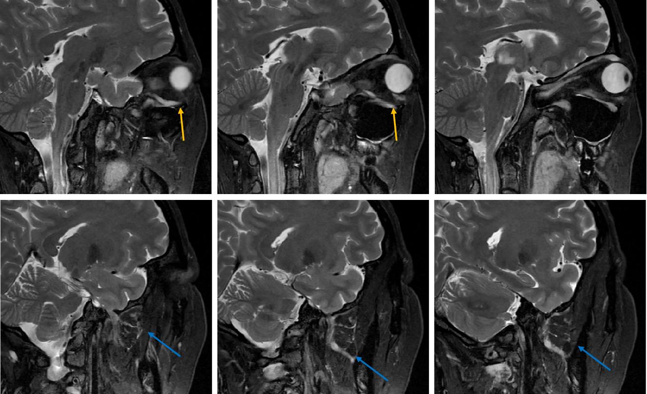
The perineurioma of ophthalmic branch passes the skull through the lateral side of the cavernous sinus and the supraorbital fissure, enters the orbit and extends to the supraorbital foramen, running between the superior rectus muscle and the supraorbital wall. The perineurioma of maxillary branch passes through the lateral circular foramen of the cavernous sinus and enters the pterygopalatine fossa, continuing through the suborbital foramen to form the suborbital nerve .The perineurioma of mandibular branch passes through the foramen ovale and enters the space between the masticatory muscles, extending downwards to the inferior alveolar nerve and the mandibular nerve (Figure 7) .
Discussion:MRI and CT can clearly display the growth path and morphological characteristics of peripheral trigeminal neurogenic tumors, as well as the corresponding structural bone changes. The characteristics of corresponding nerve thickening make the course of the three peripheral branches very clear, and the segmental division of the lesion is clear, which is beneficial for us to learn the anatomical course of the three peripheral branches. It is of great significance to determine the origin, shape, nature, and classification of tumors, as well as to develop corresponding surgical pathways to reduce surgical trauma and complications.
Acknowledgements
Thanks to clinical doctors and pathologists providing every informations.
References
1. Adaskova, M. and M. Sicak (2020). "Schwannoma of the extracranial part of the trigeminal nerve." Ceska a Slovenska Neurologie a Neurochirurgie 83(2): 153-+.
2.Mada, Y., et al. (2014). "[Four cases of extracranial trigeminal benign neurogenic tumors]." Nihon Jibiinkoka Gakkai Kaiho 117(11): 1356-1361.
Figures