1898
Predicting poor medical therapy efficacy in patients with trigeminal neuralgia: Insights from MRI and clinical features1radiology, first people's hospital of Foshan, Foshan, China, 2Sun yat-sen University, Guangzhou, China, 3functional neurosurgery, first people's hospital of Foshan, Foshan, China
Synopsis
Keywords: Peripheral Nerves, Treatment, trigeminal neuralgia; carbamazepine
Motivation: While MRI have proven valuable in predicting the surgical prognosis of trigeminal neuralgia (TN), studies on the prediction of medical therapy efficacy are still lacking.
Goal(s): Our goal was to identify MRI and clinical characteristics that can predict the efficacy of medical therapy for TN.
Approach: We conducted a blindly evaluation of MRI characteristics of TN. Clinical characteristics was obtained from the medical notes or phone follow-up. The correlation between MRI and clinical characteristics with medical therapy efficacy were subsequently analyzed.
Results: Vertebral artery being culprit vessel and involvement of trigeminal nerve ophthalmic division correlates with drug treatment efficacy.
Impact: The identified MRI and clinical characteristics in patients with TN, which are associated with the efficacy of medical therapy, can assist clinicians in offering informed guidance to patients when deciding between surgical and medical therapy.
Introduction
Trigeminal neuralgia(TN) is a chronic neuropathic pain disorder characterized by spontaneous severe electric shock–like or stabbing pain in the face region1. While TN can have various underlying causes, it is widely recognized that neurovascular compression (NVC) at the root entry zone of trigeminal nerve is a primary contributing factor2. Microvascular decompression surgery offers an effective solution for addressing the compression caused by offending vessels near the trigeminal nerve, providing pain relief to approximately 90% of patients3, 4. High-resolution MRI advancements enable a detailed preoperative evaluation of the presence, type, and severity of NVC in the cerebellopontine cistern5, 6. Furthermore, an increasing number of research have demonstrated that certain preoperative MRI characteristics of TN can aid in predicting the prognosis of microvascular decompression surgery7-11.In fact, in the initial treatment of TN, medication typically serves as the first-line approach and is generally considered highly effective, alleviating pain in nearly 90% of patients12. However, there is a notable absence of studies examining the factors associated with the poor efficacy of medical therapy. We hypothesized that certain factors could predict the limited effectiveness of medical therapy for TN and, therefore, conducted this study to help clinicians develop early, tailored treatment plans, thereby reducing patient suffering and mitigating the financial burden of unnecessary treatments.Methods
We conducted a retrospective study in our hospital from December 2019 to April 2023, involving patients diagnosed with TN based on the International Classification of Headache Disorders (3rd edition). Our study comprised 112 patients, including 44 men and 68 women. We performed a blind evaluation to assess the presence, location, and severity of NVC, as well as the type of offending vessel. Additionally, we collected demographic and clinical data, such as sex, age at the initial visit, history of hypertension and diabetes mellitus, duration of the disease, treatment methods, and efficacy of medical therapy, either from electronic medical records or through telephone follow-up. Logistic regression analysis was performed to identify the factors associated with poor medical therapy efficacy.Results
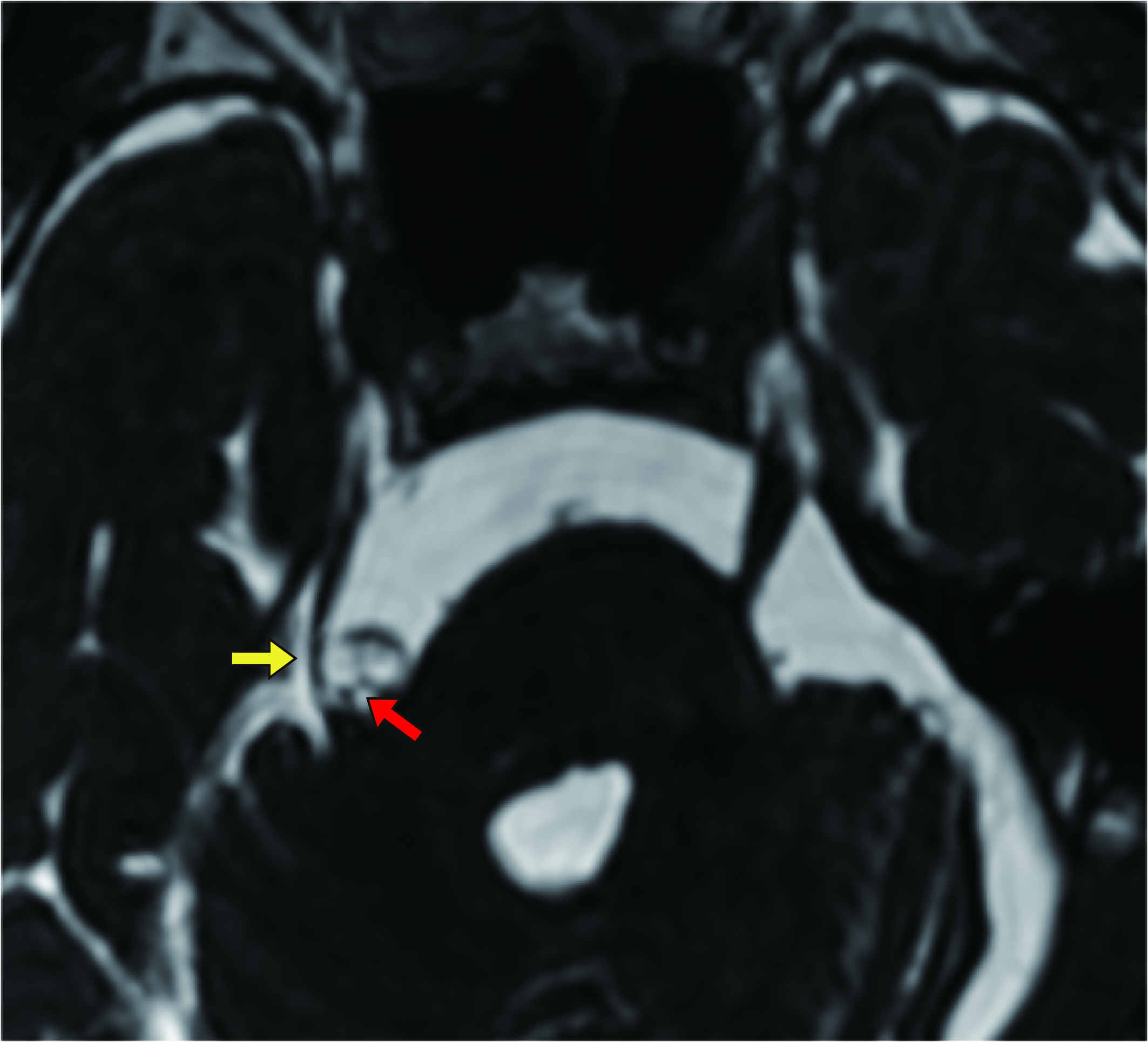
At the follow-up, 35 patients (31.3%) reported failure results. 30 patients (26.8%) reported fair. 19 patients (17.0%) reported good. Only 28 patients (25%) reported excellent. In total, 47 patients achieved a satisfactory response to medical therapy, while 65 patients experienced poor medical therapy efficacy. Logistic regression analysis identified that two independent factors significantly associated with poor medical therapy efficacy. Patients with the vertebral artery as the culprit vessel were 6.343 times more likely to experience a poor with poor medical therapy efficacy compared to those without vertebral artery involvement (95% confidence interval[CI]: 1.317-30.553, p = 0.021). Conversely, patients with trigeminal nerve branch V1 involvement were 0.395 times less likely to experience a poor with poor medical therapy efficacy compared to those without ophthalmic division (V1) of trigeminal nerve involvement (95%CI: 0.173-0.902, p = 0.027).Discussion
In clinical practice, TN commonly involves the superior cerebellar artery as the primary offending vessel11, 13. However, our study revealed a significant association between poor medical therapy efficacy and the vertebral artery, rather than the superior cerebellar artery. The vertebral artery typically forms the basilar artery at the lower border of the pons, situated beneath the trigeminal nerve14. When the vertebral artery assumes responsibility as the offending vessel, it is often considered an abnormal condition. In this case, an overly thick or tortuous vertebral artery may cause severe and wide-ranging compression, which results in a wider range of nerve involvement. We hypothesize that it is the larger involvement of the trigeminal nerve caused when the trigeminal nerve is compressed by the vertebral artery that leads to the possibility of poor medical therapy efficacy. Additionally, our findings indicated that involvement of the ophthalmic division (V1) of the trigeminal nerve was negatively correlated with poor medical therapy efficacy. These results align with the positive response to carbamazepine observed in a study by Sjaastad et al.15, suggesting that patients with V1 involvement may be better suited for drug therapy.Conclusion
In conclusion, our study explored the relationship between pre-therapeutic MRI and clinical characteristics and poor medical therapy efficacy in TN patients. Our results suggest that patients with the vertebral artery as the culprit vessel are at a higher risk of experiencing a poor efficacy to medical therapy compared to those without such involvement. Conversely, the involvement of the ophthalmic division of the trigeminal nerve is associated with a lower likelihood of a poor medical therapy efficacy. These findings can assist clinicians in making more informed early treatment decisions.Acknowledgements
The authors thank all colleagues in the Department of Radiology and the Department of Functional Neurosurgery of the First People's Hospital of Foshan for facilitating this study.References
1. Cruccu G, Ropper AH, Di Stefano G, et al. Trigeminal Neuralgia. New England Journal of Medicine 2020;383(8):754-762.
2. Liao Z, Zou L, Peng W, et al. Improving the accuracy of pre-operative evaluation of neurovascular conflict in trigeminal neuralgia using magnetic resonance subtraction. Neuroradiology 2021;63(3):295-303.
3. Tai AX, Nayar VV. Update on Trigeminal Neuralgia. Current Treatment Options in Neurology 2019;21(9).
4. Bendtsen L, Zakrzewska JM, Heinskou TB, et al. Advances in diagnosis, classification, pathophysiology, and management of trigeminal neuralgia. Lancet Neurol 2020;19(9):784-796.
5. Seeburg DP, Northcutt B, Aygun N, et al. The Role of Imaging for Trigeminal Neuralgia: A Segmental Approach to High-Resolution MRI. Neurosurg Clin N Am 2016;27(3):315-326.
6. Hu M, Zhou W, Shen W, et al. A Combination of 3D TOF MRA and FIESTA Predicts Surgery-Needed Primary Trigeminal Neuralgia and Specific Offending Vessels. J Integr Neurosci 2022;21(6):169.
7. Zhao Y, Chen J, Jiang R, et al. MRI features of responsible contacts in vascular compressive trigeminal neuralgia and prediction modeling. Acta Radiol 2022;63(1):100-109.
8. Heinskou TB, Rochat P, Maarbjerg S, et al. Prognostic factors for outcome of microvascular decompression in trigeminal neuralgia: A prospective systematic study using independent assessors. Cephalalgia 2019;39(2):197-208.
9. Hughes MA, Jani RH, Fakhran S, et al. Significance of degree of neurovascular compression in surgery for trigeminal neuralgia. J Neurosurg 2019:1-6.
10. Wang Z, Zhao Z, Song Z, et al. The application of magnetic resonance imaging (MRI) for the prediction of surgical outcomes in trigeminal neuralgia. Postgrad Med 2022;134(5):480-486.
11. Hardaway FA, Gustafsson HC, Holste K, et al. A novel scoring system as a preoperative predictor for pain-free survival after microsurgery for trigeminal neuralgia. J Neurosurg 2019:1-8.
12. Bendtsen L, Zakrzewska JM, Abbott J, et al. European Academy of Neurology guideline on trigeminal neuralgia. Eur J Neurol 2019;26(6):831-849.
13. Brinzeu A, Drogba L, Sindou M. Reliability of MRI for predicting characteristics of neurovascular conflicts in trigeminal neuralgia: implications for surgical decision making. J Neurosurg 2018:1-11.
14. Tay KY, JM UK-I, Trivedi RA, et al. Imaging the vertebral artery. Eur Radiol 2005;15(7):1329-1343.
15. Sjaastad O, Pareja JA, Zukerman E, et al. Trigeminal neuralgia. Clinical manifestations of first division involvement. Headache 1997;37(6):346-357.
Figures