1897
Exploring the diagnostic performances of CISS and DESS in visualizing extracranial nerves and localizing parotid tumors1Department of Radiology, Shengjing Hospital of China Medical University, Shenyang, China, 2MR Research Collaboration, Siemens Healthineers, Beijing, China, 3MR Clinical Application, Siemens Healthineers, Shenyang, China
Synopsis
Keywords: Head & Neck/ENT, Head & Neck/ENT
Motivation: Identification of the spatial relationship between the parotid tumor and the extracranial segment of the facial nerve can facilitate preoperative planning.
Goal(s): We compared image quality and diagnostic efficacy between double-echo steady-state with water (DESS) and constructive interference in steady-state (CISS) sequences.
Approach: DESS and CISS sequences were used to determine the visibility of the extracranial segment of the facial nerve in healthy volunteers, then locate parotid tumors and facilitate patient diagnosis.
Results: Compared with DESS images, CISS images more reliably showed the location of the facial nerve relative to the lesion in patients with parotid tumors
Impact: CISS demonstrates good visualization of the parotid branches (temporofacial and cervicofacial trunks) compared with DESS. Moreover, CISS is a reliable and direct method for identifying the locations of parotid tumors.
Introduction
Thus far, reliable and direct observation of the extracranial segment of the facial nerve remains challenging. The three-dimensional (3D) double-echo steady-state with water (DESS) sequence can reliably and directly depict morphological changes in the extracranial segment of the facial nerve. They can also show the relationships of parotid tumors with the facial nerve, exhibiting high diagnostic efficacy in tumor localization 1-3. Additionally, Guenette et al. showed that the constructive interference in steady-state (CISS) sequence is superior for facial nerve tracing, can clearly show the main trunk of the parotid nerve and the two main branches of the facial nerve in parotid tumors (i.e., temporal and cervical trunks), and has good efficacy in terms of localizing parotid tumors4. However, that study included a low number of patients (three) with surgically confirmed parotid tumors. Here, we compared the abilities of 3D DESS and 3D CISS to distinguish the extracranial segment of the facial nerve and explored their diagnostic performances in parotid tumor localization.Methods
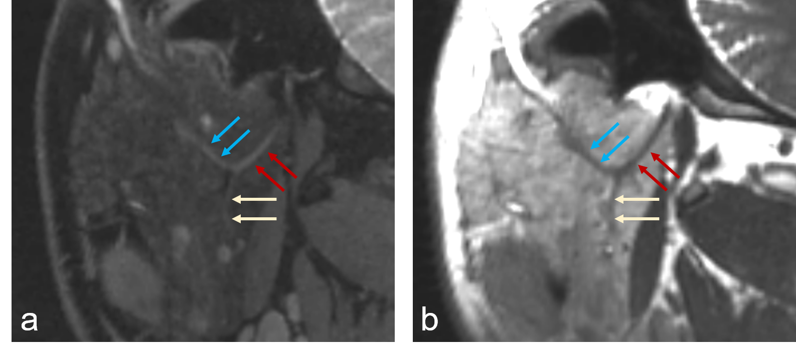
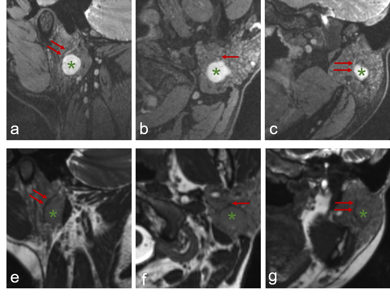
Between June 2022 and July 2023, this prospective study included 32 facial nerves in 16 healthy volunteers (Fig. 1) and 25 facial nerves in 25 patients with parotid tumors (Fig. 2) who underwent noncontrast-enhanced extracranial facial nerve MRI with both DESS and CISS sequences. All imaging examinations were performed on a 3T MR scanner (MAGNETOM Prisma, Siemens Healthcare, Erlangen, Germany) equipped with a 64-channel head and neck coil. All participants provided written informed consent to take part in the study. The imaging parameters for 3D DESS were as follows: retention time (TR), 13.64 ms; echo time (TE), 4.68 ms; field of view (FOV), 160 mm; matrix size, 320×320; slice thickness, 0.5 mm; and parallel imaging acceleration factor, 3. The imaging parameters for 3D CISS were as follows: TR, 5.83 ms for all frequencies; TE, 2.57 ms for each frequency; FOV, 160 mm; matrix size, 320×320; slice thickness, 0.5 mm; and parallel imaging acceleration factor, 3. The overall scan time for parotid MRI was approximately 10–15 minutes. All images were independently reviewed by two radiologists, who subjectively evaluated the image quality score (IQS) using a 5-point Likert scale (IQS ≥3 for diagnostic quality) (Table 1). Levels of inter- and intrarater agreement were assessed using Cohen’s kappa coefficient (κ). Receiver operating characteristic analysis was conducted, and the diagnostic performances of DESS and CISS images in localizing parotid tumors were calculated.Results
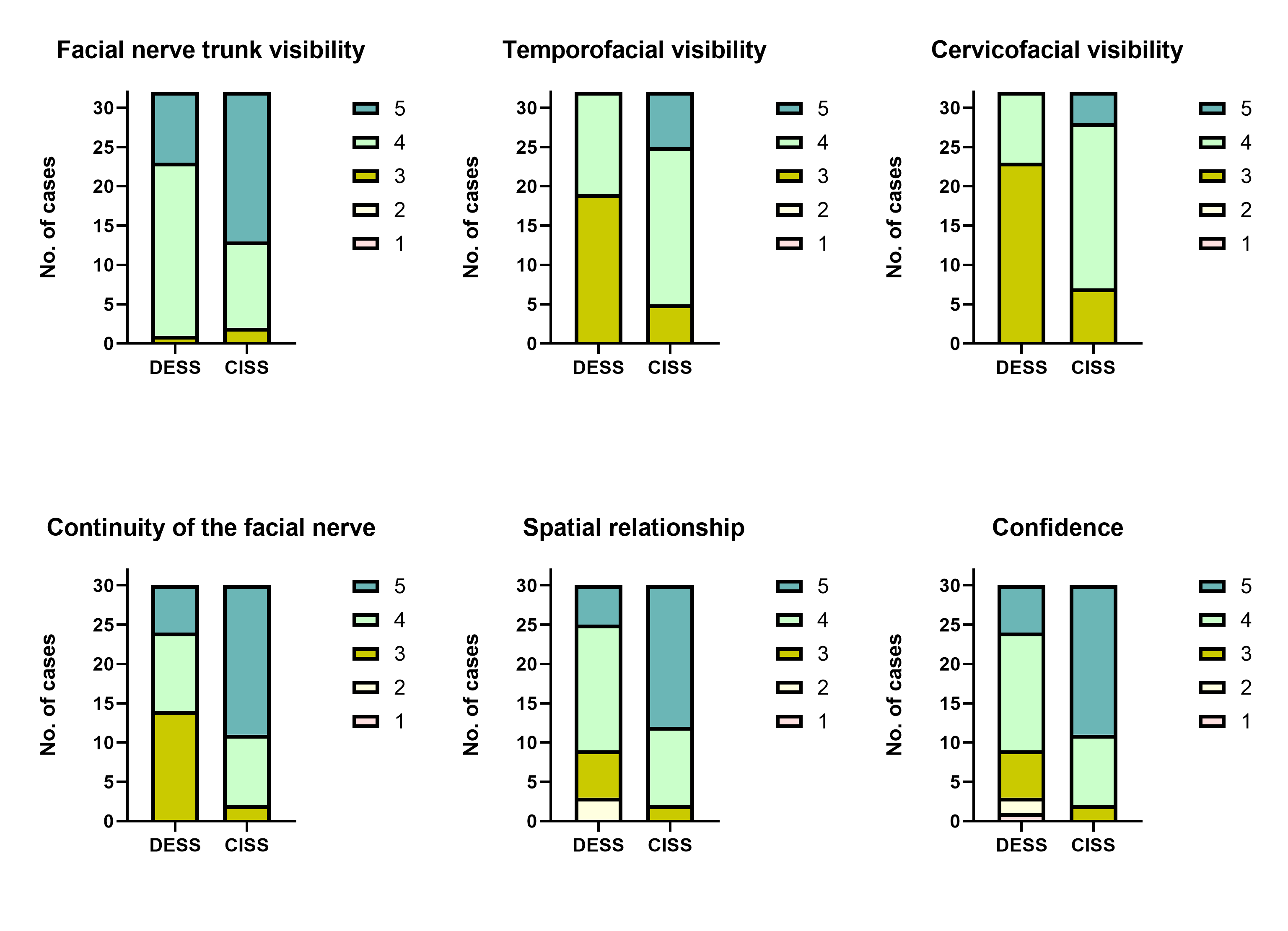
In healthy volunteers, good inter- and intrareader agreement for IQS (κ = 0.724–0.816) was observed. Although both experts could distinguish the facial nerve trunk structures well on DESS and CISS (Fig.3a), but no statistical difference was found in the identifying facial nerve trunk (p=0.061) (Fig. 3). Taking the level of facial nerve bifurcation as a reference, both experts were able to clearly distinguish the two main branches (temporal and cervical trunks) on both sequences; however, the CISS sequence scored slightly higher than DESS for the degree of clarity of the temporal and cervical trunks display (p < 0.001 for both) (Fig. 3). In patients with parotid tumor, CISS also performed better in the continuity of facial nerve to tumor, spatial relationship, and diagnostic confidence than DESS (p<0.001 for all) (Fig. 3). In terms of parotid tumor localization, CISS demonstrated excellent performance, similar to DESS (area under the curve, 1.000 vs. 0.958, p=0.1482) (Table 2).Discussion
This study showed that CISS outperforms DESS in terms of identifying the extracranial facial nerve and localizing parotid tumors. Specifically, CISS was able to visualize the spatial relationship between the facial nerve and the tumor in a manner that displayed excellent agreement with intraoperative findings. Furthermore, CISS was more accurate than DESS for parotid localization and diagnosis, and it could satisfy preoperative surgical planning requirements. Previous studies showed that DESS images could trace the facial nerve trunk and had high accuracy (>90%) for deep lobe lesions in patients with parotid tumors1, 2. Similarly, we found that DESS images enabled clear identification of the facial nerve trunk in healthy volunteers and had high diagnostic accuracy (92.6%) for deep lobe lesions in patients with parotid tumors. Accuracy was slightly lower in the present study than in the study by Fujii et al2 (97.8%), possibly because the previous study included more deep lobe lesions (25.2%), whereas 11.1% (3/27) of patients in the present study had deep lobe lesions, consistent with the incidence of deep lobe lesions in the parotid gland (10%)5Conclusion
Compared with DESS, CISS achieved comparable diagnostic performance in parotid tumor localization; it also demonstrated favorable image quality and more reliable morphological visualization of the facial nerve.Acknowledgements
NoneReferences
1. Kim Y, Jeong HS, Kim HJ, et al. Three-dimensional double-echo steady-state with water excitation magnetic resonance imaging to localize the intraparotid facial nerve in patients with deep-seated parotid tumors. Neuroradiology. 2021;63:731-739.
2. Fujii H, Fujita A, Kanazawa H, et al. Localization of Parotid Gland Tumors in Relation to the Intraparotid Facial Nerve on 3D Double-Echo Steady-State with Water Excitation Sequence. AJNR Am J Neuroradiol. 2019;40:1037-1042.
3. Jeong HS, Kim Y, Kim HJ, et al. Imaging of Facial Nerve With 3D-DESS-WE-MRI Before Parotidectomy: Impact on Surgical Outcomes. Korean J Radiol. 2023;24:860-870.
4. Guenette JP, Ben-Shlomo N, Jayender J, et al. MR Imaging of the Extracranial Facial Nerve with the CISS Sequence. AJNR Am J Neuroradiol. 2019;40:1954-1959.
5. Harney MS, Murphy C, Hone S, et al. A histological comparison of deep and superficial lobe pleomorphic adenomas of the parotid gland. Head Neck. 2003;25:649-653.
Figures