1887
High-resolution MR neurography of the wrist with diffusion-sensitized driven-equilibrium with deep learning constrained Compressed SENSE1Department of Radiological Services, Tokyo Women's Medical University Hospital, Tokyo, Japan, 2Department of Diagnostic imaging & Nuclear Medicine, Tokyo Women's Medical University Hospital, Tokyo, Japan, 3Philips Japan, Tokyo, Japan
Synopsis
Keywords: Peripheral Nerves, Nerves
Motivation: The median and ulnar nerves are fine anatomical structures, and these small nerves could not be adequately visualized on conventional MR neurography.
Goal(s): The purpose of this study is to realize high-resolution MR neurography for the diagnosis of peripheral neuropathy of the median and ulnar nerves.
Approach: We performed visualization of the detailed anatomy of the median and ulnar nerves by high-resolution diffusion sensitized-driven equilibrium (DSDE) with deep learning constrained Compressed SENSE (CS-AI) with deep learning constrained Compressed SENSE (CS-AI).
Results: MR neurography with DSDE with CS-AI could clearly visualize the median and ulnar nerves at high resolution within a feasible scan time.
Impact: High-resolution MR neurography with DSDE with CS-AI clearly visualizes the entire median and ulnar nerves, including the small common palmar digital nerves. This may be a useful technique in the diagnosis of peripheral neuropathy of the wrist.
Introduction
Assessment of median and ulnar nerves is important when deciding surgical procedures, such as carpal tunnel syndrome and Guion's tunnel syndrome. Since the median and ulnar nerves are the fine anatomy, such small nerves cannot be visualized well by the conventional MRI1-3. Previously, so-called diffusion-sensitized driven equilibrium (DSDE), a variant of motion-sensitive drive equilibrium (MSDE), has been introduced, it is an optimized combination of the pre-pulse and readout sequence for enabling higher b-value and has been applied for high resolution MR neurography of the wrist4. Recently, AI Compressed SENSE, which combines Compressed SENSE with AI, has been introduced, it enables efficient denoising by replacing the conventional wavelet transform by Adaptive-CS-Net, resulting in further accelerating the scan time. In this study, we evaluated the feasibility of accelerated DSDE MR neurography with SmartSpeed AI to improve the demonstration of the median and ulnar nerves within a shorter scan time.Methods
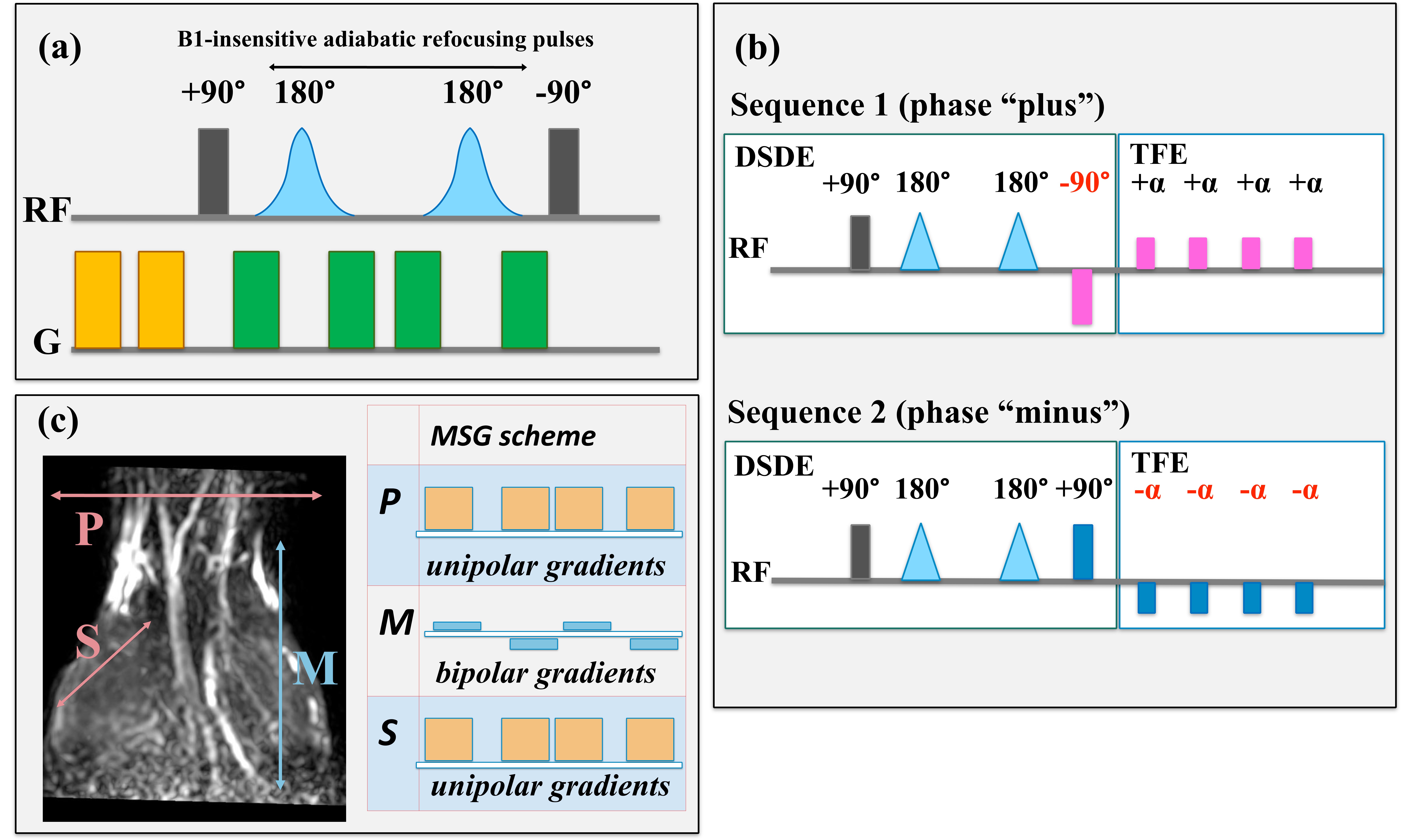
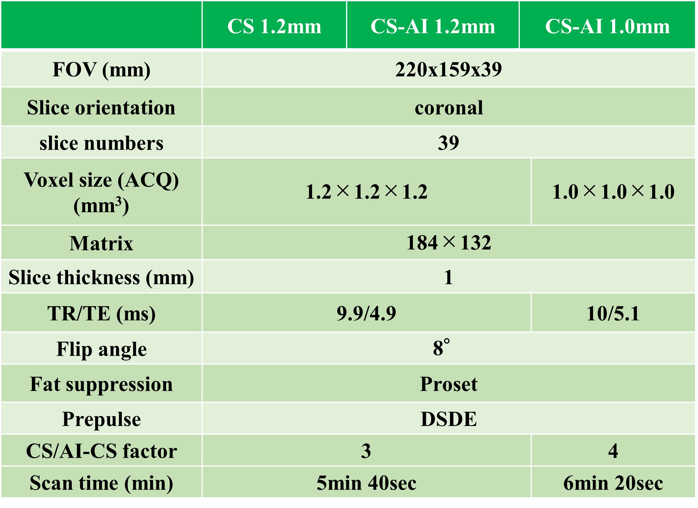
DSDE sequence has three improvement strategies. First, to improve the pulse efficiency in the of-center FOVs, B1-insensitive adiabatic pulses for refocusing pulses are applied. Second, to effectively suppress the veins while preventing signal loss of nerves which are running parallel to veins, orthogonally combined motion- and diffusion-sensitized gradient scheme5.6 is adopted. Because of the diffusion anisotropy of the human nervous system, two directions of strong unipolar MPGs were placed in both the anterior-posterior and right-left directions, which are most perpendicular to the trajectories of the nerves in the wrist. On the other hand, to suppress the vessel signals (veins) that flow in the same direction as the nerves, we added a weak bipolar MPG parallel to the course of the nerves (superior-inferior direction). Third, to eliminate the T1-effects, phase-cycling scheme is applied. Scheme of phase-cycling DSDE-TFE sequence is shown in Fig.1. Five healthy volunteers (4 males, 1 female, age 25-45 years old) were examined by 3.0T MRI (Ingenia Elition X, Philips Healthcare). The study was approved by the local IRB (Clinical Trial Review Board) and all subjects gave written informed consent. The median and ulnar nerves were visualized by DSDE with CS or SmartSpeed AI. The 3D voxel size for DSDE with SmartSpeed AI was set to 1.0 mm3 and 1.2 mm3. The 3D voxel size for DSDE with CS was set to 1.2 mm3. The imaging parameters were shown in Table 1. Evaluation of visualization of the median and ulnar nerves was performed by visual evaluation. A visual evaluation (Visualization of common palmar digital nerve, median nerves, ulnar nerves, overall image quality) was performed by three radiological technologists using a 5-point scale: 0, non-evaluative; 4, excellent quality.Results
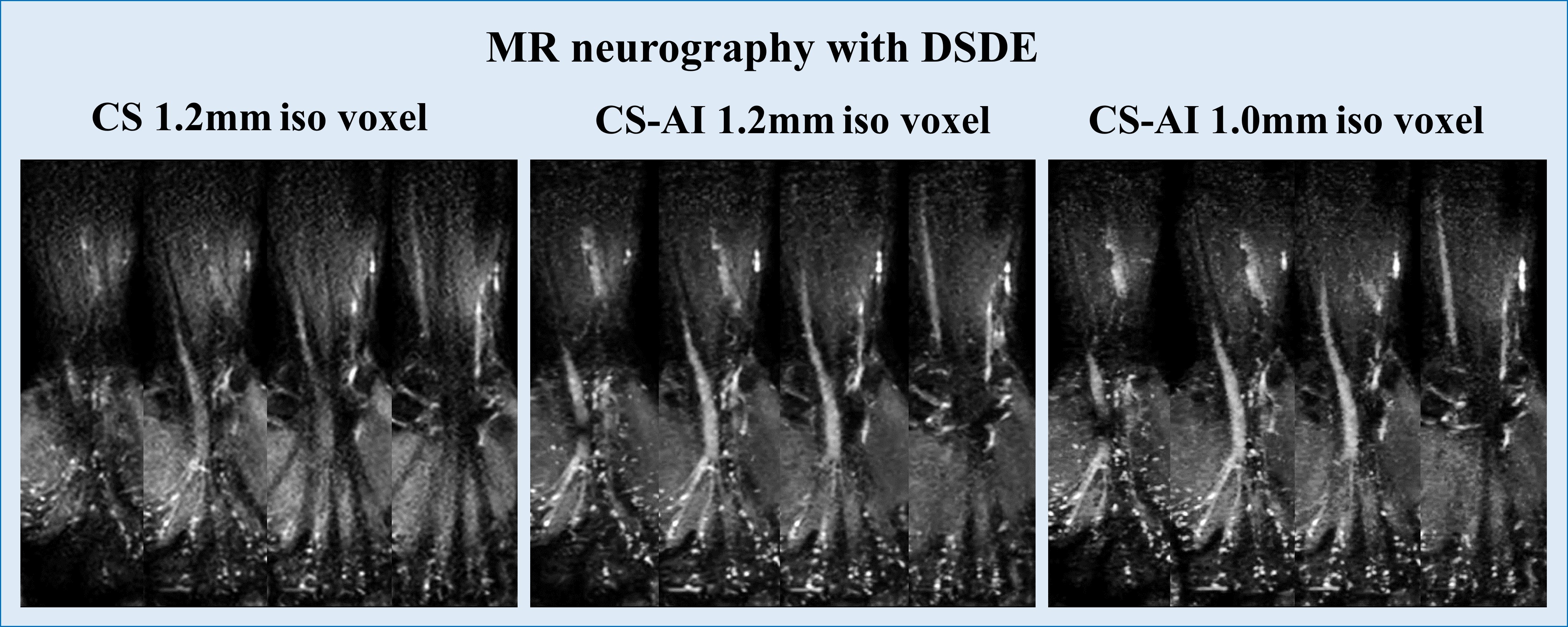
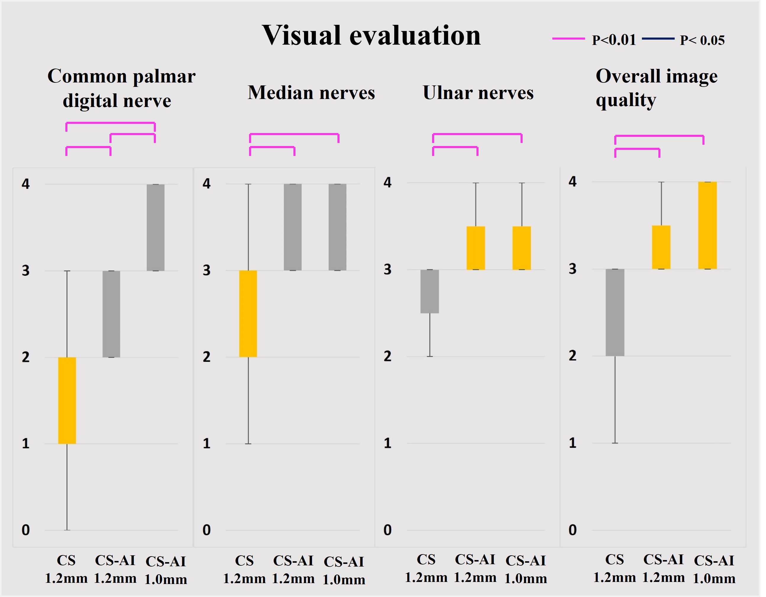
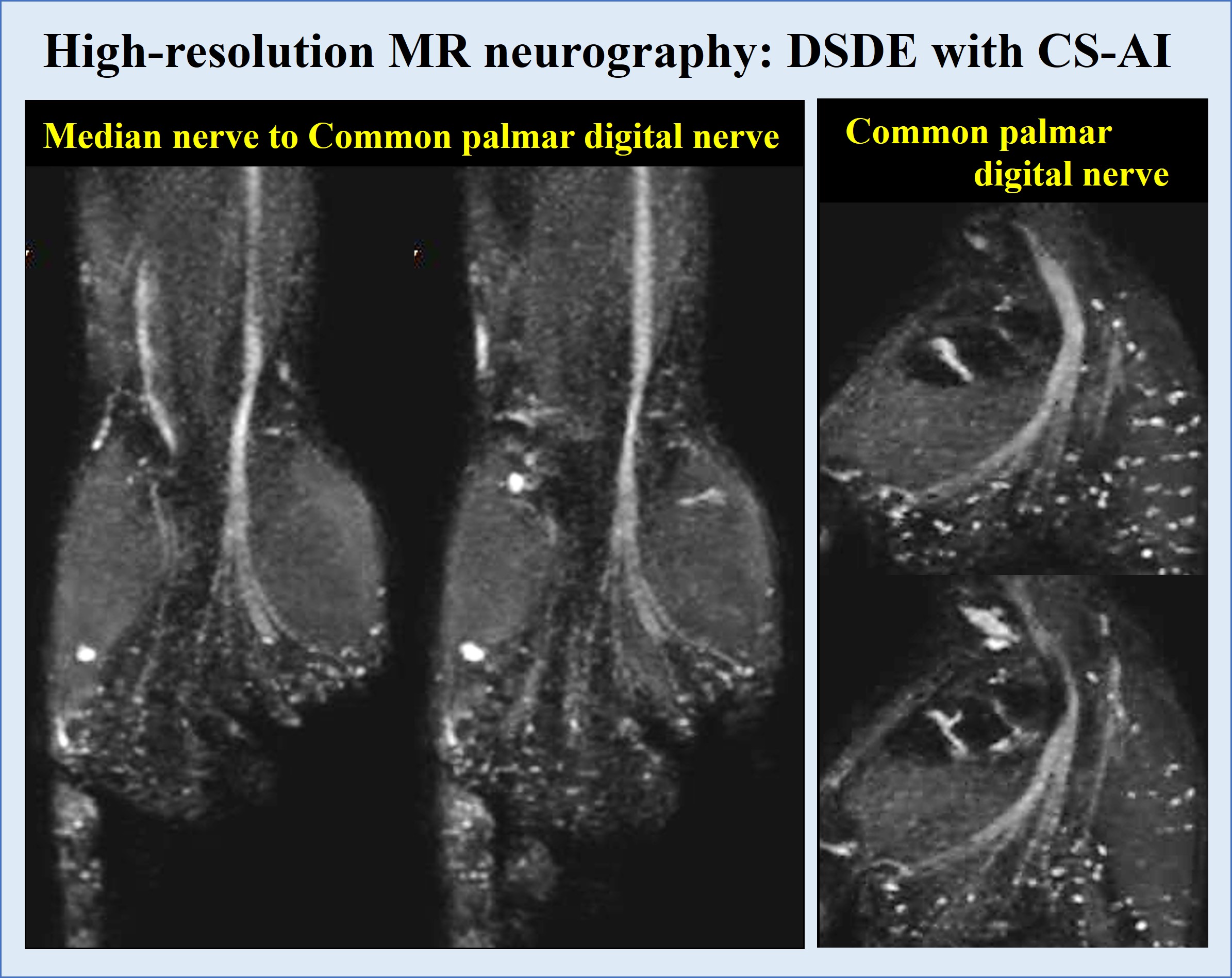
MR neurography images of 1.2 mm and 1.0 mm iso voxel DSDE with SmartSpeed AI and 1.2 mm iso voxel DSDE with CS were shown in Figure 2. The results of the visual evaluation were shown in Figure 3. DSDE with SmartSpeed AI of 1.0 mm and 1.2 mm iso voxels clearly visualized the median and ulnar nerves. Furthermore, DSDE with SmartSpeed AI of 1.0 mm iso voxels clearly improved visualization of common palmar digital nerves. DSDE with SmartSpeed AI showed better visualization of median and ulnar nerve compared to DSDE with CS. Representative wrist MR neurography using DSDE with SmartSpeed AI with 1.0 mm isotropic acquisition is shown in figure 4.Discussion
DSDE with CS had difficulty visualizing the median and ulnar nerves within a feasible time frame due to low SNR and image blurring caused by increased reduction factor and over denoising. DSDE with SmartSpeed AI could clearly visualize the median and ulnar nerves with higher SNR without blurring thanks to its efficient denoising. High-resolution images are needed to confirm the detailed structure of common palmar digital nerves. DSDE with SmartSpeed AI with 1.0mm isotropic acquisition was the best to demonstrate the detailed anatomy of the common palmar digital nerves.Conclusion
DSDE with SmartSpeed AI clearly visualized the whole median and ulnar nerves including small common palmar digital nerve. It could be a helpful sequence to contribute the diagnosis of peripheral neuropathy of the median and ulnar nerves and their blanches.Acknowledgements
No acknowledgement found.References
1. Swati Deshmukh et al. Pins and Needles From Fingers to Toes: High-Resolution MRI of Peripheral Sensory Mononeuropathies. AJR:208, January 2017. 2. Neda Faridian-Aragh et al. High-resolution 3T MR neurography of radial neuropathy. Journal of Neuroradiology (2011) 38, 265—274. 3. Alissa J. Burge et al. High-Resolution Magnetic Resonance Imaging of the Lower Extremity Nerves. Neuroimag Clin N Am 24 (2014) 151–170. 4. Masami Yoneyama et al. High-Resolution 3D MR Neurography of the Wrist using Phase-Cycling Diffusion-Sensitized Driven-Equilibrium (pcDSDE). ISMRM 2015: 0313. 5. Barbara Cervantes et al. Orthogonally Combined Motion- and Diffusion-Sensitized Driven Equilibrium (OC-MDSDE) Preparation for Vessel Signal Suppression in 3D Turbo Spin Echo Imaging of Peripheral Nerves in the Extremities. Magnetic Resonance in Medicine 79:407–415 (2018) 6. Takayuki Sakai et al. Improvement of 3D difusion-prepared MR neurography in the extremities using improved difusion-sensitized driven-equilibrium (iDSDE) with phase-cycling turbo field echo sequence. ISMRM 2017: 5027.Figures