1885
Myelin-sensitive inversion recovery (MySIR) for quantification of myelin in the peripheral nerve1Department of Radiology, Chiba University Hospital, Chiba, Japan, 2Diagnostic Radiology and Radiation Oncology, Graduate School of Medicine, Chiba University, Chiba, Japan, 3Philips Japan, Tokyo, Japan, 4Philips Canada, Mississauga, ON, Canada
Synopsis
Keywords: Peripheral Nerves, Neurography
Motivation: Quantification of myelin in peripheral nerves remains challenging.
Goal(s): To evaluate new imaging techniques that quantify myelin in the peripheral nerve.
Approach: Myelin-sensitive inversion recovery (MySIR) was compared with inhomogeneous magnetization transfer (ihMT) for its ability to reflect myelin value and spatial resolution in the peripheral nerve.
Results: Myelin values of MySIR and ihMT were strongly correlated. MySIR had a better ability to visualize nerve fascicles than ihMT.
Impact: MySIR could quantify myelin while maintaining high spacial resolution in peripheral nerves.
Introduction
Myelin-specific MR imaging techniques remain an active area of research where new approaches are being explored. This technology has been studied extensively in the brain and spinal cord and recently applied to the peripheral nerve. Many methods for imaging myelin have been reported, including ultrashort echo time (UTE) 1, phase-sensitive inversion recovery (PSIR) 2, short-TR adiabatic inversion recovery (STAIR) 3 and inhomogeneous magnetization transfer (ihMT). We reported that myelin of the sciatic nerve measured by ihMT was significantly lower in CIDP patients than in healthy controls and could be used as a new biomarker for diagnosing CIDP 4. However, ihMT quantifies myelin from multiple time-phase images, which causes image misalignment problems.We propose myelin-sensitive inversion recovery (MySIR) sequence as a new scan sequence to quantify myelin. MySIR combines the STAIR, an IR sequence using short TR, and the PSIR, which acquires intensity image and reference images and reconstructs corrected real images. MySIR has the potential to quantify myelin while maintaining high spatial resolution. In this study, we evaluated the feasibility of MySIR as a new method for quantifying myelin.Methods
Ten volunteers, one woman and nine men (mean age 27.0 ± 3.4 years) with electrophysiological studies demonstrating the absence of peripheral neuropathy were examined on 3T MR unit (Ingenia 3T, Philips Healthcare). MySIR, ihMT, UTE, and T2-weighted images (T2WI) of the distal right thigh (up to 16 cm proximal to the patella) were taken using a torso coil. The scan parameters are shown in Figure 1.1. Correlation between MySIR ratio and ihMTR
The myelin value of MySIR (MySIR ratio) were calculated as follows.(Figure 2)
MySIR = PSIR image / T1 contrast image
The quantitative value of ihMT was calculated by ihMT ratio (ihMTR) defined in Figure 2. The irregularly shaped region of interest (ROI) was set to match the contour of the sciatic nerve from the slice in which the nerve was most clearly delineated with reference to T2WI . We evaluated the correlation between the calculated MySIR ratio and ihMTR.Spearman's rank correlation coefficient was used for statistical analysis.
2. Visual assessment of the ability to separate nerve fascicles
The spatial resolution of the four images (T2WI, MySIR, ihMTR and UTE) was assessed by visual evaluation for the ability to separate nerve fascicles. Scheffe's pairwise comparison method (Nakatani's variant) was used for the evaluation. A five-point scoring system (2: B very better than A, 1: B better than A, 0: B equal to A, -1: B poor, -2: B very poor than A) was used to assess whether sciatic nerve fibers could be observed in isolation. Three blinded independent readers compared the results.
Results
1. The correlation coefficient between MySIR and IHMTR was 0.82 (P<0.001), indicating a strong correlation (Figure 3).2. The visual assessment showed that T2WI, MySIR, UTE and ihMT had the highest resolution in that order (Figure 4). T2WI and MySIR were significantly higher than ihMT and UTE (P<0.05). T2WI and MySIR were not significantly different.
Discussion
Previous studies have reported that ihMTR can quantify myelin precisely.5 6 The strong correlation between MySIR and ihMT suggests that MySIR can quantify myelin as well as ihMT. Visual assessment showed that ihMTR and UTE resulted in lower special resolution than T2WI and MySIR. Each image is shown in Figure 5. ihMTR was calculated from multiple time-phase images, which may have reduced the ability to separate nerve fascicles of the sciatic nerve due to misalignment of the slice positions. UTE had good contrast between the sciatic nerve and background signal and was highly visible, but failed to reproduce resolution comparable to that of T2WI. Therefore, ihMT and UTE might not be suitable for quantifying myelin in peripheral nerves. On the other hand, MySIR had the ability to separate nerve fascicles with high resolution comparable to T2WI. MySIR might be expected to be a useful sequence for myelin quantification in peripheral nerves.Limitation
The subjects in this study were healthy volunteers. Future studies should be needed to evaluate whether myelin values in the diseased group can be reflected.Conclusion
MySIR had the potential to quantify myelin in peripheral nerves.Acknowledgements
No acknowledgement found.References
1. Yj Ma , et al .Quant Imaging Med Surg. 2020 Jun; 10(6): 1186–1203.doi: 10.21037/qims-20-541
2. Papinutto N, et al . 2D phase-sensitive inversion recovery imaging to measure in vivo spinal cord gray and white matter areas in clinically feasible acquisition times. J Magn Reson Imaging. 2015 Sep;42(3):698-708. doi: 10.1002/jmri.24819. Epub 2014 Dec 8. PMID: 25483607; PMCID: PMC5953416.
3. Yj Ma , et al . Myelin water imaging using a short-TR adiabatic inversion-recovery (STAIR) sequence , Magnetic Resonance in MedicineVolume 88, Issue 3 p. 1156-1169
4. Ryuna K et al , inhomogeneous Magnetization Transfer of peripheral nerve in patients with chronic inflammatory demyelinating polyradiculoneuropathy, ISMRM., 2023; 3542.
5. Varma, G. et al ,Magnetization transfer from inhomogeneously broadened lines: a potential marker for myelin: Magn. Reson. Med.
6. Varma, G. et al ,In vivo measurement of a new source of contrast, the dipolar relaxation time, T1D ,using a modified inhomogeneous magnetization transfer (ihMT) sequence. Magn.Reson. Med. 78, 1362–1372
Figures
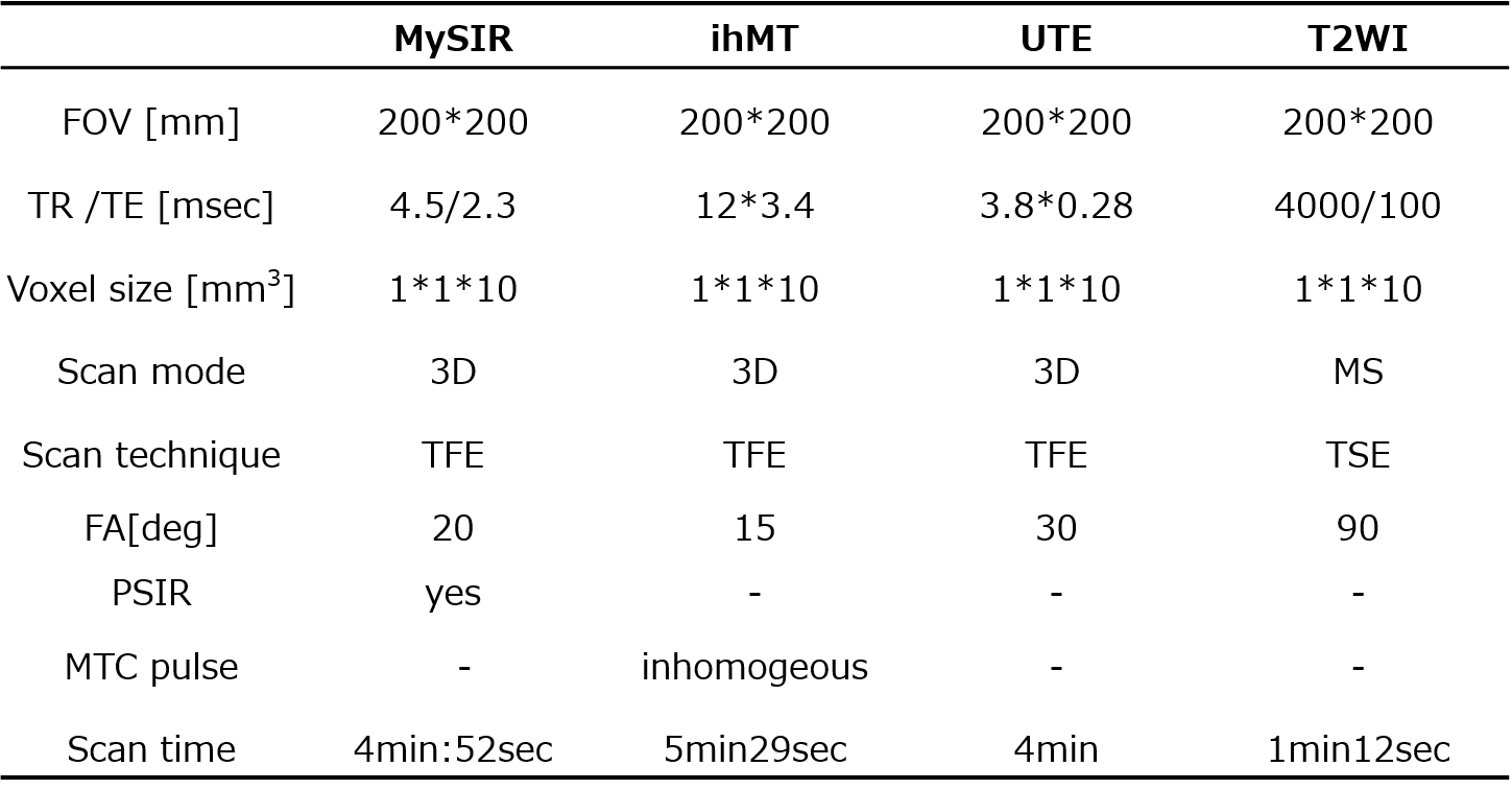
Figure 1: Scan Parameters
The scan parameters were set so that the voxel size was the same.
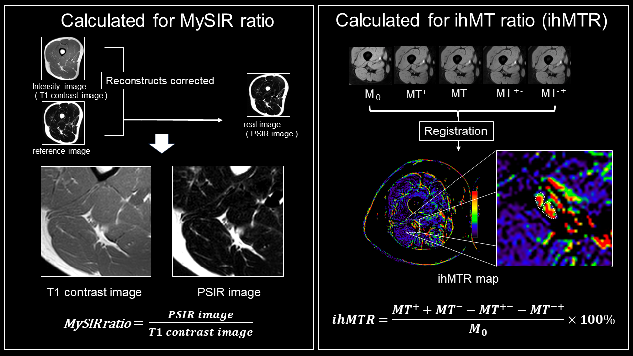
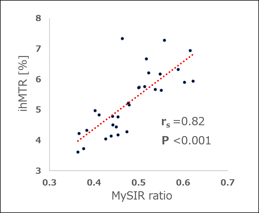
Figure 3 : Correlation between MySIR ratio and ihMTR
Spearman's correlation coefficient was 0.82 (P < 0.01), indicating a strong correlation between MySIR and ihMT.
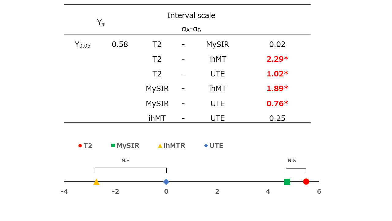
Figure 4 : Results of scheffe's pairwise comparison
T2WI, MySIR, UTE, and ihMT had better ability to separate nerve fascicles in that order. No significant differences were found between T2WI and MySIR, indicating that the spatial resolution of the two sequences is equivalent.
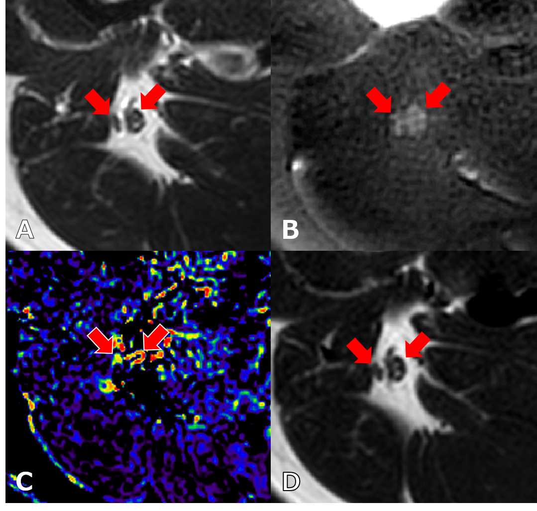
Figure 5 : Images of the sciatic nerve taken during each sequence ( A : T2WI , B : UTE , C : ihMTR , D : MySIR )
UTE had good contrast between the sciatic nerve and background signal but failed to reproduce the ability to separate nerve fascicles comparable to that of T2WI. ihMTR had poor sciatic nerve delineation and is difficult to identify without T2WI reference.