1884
MOTIF-CORD: Motion Integrated Forward Model with Co-Estimated Coil Sensitivity and Regularization by Denoiser for Free Breathing Liver DCE-MRI1Washington University in St. Louis, St. Louis, MO, United States
Synopsis
Keywords: Image Reconstruction, Motion Correction
Motivation: Dynamic contrast-enhanced (DCE) MRI faces challenges from respiratory motion and sub-optimal DCE contrast timing. Free-breathing DCE with high temporal resolution is desirable.
Goal(s): We aim to reconstruct respiratory motion-free and high temporal resolution DCE-MRI.
Approach: We proposed a Motion Integrated Forward model using motion vector fields and jointly estimated coil sensitivity to reconstruct severely under-sampled DCE data. Furthermore, we utilized a model-based deep learning framework to amalgamate the knowledge of the measurement model and the denoising prior.
Results: The proposed method provided deformable motion vector fields, coil-sensitivity maps, and sharp motion-free DCE images without artifacts using highly under-sampled data.
Impact: This method provides good quality free-breathing liver DCE MR images with high temporal resolution. It will eliminate the need for breath-holding. Moreover, continuous acquisition and high temporal resolution reconstruction mitigate the problem of sub-optimal DCE contrast in clinical diagnosis.
Introduction
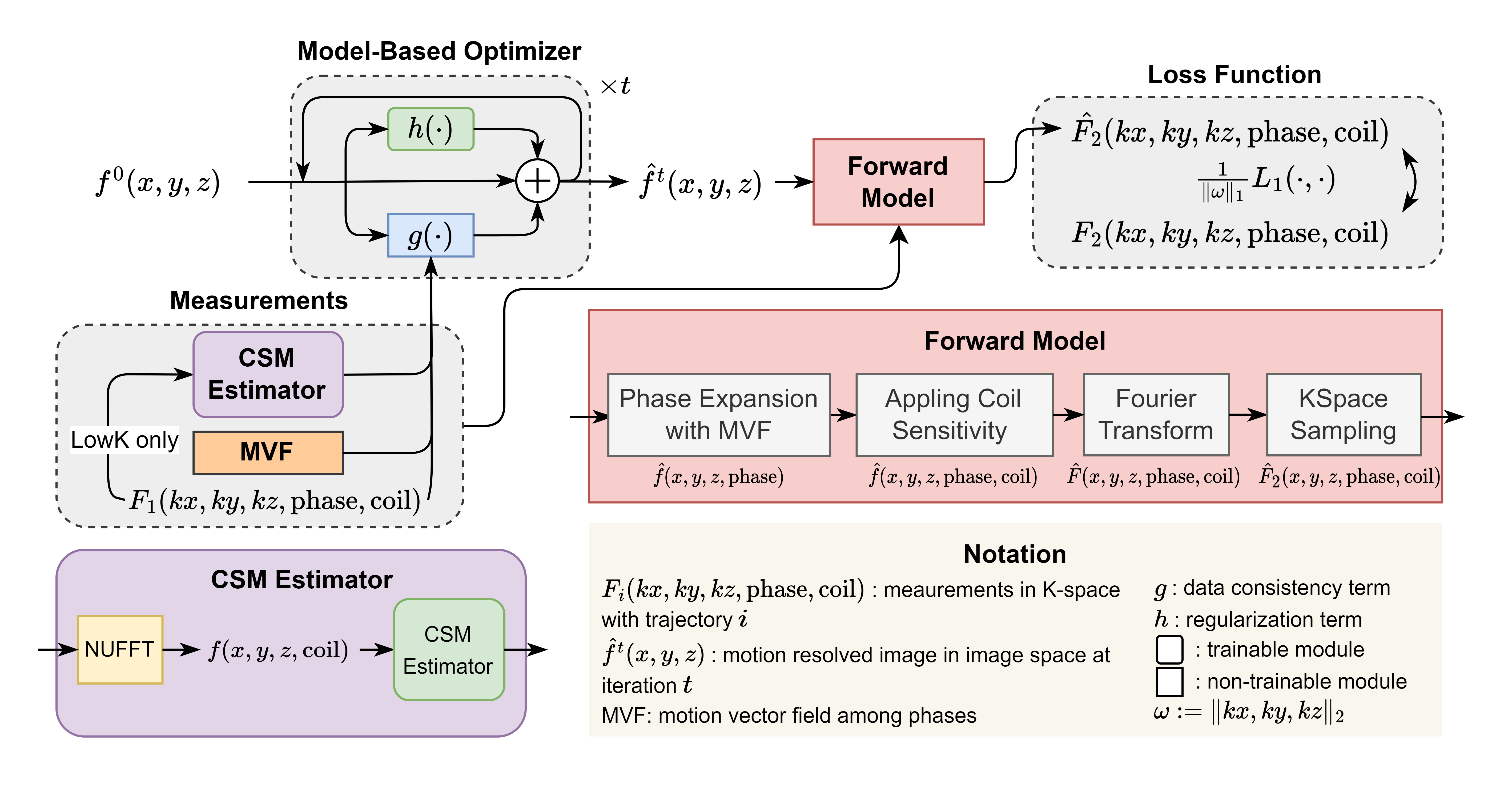
The standard-of-care (SOC) for DCE-MRI typically involves acquiring multiple breath-hold images after contrast injection 1. Breath hold acquisition usually takes about 15-25 seconds, which is challenging for patients 1-3. Moreover, discrete multiple post-contrast phases (e.g. arterial phase, portal venous phases, etc) are often difficult to time appropriately. Any divergence from optimal timing (i.e., too early or too late) can impact diagnosis 1. Free-breathing DCE with continuous acquisition and high temporal DCE reconstruction provide solutions to these challenges.The primary objective in this study is to mitigate respiratory motion artifacts and improve temporal resolution for DCE-MRI. In our study, we presented an integrated method to derive deformable motion vector field (MVF), jointly estimate coil sensitivity, and integrated motion in a physics-based deep learning reconstruction with a learned denoiser prior.
Methods
Data Acquisition and Phase Binning: A self-navigated respiratory motion detection MR sequence, CAPTURE 4, was used to acquire DCE images before, during, and after contrast injection. The DCE images were divided into 34 DCE contrasts with a time interval of 10 seconds, each DCE contrast data (10 seconds) were binned into 5 respiratory phases.4D MRI and Motion Estimation: Phase2Phase (P2P) 5 was employed to reconstruct 4D motion-resolved MRI images from under-sampled k-space data for each DCE contrast. A registration method NODEO 6 was used to estimate 3D deformable MVFs from the 4D MRI images.
Image Reconstruction with Motion Correction: MOTIF-CORD is a physics model-based deep learning (MBDL) algorithm that integrates MVF and uses denoiser as a prior. Briefly, we can formulate our reconstruction as an optimization problem:
$$
\boldsymbol{x}^*=\underset{\boldsymbol{x}}{\arg \min } \ g(\boldsymbol{x})+h(\boldsymbol{x}) ,
$$
where $$$g$$$ is data consistency (DC) term and $$$h$$$ is the regularization term.
The DC is based on the forward model of MR imaging:
$$
\boldsymbol{H}_i^t=\boldsymbol{S}^t \boldsymbol{F} \boldsymbol{C}_i \boldsymbol{M}^t ,
$$
where $$$\boldsymbol{S}^t$$$ is the sampling function of phase $$$t$$$, $$$\boldsymbol{F}$$$ is the forward-Fourier (FFT) operator, $$$\boldsymbol{C}_i$$$ is the coil sensitivity for Coil $$$i$$$ which is also jointly estimated from measurements, $$$\boldsymbol{M}^t$$$ are the MVFs generated from P2P that transform phase $$$1$$$ to the target phase $$$t$$$. Then we can define our DC term:
$$
g(\boldsymbol{x})=\sum_t \sum_i \frac{1}{2}\left\|\boldsymbol{H}_i^t \boldsymbol{x}-\boldsymbol{y}_i^t\right\|_1 ,
$$
then we will define our explicit regularization term:
$$
h(\boldsymbol{x})=\frac{\tau}{2} \boldsymbol{x}^{\top}\left(\boldsymbol{x}-\mathrm{D}_\sigma(\boldsymbol{x})\right) ,
$$
where $$$\mathrm{D}_\sigma$$$ is a learned image denoiser. The gradient of the explicit regularizer has the form of:
$$
\nabla_{\boldsymbol{x}} h(\boldsymbol{x})=\tau\left(\boldsymbol{x}-\mathrm{D}_\sigma(\boldsymbol{x})\right) .
$$
The gradient of the loss function is:
$$
\mathrm{G}(\boldsymbol{x})=\nabla g(\boldsymbol{x})+\tau\left(\boldsymbol{x}-\mathrm{D}_\sigma(\boldsymbol{x})\right) .
$$
We can apply RED7,12 variant of deep unrolling8,9 (DU) as an optimizer, and the update step of each unrolling layer is:
$$
\boldsymbol{x}^k=x^{k-1}-\gamma \mathrm{G}\left(x^{k-1}\right).
$$
Training of Denoiser and CSM Estimator: The training was conducted using an end-to-end approach with the DU structures. A Noise2Noise10 (N2N) was employed as training strategy, which allows for training with only noisy measurements.
Results
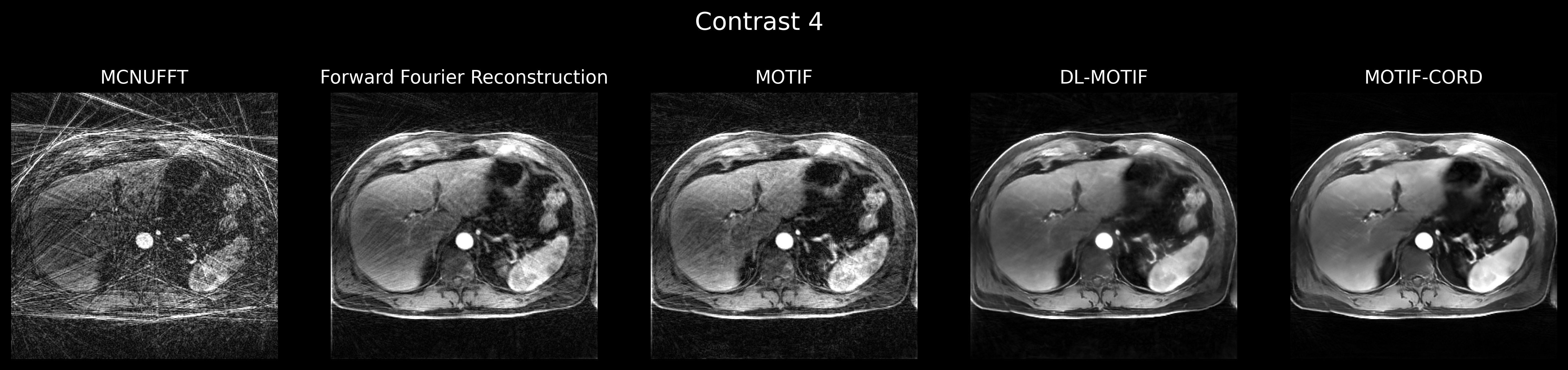
For comparative, we selected several reconstruction methods:MCNUFFT: Using end-of-expiration phase acquisition data, which constitutes 1/5 of the total acquired data.
Fourier-Forward11 (FF): Utilizing all the acquired data. The reconstruction is based on gradient descent to minimize the DC, without integrating MVF.
MOTIF: Similar to FF, except the forward model integrates the MVF.
DL-MOTIF: Similar to MOTIF, except the loss consists of both the DC and regularizer.
MOTIF-CORD: Using all the acquired data and utilizing DU structure to optimize the loss function and jointly estimate CSMs.
As depicted in Figure 2, the reconstructed images at Contrast 4 (corresponding to the early arterial phase) are presented. MCNUFFT is characterized by streaking artifacts. Although the FF, MOTIF and DL-MOTIF method reduces these artifacts. MOTIF-SEIR presents the best result compared to previous methods.
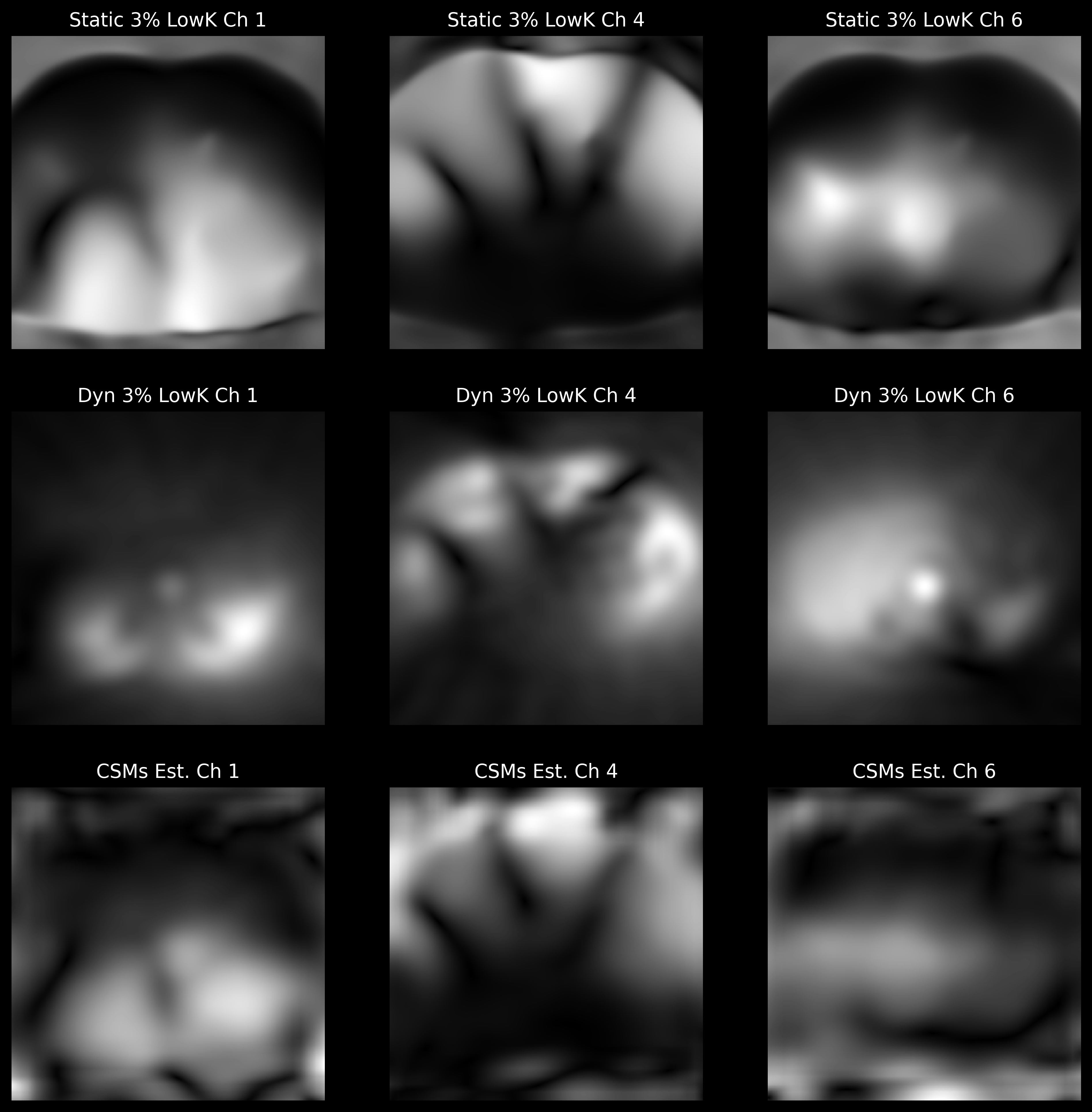
Figure 3 presents a comparison of the estimated CSMs with the widely recognized LowK method. The difference between static and dynamic 3% LowK is to generate single set of CSMs or different CSM for each contrast. Our jointly estimated CSMs exhibit greater smoothness and resemblance to the Static 3% LowK result, particularly within the body.
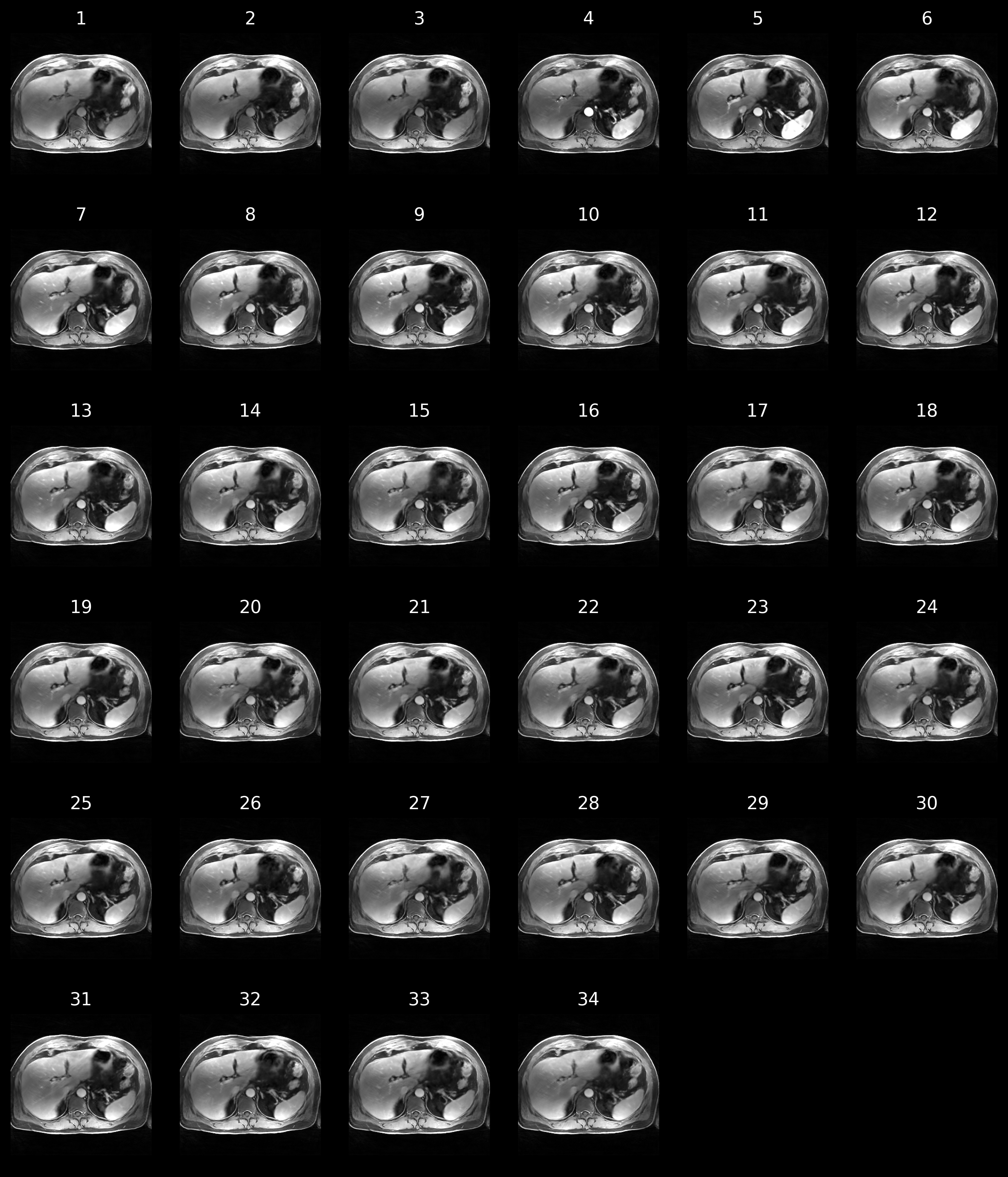
Figure 4 presents the MOTIF-SEIR images across all 34 DCE contrasts from a single free-breathing DCE scan for a patient. These images provide continuous DCE contrasts. Clinical widely-used arterial, portal venous, transitional, and 5-minute delay phases are readily available from these 34 continuous DCE sets.
Conclusion
MOTIF-CORD has improved image quality and accuracy by reconstructing sharp, artifact-free and motion-free DCE-MRI images from severely under-sampled data at a 14.9% of the Nyquist sampling rate.It corrects motion by integrating MVF and all k-space data. Furthermore, physics model based deep learning reconstruction with jointly learned coil sensitivity and denoiser prior provides high quality DCE images with a temporal resolution of 10 seconds.
Acknowledgements
This project is partly supported by funding from Siemens Healthineers.References
- Choyke PL, Dwyer AJ, Knopp MV. Functional tumor imaging with dynamic contrast-enhanced magnetic resonance imaging. Journal of Magnetic Resonance Imaging: An Official Journal of the International Society for Magnetic Resonance in Medicine 2003;17(5):509-520
- Cruz G, Atkinson D, Buerger C, Schaeffter T, Prieto C. Accelerated motion corrected three-dimensional abdominal MRI using total variation regularized SENSE reconstruction. Magnetic resonance in medicine 2016;75(4):1484-1498
- Reiner CS, Neville AM, Nazeer HK, Breault S, Dale BM, Merkle EM, Bashir MR. Contrast-enhanced free-breathing 3D T1-weighted gradient-echo sequence for hepatobiliary MRI in patients with breath-holding difficulties. European radiology 2013;23(11):3087-3093
- Eldeniz C, Fraum T, Salter A, Chen Y, Gach HM, Parikh PJ, Fowler KJ, An H. CAPTURE: Consistently Acquired Projections for Tuned and Robust Estimation: A Self-Navigated Respiratory Motion Correction Approach. Investigative radiology 2018;53(5):293-305
- Eldeniz C, Gan W, Chen S, Fraum TJ, Ludwig DR, Yan Y, Liu J, Vahle T, Krishnamurthy U, Kamilov US, An H. Phase2Phase: Respiratory Motion-Resolved Reconstruction of Free-Breathing Magnetic Resonance Imaging Using Deep Learning Without a Ground Truth for Improved Liver Imaging. Investigative Radiology 2021. doi:10.1097/rli.0000000000000792
- Wu Y, Jiahao TZ, Wang J, Yushkevich PA, Hsieh MA, Gee JC. Nodeo: A neural ordinary differential equation based optimization framework for deformable image registration. InProceedings of the IEEE/CVF Conference on Computer Vision and Pattern Recognition 2022 (pp. 20804-20813).
- Romano Y, Elad M, Milanfar P. The little engine that could: Regularization by denoising (RED). SIAM Journal on Imaging Sciences. 2017;10(4):1804-44.
- Schlemper J, Caballero J, Hajnal JV, Price AN, Rueckert D. A deep cascade of convolutional neural networks for dynamic MR image reconstruction. IEEE transactions on Medical Imaging. 2017 Oct 13;37(2):491-503.
- Aggarwal HK, Mani MP, Jacob M. MoDL: Model-based deep learning architecture for inverse problems. IEEE transactions on medical imaging. 2018 Aug 13;38(2):394-405.
- Lehtinen J, Munkberg J, Hasselgren J, Laine S, Karras T, Aittala M, Aila T. Noise2Noise: Learning image restoration without clean data. arXiv preprint arXiv:1803.04189. 2018 Mar 12.
- Chen S, Eldeniz C, Gan W, Kamilov U, Fraum T, An H. Forward-Fourier Motion-Corrected Reconstruction for Free-Breathing Liver DCE-MRI. Proc Intl Soc Mag Reson Med; 2021. p 0128.
- Liu J, Sun Y, Eldeniz C, Gan W, An H, Kamilov US. RARE: Image reconstruction using deep priors learned without groundtruth. IEEE Journal of Selected Topics in Signal Processing. 2020 May 28;14(6):1088-99.
Figures