1881
Zero-shot self-supervised deep learning reconstruction for abdominal DCE MR multitasking1UIH America, Inc., Houston, TX, United States, 2Cedars-Sinai Medical Center, Los Angeles, CA, United States, 3University of California, Los Angeles, Los Angeles, CA, United States, 4Department of Radiology, Zhongshan Hospital, Fudan University, Shanghai, China, 5United Imaging Healthcare, Shanghai, China
Synopsis
Keywords: Image Reconstruction, Machine Learning/Artificial Intelligence, MR Multitasking, DCE MRI, self-supervised learning, zero-shot learning
Motivation: Deep learning (DL) MR multitasking reconstruction can reduce the reconstruction time, but previous methods are supervised learning, which may learn artifacts from the reference images.
Goal(s): Our goal was to develop a DL reconstruction method that can improve image quality beyond supervised DL and conventional iterative reconstruction.
Approach: We developed a zero-shot self-supervised deep learning method for DCE MR multitasking reconstruction.
Results: With shorter reconstruction time than conventional iterative reconstruction, the proposed method obtained better image quality than both supervised DL and conventional iterative reconstruction methods.
Impact: With the proposed method, DCE MR multitasking can have better image quality with shorter reconstruction time than previous iterative reconstruction, which is essential for the potential clinical application of the motion-resolved and high spatial-temporal-resolution abdominal DCE MR multitasking.
Introduction
Abdominal dynamic contrast-enhanced (DCE) MRI can be used to determine liver function and detect cancer1. However, conventional techniques often suffer from respiratory motion and the trade-off among spatial resolution, temporal resolution and spatial coverage.Recently, MR multitasking2 has shown promising results for motion-resolved and high spatial-temporal-resolution abdomen DCE MRI3. However, its iterative reconstruction is long and subject to intense hyperparameter tuning. Deep learning (DL) reconstruction has shown great potential for reducing MR reconstruction time4, including MR Multitasking5. Most prior deep learning methods are supervised, limiting them from exceeding the imaging quality of their training reference images. Especially in MR multitasking reconstruction, using iterative reconstruction as the training reference due to the lack of ground truth can lead supervised DL to learn their artifacts. To address this, we propose the first zero-shot self-supervised DL reconstruction approach for MR multitasking. We apply this method to abdominal DCE MR multitasking, aiming to improve image quality beyond supervised DL and conventional iterative reconstruction.
Methods
Acquisition and dataset15 abdominal DCE MR Multitasking scans6 were collected from 3T scanners (uMR 780 and 790, United Imaging Healthcare, Shanghai, China), with a 10/1/4 training/validation/testing allocation. The FOV for acquisition was 380*280*210 mm3. The matrix size of spatial factors $$$[N_x,N_y,N_z,L]$$$ was [256,89,51,15], where $$$L$$$ is the number of ranks. The phase-encoding plane $$$[N_y,N_z]$$$ was treated as slice in the 2D network, and the $$$L$$$ dimension was treated as channels in the network.
Reconstruction methods
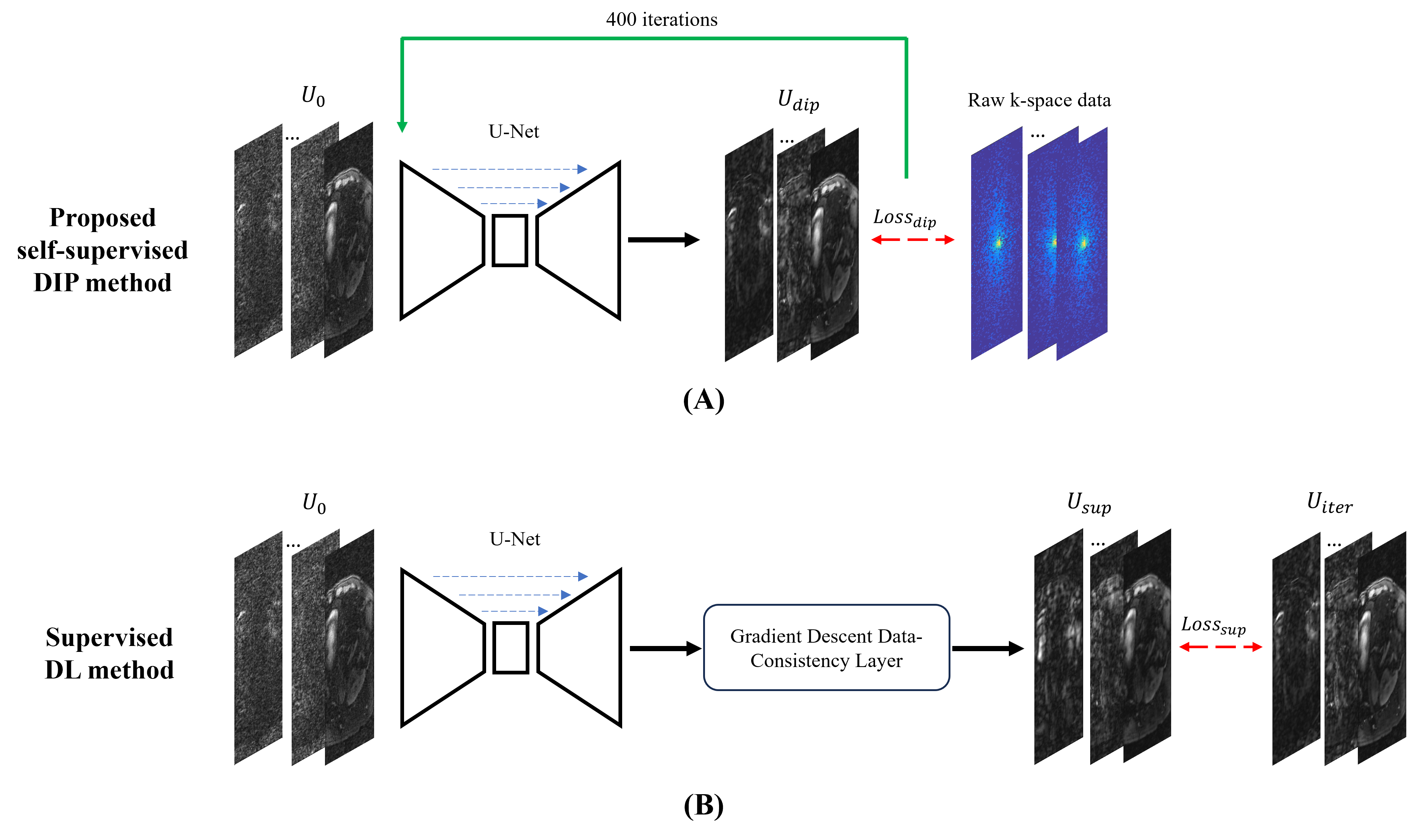
The proposed method applied deep image prior (DIP)7,8 for self-supervised MR multitasking reconstruction. It did not require pre-training but was iteratively trained with a single scan that needs to be reconstructed.
For comparison, three reconstruction methods were used to determine the spatial factor U in MR Multitasking:
a) The proposed self-supervised DIP reconstruction $$$U_{dip}$$$ (Figure 1A).
b) Supervised DL multitasking reconstruction5 $$$U_{sup}$$$ (Figure 1B).
c) Conventional iterative reconstruction $$$U_{iter}$$$ that uses 10 ADMM Total Variation (TV)-thresholding + CG-DC iterations2.
The inputs for all methods were zero-filled reconstruction $$$U_{0}$$$. Loss function for the proposed DIP method is the L2 loss in k-space:
$$Loss_{dip}=\left\|AU_{dip}-b\right\|_2^2=U_{dip}^HA^HAU_{dip}-2Re(U_{dip}^HA^Hb)+b^Hb$$
Where $$$U_{dip}$$$ is the spatial factor, $$$b$$$ is acquired k-space, and $$$A$$$ is the encoding operator that includes sensitivity maps, Fourier transform, undersampling and temporal factors5. In each DIP reconstruction, $$$Loss_{dip}$$$ is optimized for 400 iterations.
Loss function for supervised DL reconstruction is L2 loss of spatial factors compared to iterative reconstruction references:
$$Loss_{sup}=\left\|U_{sup}-U_{iter}\right\|_2^2$$
Note that only supervised DL uses the training and validation sets. The DIP method is a zero-shot method, which does not need pre-training and is only applied to reconstruct the testing set.
Evaluation methods
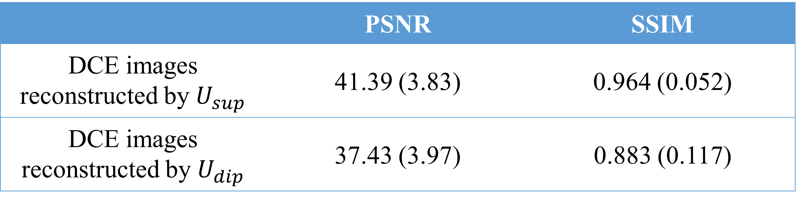
The DCE images reconstructed by $$$U_{dip}$$$ and $$$U_{sup}$$$ were compared against the iterative reconstruction $$$U_{iter}$$$ using PSNR and SSIM.
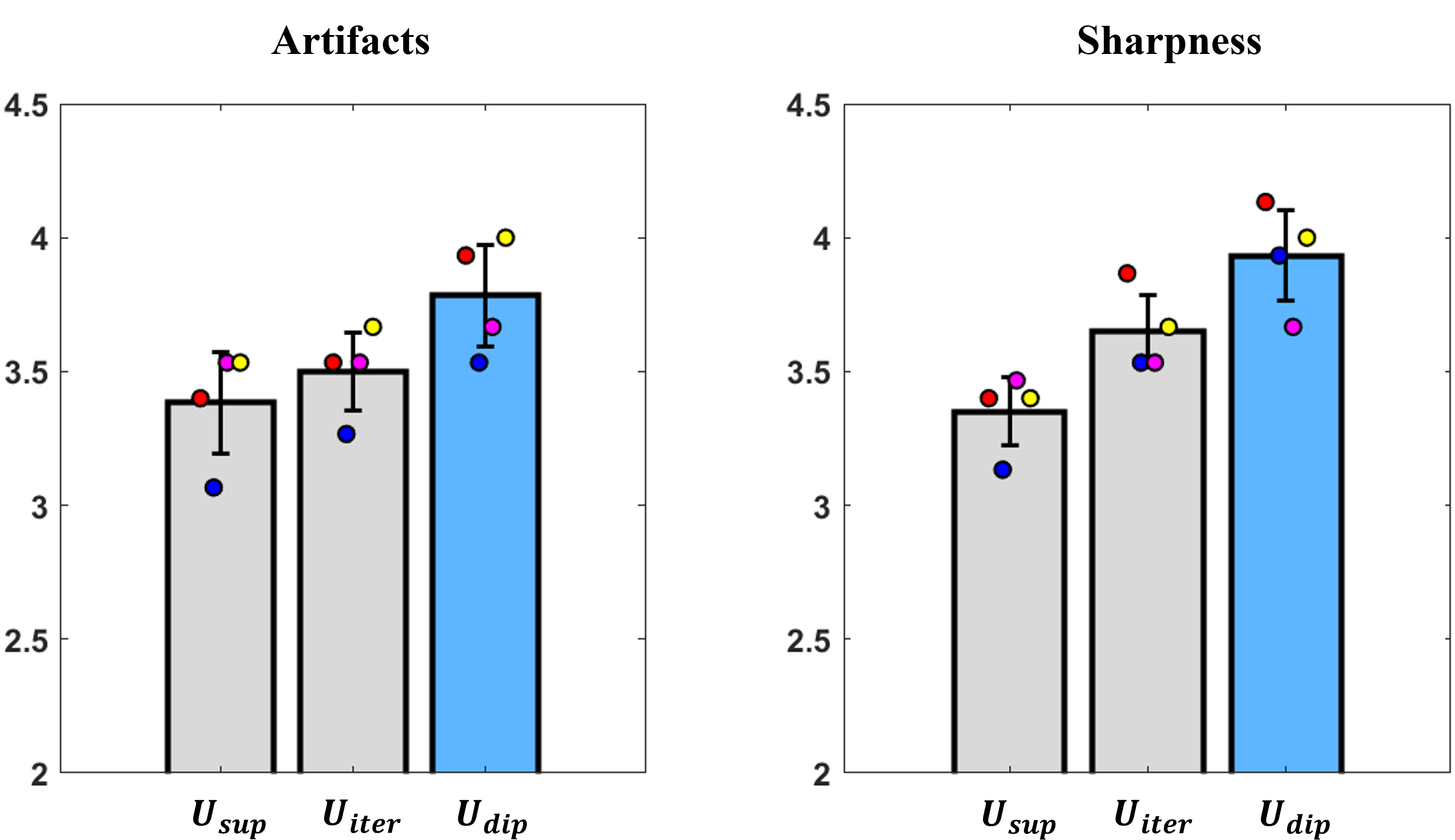
However, it is not necessary to resemble iterative reconstruction for a good-quality DL reconstruction since iterative reconstruction is not ground truth. Therefore, we also performed blind imaging quality ratings. Artifact level (1, nondiagnostic; 2, severe; 3, moderate; 4, mild; 5, minimum) and image sharpness (1, nondiagnostic; 2, poor; 3, adequate; 4, good; 5, excellent) were evaluated by three experienced doctors and then averaged.
Results
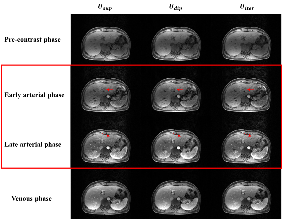
For each scan, reconstruction time of $$$U_{sup}$$$, $$$U_{dip}$$$ and $$$U_{iter}$$$ are 30s, 20min and 30min respectively. Figure 2 shows the averaged PSNR and SSIM of DCE images. All the calculations are done slice-by-slice, and there are about 80 DCE time frames for each slice. Supervised learning method surpasses the proposed DIP method in PSNR and SSIM. However, in the imaging quality ratings (Figure 3), the proposed method outperforms both supervised DL method and iterative reconstruction, for both artifact level and sharpness. Figure 4 shows example DCE images reconstructed by $$$U_{sup}$$$, $$$U_{dip}$$$ and $$$U_{iter}$$$ at different enhancement phases. The proposed DIP method shows the least artifacts, especially in the early and late arterial phases (red box in Figure 4).Discussion
Although $$$U_{dip}$$$ has lower PSNR and SSIM compared to $$$U_{sup}$$$, it achieved higher image quality ratings by doctors. It suggests that quantitative image metrics may conflict with qualitative image rating when the reference image is not ground truth, and it is important to obtain doctors’ opinions for evaluation of deep learning methods.In this study, DIP method is faster than iterative method, but it is still long compared to supervised DL. Future work will include how to shorten the reconstruction time of DIP for clinical application.
Conclusion
We proposed a zero-shot self-supervised DL reconstruction method for abdominal DCE MR multitasking. The method obtained better image quality than both supervised DL and conventional iterative reconstruction methods.Acknowledgements
This work was partially facilitated by a non-exclusive license agreement between Cedars-Sinai Medical Center and United Imaging Healthcare.References
1. Johansson A, Balter JM, Cao Y. Abdominal DCE-MRI reconstruction with deformable motion correction for liver perfusion quantification. Med Phys. 2018;45(10):4529-4540. doi:10.1002/mp.13118
2. Christodoulou AG, Shaw JL, Nguyen C, et al. Magnetic resonance multitasking for motion-resolved quantitative cardiovascular imaging. Nat Biomed Eng. 2018;2(4):215-226. doi:10.1038/s41551-018-0217-y
3. Wang N, Gaddam S, Wang L, et al. Six‐dimensional quantitative DCE MR Multitasking of the entire abdomen: Method and application to pancreatic ductal adenocarcinoma. Magn Reson Med. 2020;84(2):928-948. doi:10.1002/mrm.28167
4. Yang G, Yu S, Dong H, et al. DAGAN: Deep De-Aliasing Generative Adversarial Networks for Fast Compressed Sensing MRI Reconstruction. IEEE Trans Med Imaging. 2018;37(6):1310-1321. doi:10.1109/TMI.2017.2785879
5. Chen Z, Chen Y, Xie Y, Li D, Christodoulou AG. Data-Consistent Non-Cartesian Deep Subspace Learning for Efficient Dynamic MR Image Reconstruction. In: 2022 IEEE 19th International Symposium on Biomedical Imaging (ISBI). IEEE; 2022:1-5. doi:10.1109/ISBI52829.2022.9761497
6. Liu Q, Liu Q, Li J, et al. 3D free-breathing renal DCE MRI with high spatial and temporal resolution using Multitasking. In: Proc. Intl. Soc. Mag. Reson. Med. 31 (2023).
7. Ulyanov D, Vedaldi A, Lempitsky V. Deep Image Prior. Int J Comput Vis. 2020;128(7):1867-1888. doi:10.1007/s11263-020-01303-4
8. Hamilton JI. A Self-Supervised Deep Learning Reconstruction for Shortening the Breathhold and Acquisition Window in Cardiac Magnetic Resonance Fingerprinting. Front Cardiovasc Med. 2022;9:928546. doi:10.3389/fcvm.2022.928546
Figures