1880
Plug and play truncated low rank optimization combined iterative soft thresholding (PNPT) for dynamic MRI reconstruction1Center for Biomedical Imaging Research, Department of Biomedical Engineering, Tsinghua University, Beijing, China
Synopsis
Keywords: Image Reconstruction, Heart, Reconstruction
Motivation: Try to obtaining high spatial and temporal resolution image in dynamic MRI.
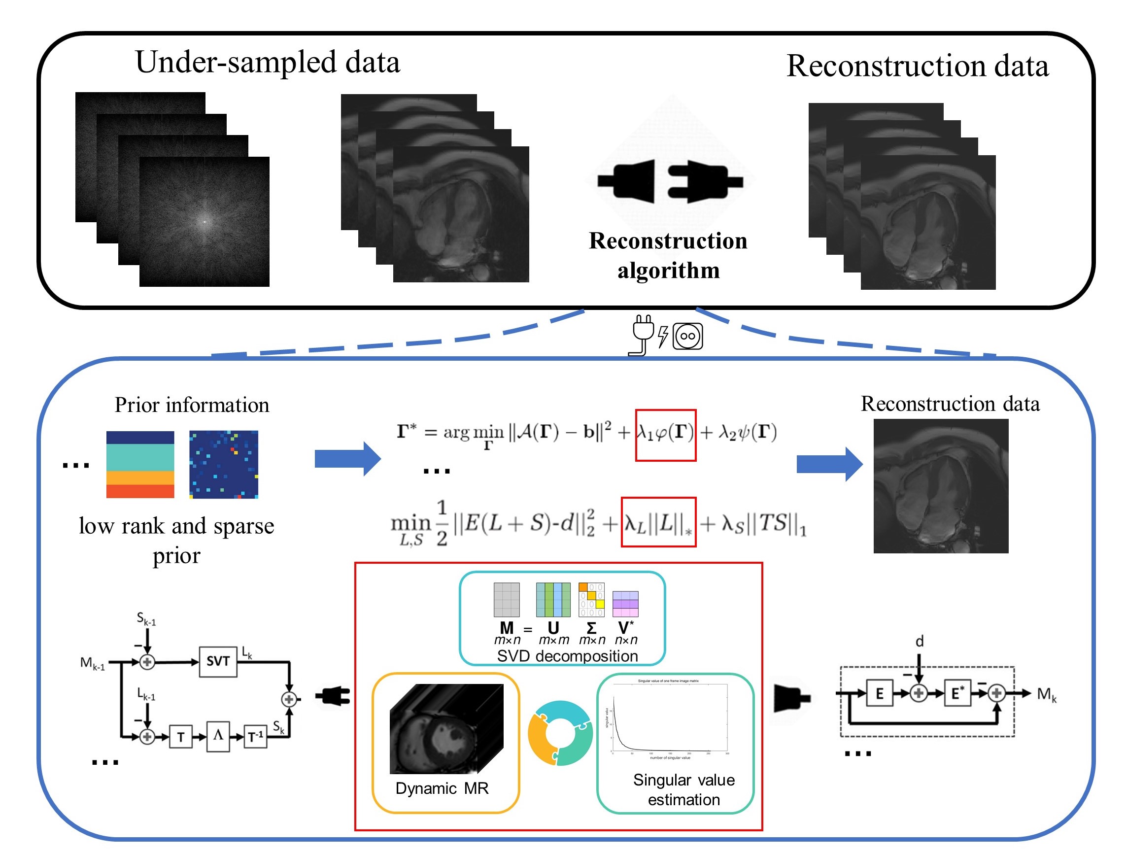
Goal(s): The plug and play truncated low rank optimization combined iterative soft thresholding(PNPT-) embedded method was proposed to reconstructe data and tested to accurately estimate the rank function for dMRI reconstruction.
Approach: The PNPT- method uses truncated nuclear norm combined iterative soft thresholding to assign different shrink values to different singular vectors, enabling the preservation of essential image information while effectively eliminating noise.
Results: The embedded method was proposed to reconstructe data and tested to accurately estimate the rank function for dMRI reconstruction.
Impact: The proposed method demonstrated reduced aliasing artifacts compared to other methods and yielded better reconstruction images in cine and perfusion data. Consequently, it provided more accurate and efficient image information, benefiting the clinical diagnosis of heart-related diseases.
Introduction
Dynamic magnetic resonance imaging (dMRI) plays an important role in clinical applications [1]. Nevertheless, achieving high spatial and temporal resolution images in dynamic MRI poses a significant challenge due to the constraints imposed by the Nyquist criterion [2,3]. One primary approach involves under-sampling the raw data acquisition and utilizing advanced reconstruction techniques to enhance the quality of the image, such as kt-SLR[4] and L+S [5]. However, both approaches encounter imprecise challenges in solving singular values with low rank constraints, potentially resulting in inaccurate suboptimal outcomes. Truncated nuclear norm [6,7] to assign different shrink values to different singular vectors, enabling the preservation of essential image information while effectively eliminating noise. It can be embedded into various reconstruction methods using low rank constraints. Iterative Soft Thresholding (IST) algorithm [5] exhibits a reduced parameter count and superior solving efficiency. Hence, the plug and play truncated low rank optimization combined iterative soft thresholding (PNPT-) embedded method was proposed to reconstructed data and tested to accurately estimate the rank function for dMRI reconstruction.Method
Theory: Generally, the discretization form of the dynamic MRI method can be presented as:$$d = E\left( X \right) + n \tag{1}$$ where $$$X \in {\mathbb{C}^{{N_x}{N_y} \times {N_t}}}$$$ denotes the image series that expands along the time dimension,$$$\mathbb{C}$$$ denotes the complex set, $$${N_x},{N_y}$$$ and $$${N_t}$$$ denote the width, height and the total time frames, respectively;$$$d$$$ denotes the stacked $$$\left( {{\mathbf{k}},t} \right)$$$ space acquired data vector;$$$E$$$ denotes the MRI encoding operator modeling the under-sampling mask, Fast Fourier Transform (FFT) and coil sensitivity,$$$n$$$ denotes the noise.Since the $$$X$$$ in dMRI usually can be assumed to have a low-rank structure [4,5] , given $$$l = \min \left( {{N_x} \times {N_y},{N_t}} \right)$$$.the rank’s truncated parameter $$$t$$$.The truncated nuclear norm is defined as follows:$${\left\| {{X}} \right\|_{t,*}} = \mathop \sum \limits_{i = t + 1}^l {\sigma _i}\left( X \right) \tag{2} $$where $$${\sigma _i}\left( \cdot \right)$$$ is the $$$i$$$th largest eigenvalue of $$$X$$$. To solve the truncated nuclear norm minimization problem, the von Neumann’s lemma [8-10] were used. And Iterative Soft Thresholding (IST) algorithm is used in combination to improve the solving efficiency.The truncated nuclear norm can be embedded into kt-SLR and L+S methods. Therefore, PNPT-SLR and PNPT-L+S method can be obtained as follows respectively:$$\mathop {\min }\limits_X {\left\| X \right\|_{t,*}} + {\mu _x}{\left\| {TX} \right\|_1}\;{\rm{s}}.{\rm{t}}.\;E\left( X \right) = d\tag{3}$$ $$\mathop {\min }\limits_{L,S} {\left\| L \right\|_{t,*}} + {\mu _S}{\left\| {TS} \right\|_1}\;{\rm{s}}.{\rm{t}}.\;X = L + S,E\left( {L + S} \right) = d\tag{4}$$where $$${\mu _x}$$$ and $$${\mu _s}$$$ are constraint operators.Retrospective experiments: A cardiac cine dataset (CineSet) [11] and a perfusion dataset (PerfusionSet) [4,12] acquired with three simulated undersampling factors (R) of 2, 4 and 6, were used to verify the method. The CineSet contains cardiac cine MR images of randomly selected 8 cases from the OCMR dataset [11]. The PerfusionSet contains 2 public cardiac perfusion MR data from [4] and [12].The Cartesian masks with varying sampling ratios [13] were used in experiments.Reconstruction performance was evaluated using the Peak Signal to Noise Ratio (PSNR) and Root Mean Squared Error (RMSE) .The regions of interest (ROI) for the blood pool and myocardium in perfusion data were manually sketched.
Results
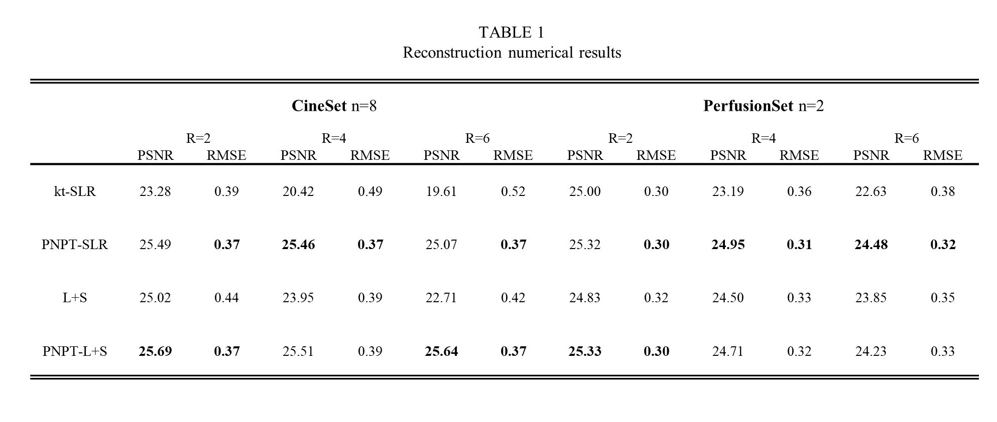
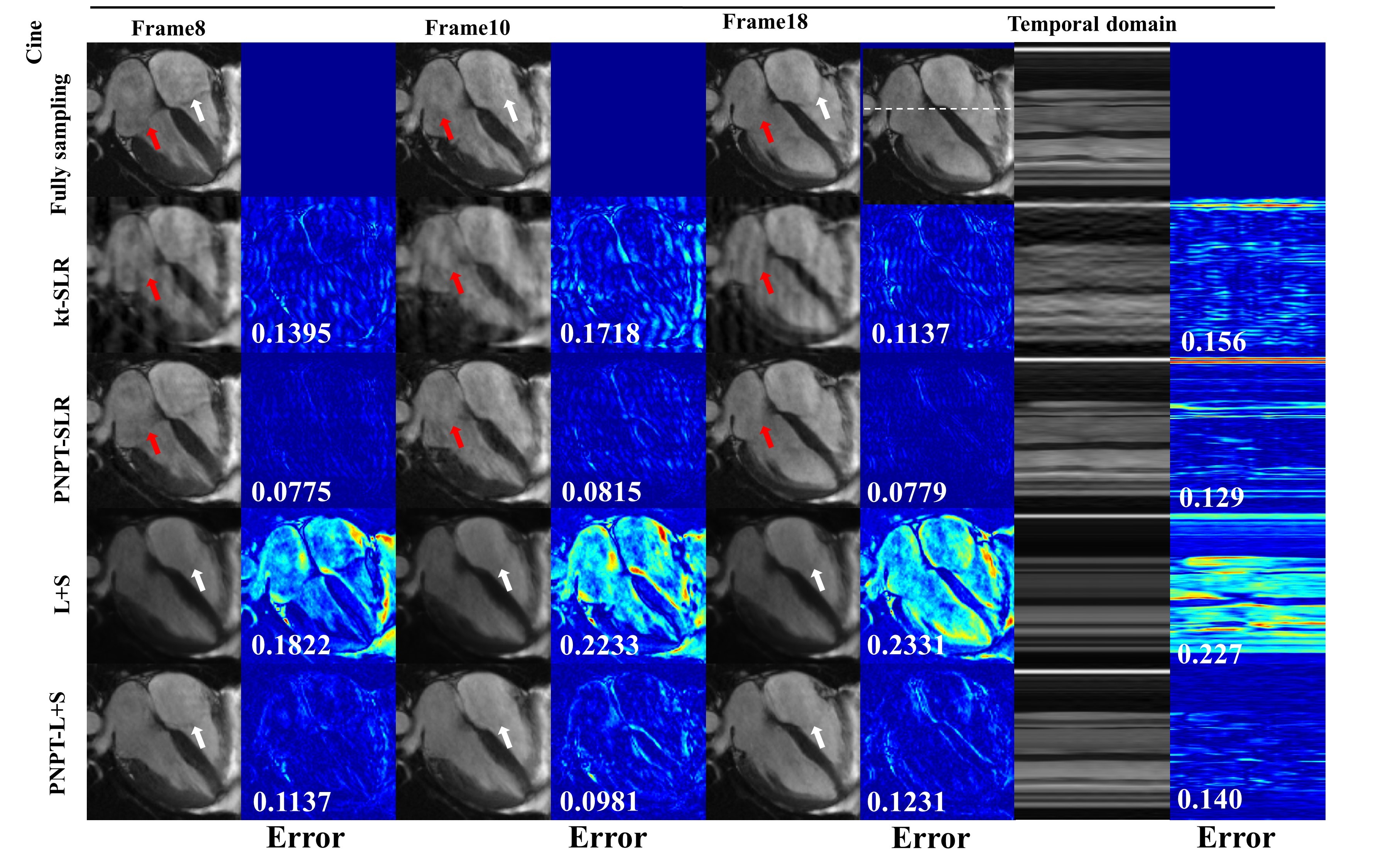
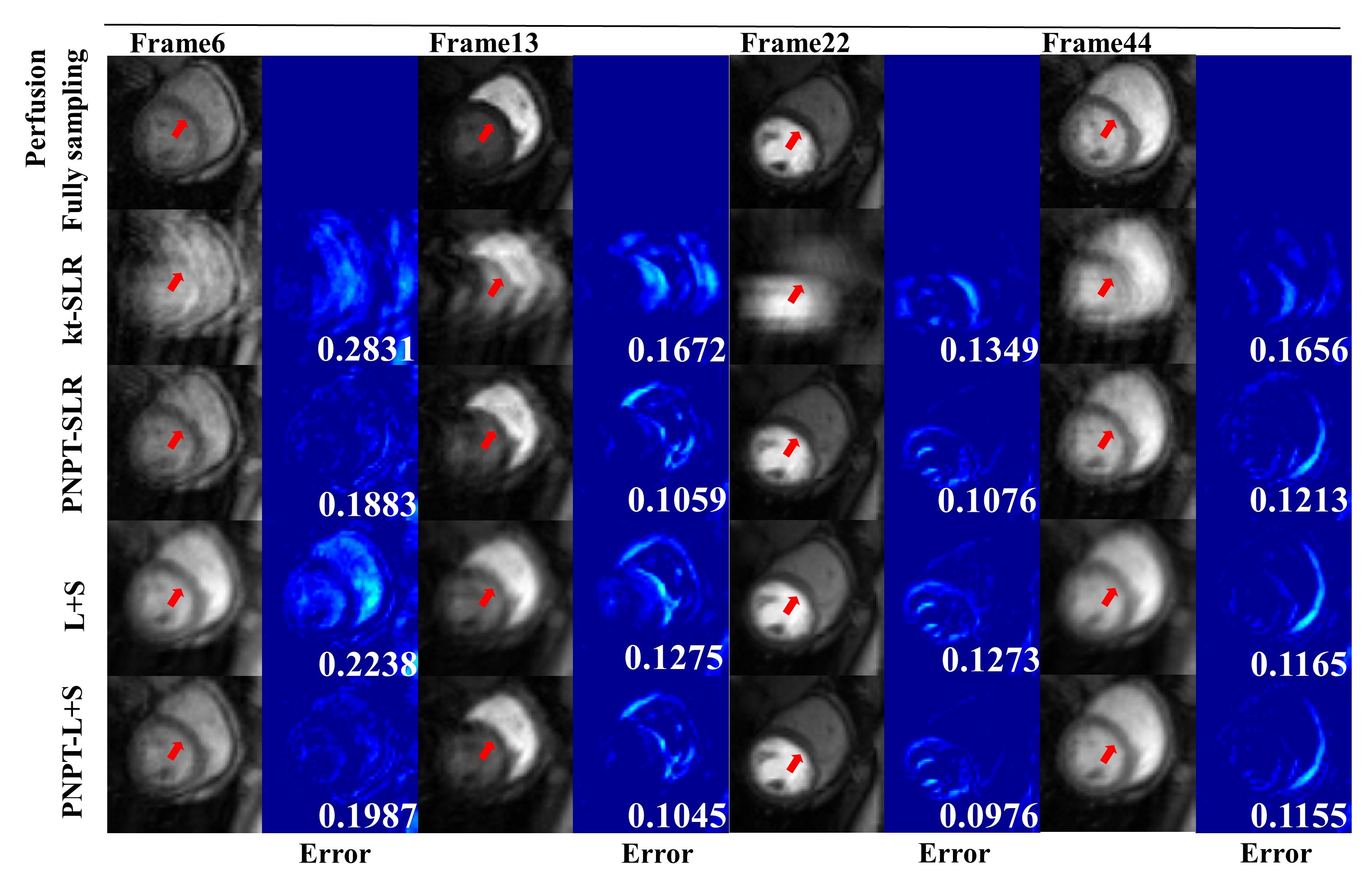
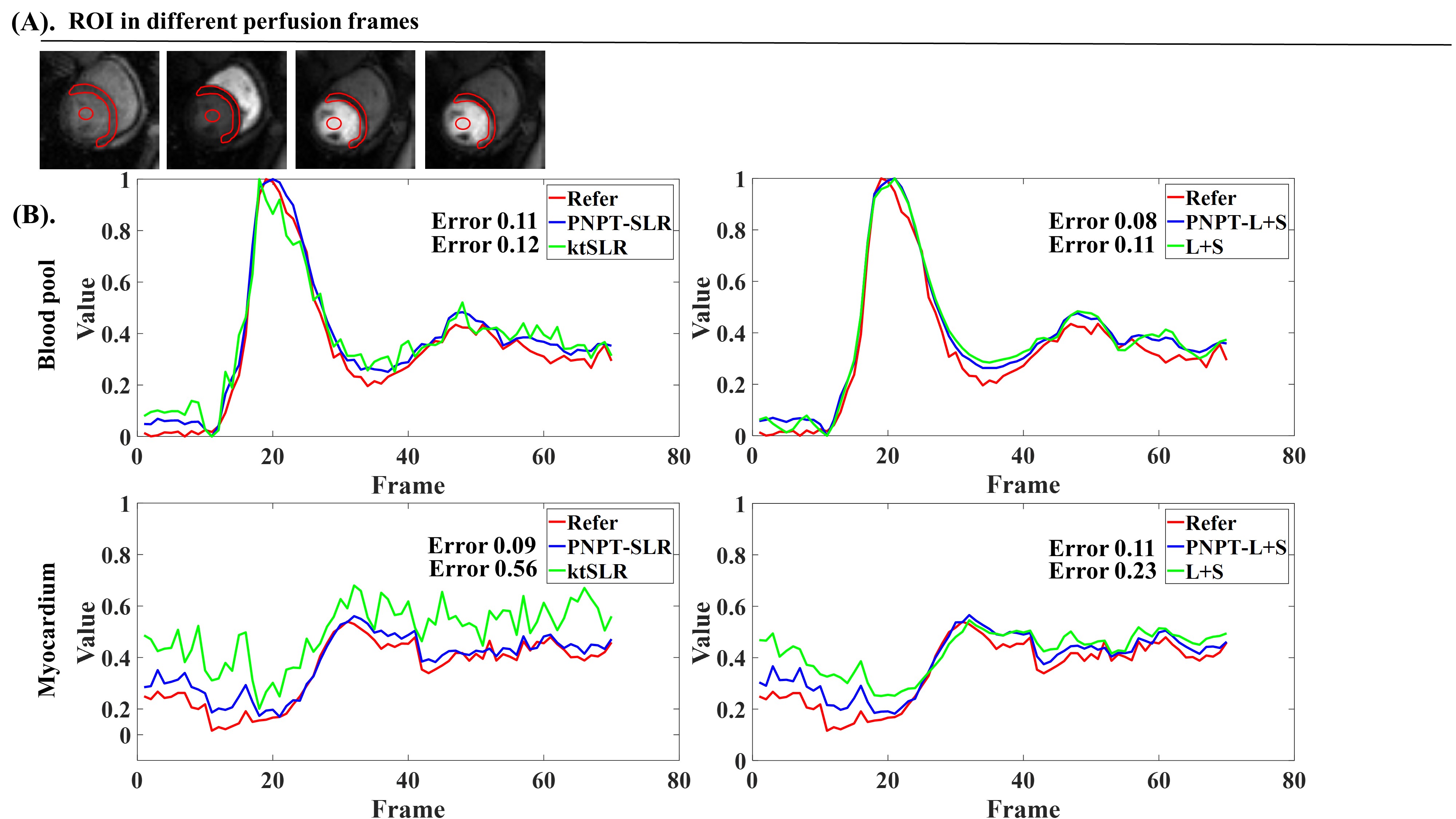
Firstly, the proposed embedded methods consistently attained the highest PSNR and the lowest RMSE as shown in Table 1. In Figure 2, the reconstructed images obtained from kt-SLR reveal noticeable noise in spatial domain, contrasting with the PNPT-SLR that exhibits significantly reduced noise levels in the corresponding images as shown by the red arrows. L+S offers a smoother image, whereas PNPT-L+S renders the heart's mitral valve more distinctly as shown by the white arrows. In general, the proposed embedded methods have smaller errors in both spatial and temporal dimensions. From Figure. 3, the images reconstructed by kt-SLR and L+S methods had strong blurring artifacts, especially in the boundary area of the interventricular septum due to inaccuracy utilization of the low-rank prior. In contrast, the proposed embedding method effectively highlighted the boundary of the interventricular septum over time as shown by the red arrows, surpassing the performance of the other methods. Due to improve the low rank constraints, the proposed method can more accurately depict the intensity change of contrast agent over time as show in Figure. 4. Notably, as the contrast agent concentration increased, the curve derived from the proposed method closely matched the ground truth and exhibited fewer fluctuations compared to other methods.Conclusions
The plug and play truncated low rank optimization combined iterative soft thresholding embedded method was proposed to reconstruct data. The proposed model has an improved approximation effect for low-rank prior.Acknowledgements
None
References
[1].Z. Liang, H. Jiang, C. Hess, and P. Lauterbur, “Dynamic imaging by model estimation,” Int. J. Imag. Syst. Technol., vol. 8, no. 6, pp. 551–557, 1997
[2]Sodickson, D.K. , Manning, W.J. , Simultaneous acquisition of spatial harmonics (smash): fast imaging with radiofrequency coil arrays. Magnetic Resonance in Medicine. 1997, 38 (4), 591–603.
[3]Yan L, Wang S, Zhuo Y, Wolf RL, Stiefel MF, An J, Ye Y, Zhang Q, Melhem ER, Wang DJ. Unenhanced dynamic MR angiography: high spatial and temporal resolution by using true FISP-based spin tagging with alternating radiofrequency. Radiology. 2010 Jul;256(1):270-9.
[4]Lingala S G , Hu Y , Dibella E , et al. "Accelerated Dynamic MRI Exploiting Sparsity and Low-Rank Structure: k-t SLR. " IEEE Transactions on Medical Imaging, 2011, 30(5):1042-1054
[5]Otazo R , Candès, Emmanuel, Sodickson D K. "Low‐rank plus sparse matrix decomposition for accelerated dynamic MRI with separation of background and dynamic components. " Magnetic Resonance in Medicine, 2015, 73(3):1125-1136.
[6]Cai J-F, Cande`s E, Shen Z. A singular value thresholding algorithm for matrix completion. SIAM J Optim 2010;20:1956–1982.
[7]Daubechies I, Defrise M, De Mol C. An iterative thresholding algorithm for linear inverse problems with a sparsity constraint. Comm Pure Appl Math 2004;57:1413–1457.
[8] Le T , Tao P D . The DC (Difference of Convex Functions) Programming and DCA Revisited with DC Models of Real World Nonconvex Optimization Problems. Annals of Operations Research, 2005, 133:23-46
[9] J. Von Neumann, Some matrix-inequalities and metrization of matric space, 1937.
[10] L. Mirsky, A trace inequality of John von Neumann, Monatshefte fur mathematik ,1975, 79 (4) (1975) 303–306.
[11] Partridge M., and Jabri M., Robust Principal Component Analysis,Neural Networks for Signal Processing X, IEEE Signal Processing Society Workshop IEEE, 2002.
[12] Otazo R, Kim D, Axel L, Sodickson DK. Combination of compressed sensing and parallel imaging for highly accelerated first-pass cardiac perfusion MRI. Magn Reson Med. 2010 Sep;64(3):767-76.
[13]. Yao J, Xu Z, Huang X, Huang J. An efficient algorithm for dynamic MRI using low-rank and total variation regularizations. Med Image Anal. 2018 Feb;44:14-27.
Figures