1878
MULTI-PHASE SPATIAL RECONSTRUCTION METHOD FOR ACCELERATED DYNAMIC CONTRAST-ENHANCED MRI1The Edward S. Rogers Sr. Department of Electrical and Computer Engineering, University of Toronto, Toronto, ON, Canada, 2Institute of Biomedical Engineering, Toronto, ON, Canada, 3Ted Rogers Centre for Heart Research, Translational Biology & Engineering Program, University of Toronto, Toronto, ON, Canada
Synopsis
Keywords: Image Reconstruction, Perfusion, Dynamic Contrast Enhancement, Real-Time MRI
Motivation: Dynamic contrast-enhanced (DCE) MRI requires both high spatial and high temporal resolution for accurate quantification and delineation. In practice, temporal resolution is often traded for spatial resolution and volume coverage.
Goal(s): To develop a reconstruction method that offers both high spatial and temporal resolution.
Approach: We describe a two-stage reconstruction technique that consistently produces high-temporal, high-spatial resolution estimates of the ground truth data and is more accurate than current state-of-the-art methods.
Results: The proposed method achieves high quality reconstruction with as few as one acquired spoke per frame when radial sampling is used.
Impact: The theoretical and practical success of the spatial subspace method over temporal subspace methods encourages further research on the topic. Furthermore, reconstruction at 1 spoke per frame enables larger imaging volumes, thus more capable imaging tools.
Synopsis
Motivation: Dynamic contrast-enhanced (DCE) MRI requires both high spatial and high temporal resolution for accurate quantification and delineation. In practice, temporal resolution is often traded for spatial resolution and volume coverage.Goal: To develop a reconstruction method that offers both high spatial and temporal resolution.
Approach: We describe a two-stage reconstruction technique that consistently produces high-temporal, high-spatial resolution estimates of the ground truth data and is more accurate than current state-of-the-art methods.
Results: The proposed method achieves high quality reconstruction with as few as one acquired spoke per frame when radial sampling is used.
Impact
The theoretical and practical success of the spatial subspace method over temporal subspace methods encourages further research on the topic. Furthermore, reconstruction at 1 spoke per frame enables larger imaging volumes, thus more capable imaging tools.Introduction
Dynamic contrast-enhanced (DCE) MRI ideally requires both high spatial and high temporal resolution; however, hardware limitations prevent acquisitions from simultaneously achieving both. Specialized pulse sequences and retrospective reconstruction techniques can artificially create the spatial resolution at a given temporal resolution by estimating the data that is not acquired, but, ultimately, spatial details are sacrificed at very high acceleration rates. The purpose of this work is to develop an acquisition and reconstruction method based on spatial subspace estimation to enable high spatial resolution image reconstruction from as few as one spoke in k-space.$$\hat{x}_{_L} = \underset{x_{_L}}{\mathrm{argmin}} ||\mathrm{E} x_{_L} - y_{_L}||_2^2 + \lambda ||\Phi(x_{_L})||_1$$
where is the estimated low temporal resolution dataset at $$$L$$$ spokes per frame; $$$\mathrm{E}$$$ is an encoding operator that includes the Fourier transform, coil sensitivity, and undersampling; $$$x_{_L}$$$ is a candidate for the final estimate of the dataset; $$$y_{_L}$$$ are vectorized undersampled measurements; $$$\Phi$$$ is a sparsity transform; and $$$\lambda$$$ is a weighting coefficient.
Singular value decomposition is then used to determine the most significant basis vectors, which are retained and stored in $$$\mathrm{U_b}$$$. Then, the acquired data is re-organized to $$$H$$$ spokes per frame, and a high-temporal resolution reconstruction is performed by solving Eq. [2]:
$$\hat{x}_{\mathrm{H}} = \mathrm{U_b} \hat{\mathrm{A}}_{\mathrm{H}}\\
\hat{\mathrm{A}}_{\mathrm{H}} = \underset{x_{_H}}{\mathrm{argmin}} ||\mathrm{EU_bA_H} - y_{_H}||_2^2 + \lambda ||\Phi(\mathrm{U_bA_H})||_1$$
Results
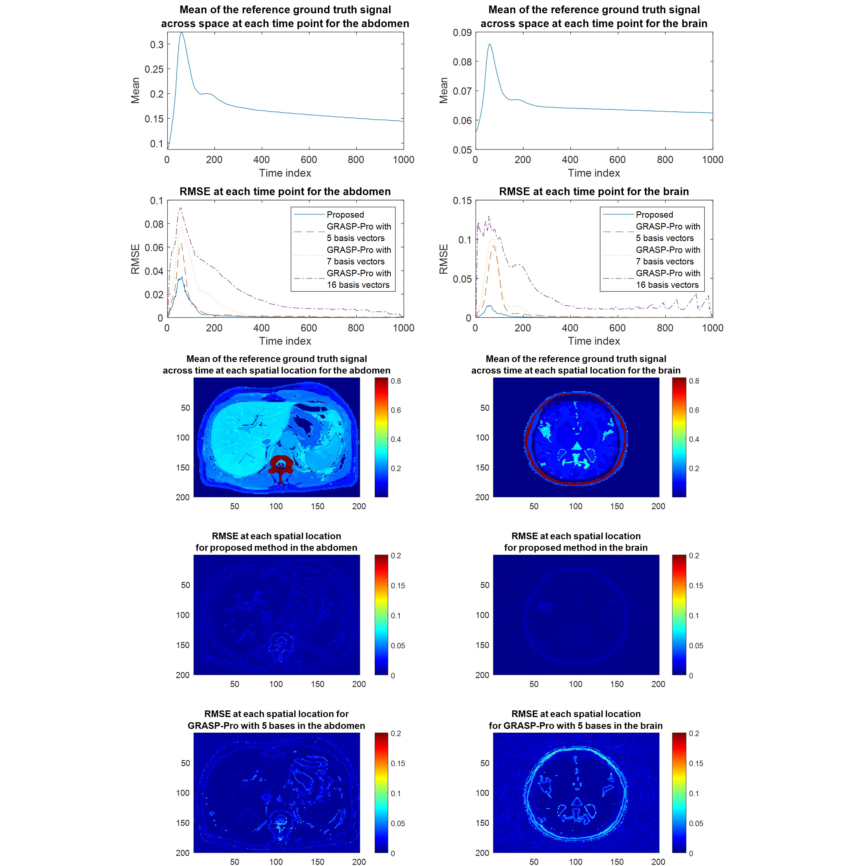
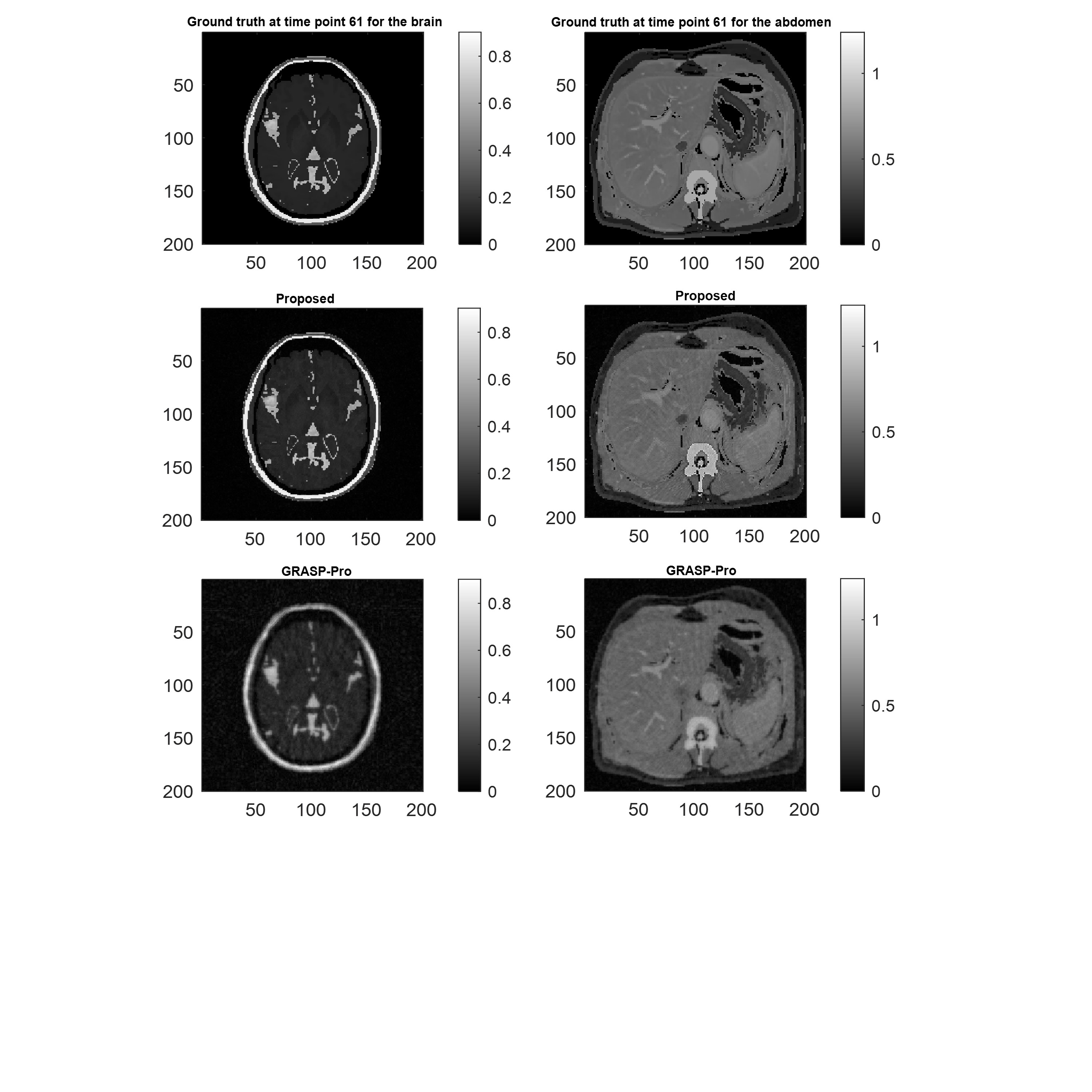
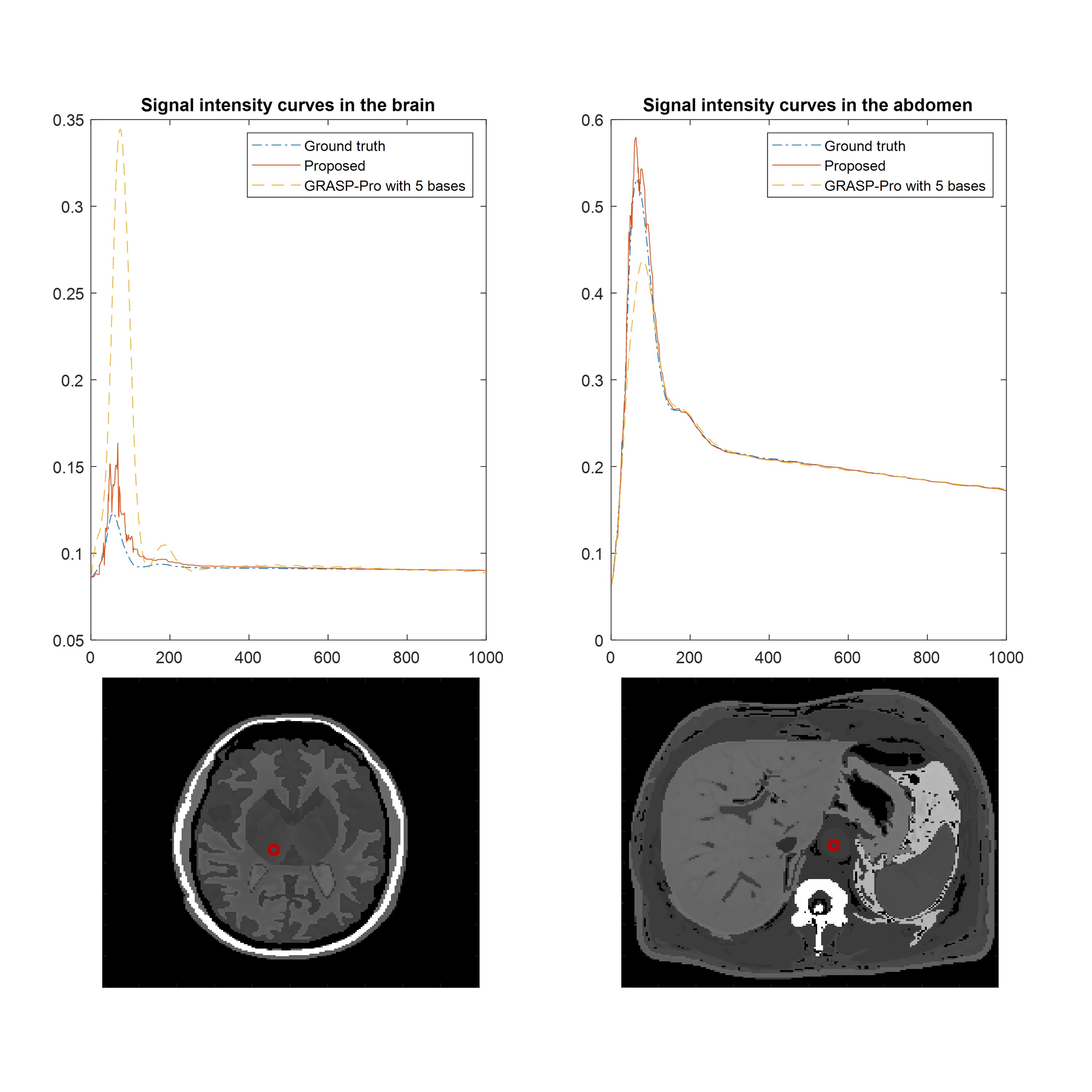
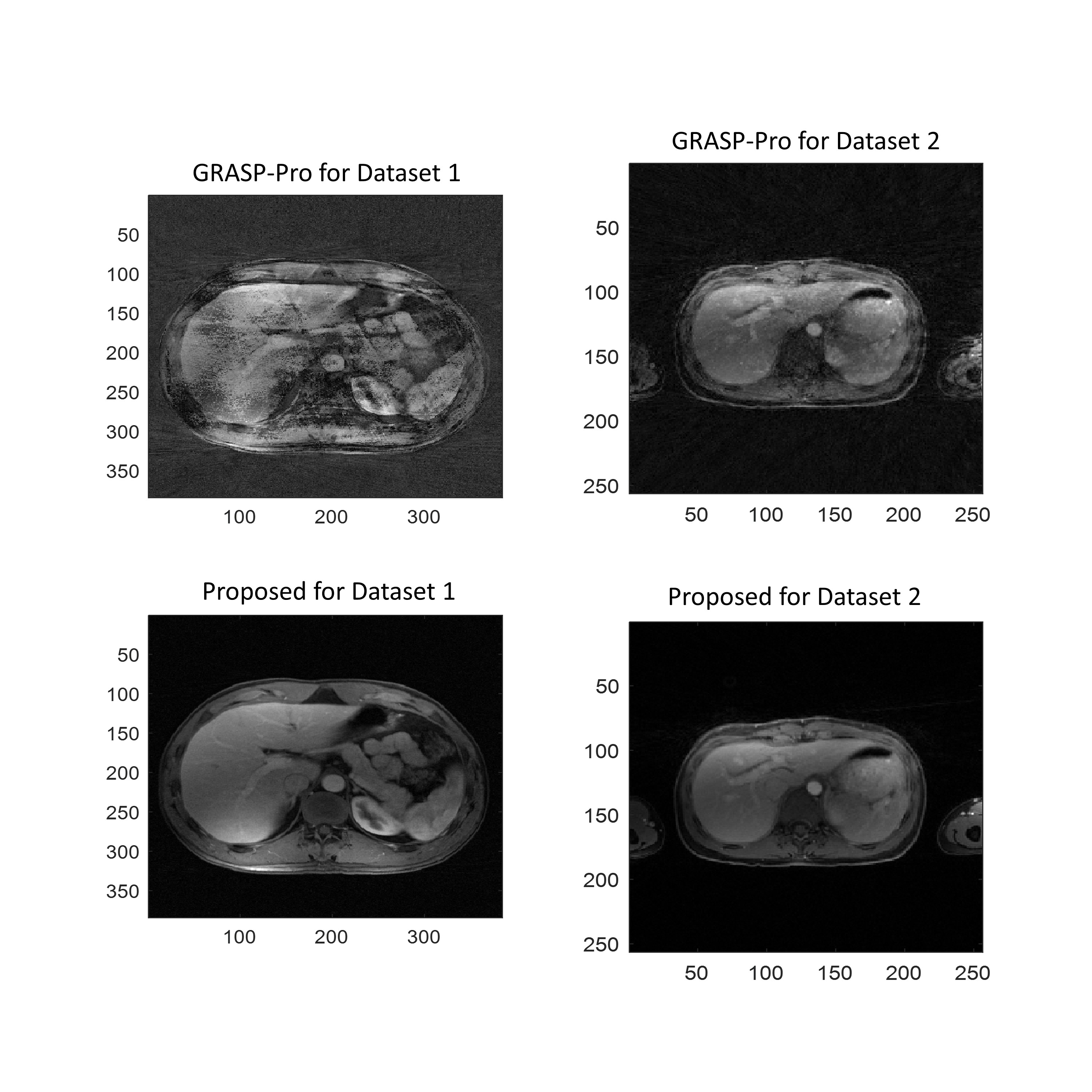
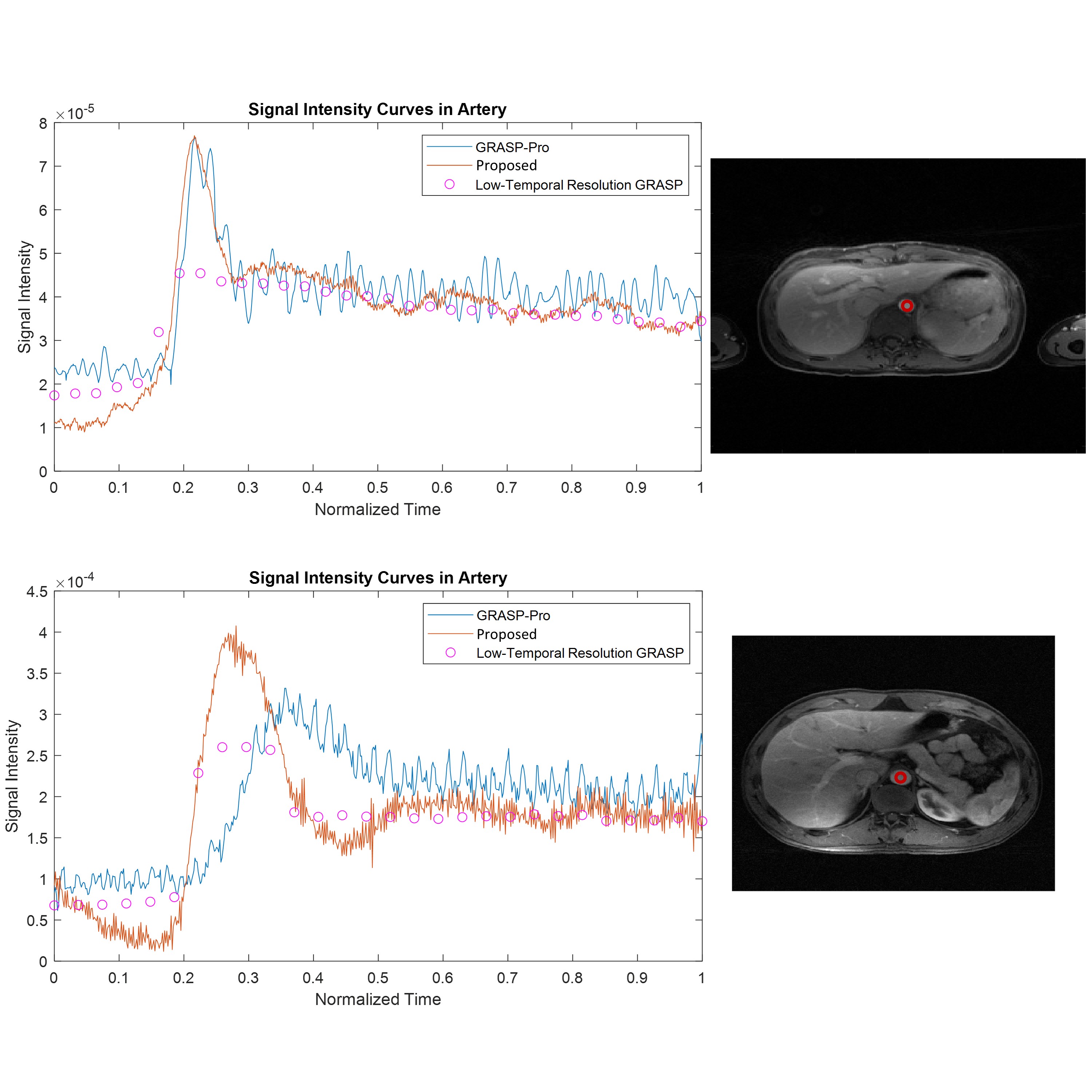
Two simulated datasets were generated and retrospectively undersampled at 1 spoke per frame, with 1000 frames total. Reconstruction quality from a mean squared error (MSE) perspective is shown in Figure 1. In both the abdomen and brain, our proposed method outperformed GRASP-Pro reconstruction that had been given perfect temporal subspaces as priors. The MSE, whether viewed over time or over space, shows that the proposed method offered more accurate reconstructions relative to GRASP-Pro1. The improvement can also be appreciated by comparing the reconstructed image with the worst MSE (via our method) against the corresponding ground-truth image (Figure 2). Figure 3 illustrates two examples of ground truth DCE-MRI signal intensity-time curves compared to estimates by our method and GRASP-Pro. Reconstruction of two in-vivo liver datasets was performed. Dataset 1 was reconstructed at 1 spoke per frame, and dataset 2 was reconstructed at 3 spokes per frame. Figure 4 shows the image reconstruction quality of the proposed method compared to GRASP-Pro for each dataset. Figure 5 shows the signal intensity over time in an artery for each of these datasets, as well as a low-temporal resolution GRASP reconstruction for reference2.Discussion
Mathematically, conventional temporal subspace reconstructions do not permit accurate modeling of temporal dynamics, as the problem is often underdetermined. In contrast, spatial subspace estimation is much likelier to produce an overdetermined optimization problem – in fact, we have the flexibility to maintain both spatial and temporal resolution over a vast range in the number of subspace vectors.Conclusion
Our new spatial subspace estimation method can reconstruct highly undersampled DCE-MRI data with higher accuracy and spatial resolution compared to the current gold standard, GRASP-Pro, using as few as one acquisition spoke per time frame.Acknowledgements
No acknowledgement found.References
[1] Feng, L. et al. GRASP-Pro: imProving GRASP DCE-MRI through self-calibrating subspace-modeling and contrast phase automation. Magn Reson Med (2020) doi:10.1002/mrm.27903.
[2] Feng, L. et al. Golden-angle radial sparse
parallel MRI: Combination of compressed sensing, parallel imaging, and
golden-angle radial sampling for fast and flexible dynamic volumetric MRI. Magn
Reson Med 72, (2014).
Figures