1876
Improving the spatial-temporal fidelity and resolution of dynamic MRI using complex-valued spatial-temporal super-resolution method1The University of Arizona, Tucson, AZ, United States
Synopsis
Keywords: Image Reconstruction, Image Reconstruction
Motivation: To improve image quality and spatial-temporal resolution of T2*-weighted dynamic MRI in applications such as MRI-guided focused ultrasound surgery, resulting in more effective diagnostic and treatment outcomes.
Goal(s): Our goal is to present an innovative complex-valued spatial-temporal multi-band super-resolution technique, enhancing spatial-temporal resolution, signal-to-noise ratio (SNR) and minimizing susceptibility artifacts in T2*-weighted dynamic MRI.
Approach: We highlight the susceptibility artifact through a hybrid simulation and design a multi-band-super-resolution sequence. We perform an experiment on phantom and the acquired data is reconstructed using our proposed method.
Results: Hybrid simulation and reconstruction on phantom data demonstrate improvements in SNR, spatial-temporal-resolution, and reduction of susceptibility artifacts.
Impact: Conventional magnitude-based super-resolution reconstruction exhibits signal loss due to susceptibility effects. Our proposed method integrating phase in reconstruction shows an improvement in SNR, spatial-temporal resolution, and a reduction of susceptibility artifacts.
Introduction:
Enhancing spatial-temporal resolution has a direct positive impact on the sensitivity and precision of dynamic phase mapping. In applications like MRI-guided focused ultrasound surgery where MRI phase mapping plays a crucial role in monitoring temperature and confirming the accuracy of targeting and heating for thermal ablation. Nevertheless, achieving a balance between high signal quality, accurate temperature variations through phase information, and superior spatial-temporal resolution is challenging due to the inherent trade-offs among resolution, acquisition time, and SNR.To address this challenge, one approach utilizes super-resolution1 technique to reconstruct high-resolution images by processing low-resolution images with sub-voxel-shift2, 3 along slice-selection direction. Recent studies have demonstrated that the combination of super-resolution and multi-band method (like SLIDER-SMS4, 5) has the potential to enable the reconstruction of high spatial-temporal-resolution MRI.
However, existing magnitude-only super-resolution techniques encountered challenges in effectively reconstructing phase variations and eliminating susceptibility-induced signal loss in dynamic MRI temperature mapping data acquired with thicker slices. Therefore, we develop a complex-valued spatial-temporal-super-resolution approach to address the limitations of magnitude-only super-resolution.
Methods:
I). Evaluation on susceptibility effect with hybrid simulations and super-resolution reconstruction on BigBrain data in spatial domainUltra-high-resolution (100 micron) complex 3D BigBrain MR data6 (TE = 13 ms and 80 ms) was firstly down-sampled to 1 mm isotropic high-resolution volumes as ground truth. Subsequently, these 1 mm isotropic volumes were further down-sampled to 1 mm x 1 mm x 3 mm low-resolution volumes to simulate various acquisition perspectives. Finally, conventional magnitude-based super-resolution and our proposed complex-valued super-resolution were applied to reconstruct low-resolution data into volumes with an isotropic resolution of 1 mm.
II). Evaluation on susceptibility effect with hybrid simulations and super-resolution reconstruction in temporal-domain
Artificially dynamic linear phase variations were added into the phase(s) of single or multiple target voxels in a high-resolution 1D complex image vector. This dynamic 1D image vector (served as a ground truth) underwent spatially down-sampling by averaging every 3 voxels. Subsequently, spatial-temporal sub-voxel shifts of 0, 1, or 2 high-resolution voxels were applied, aligning with respective time points, to generate a low-resolution dynamic image vector. Finally, our proposed complex-valued super-resolution was employed for dynamic phase-variation reconstruction. Conventional magnitude-based super-resolution was not involved since it does not incorporate phase in reconstruction.
III). Complex-valued super-resolution for SNR and spatial-temporal resolution improvement on real dynamic data acquired from a customized phantom
Dynamic low-resolution images (2 mm x 2 mm x 7.5 mm x 18 time points) were acquired from our customized phantom containing heated (78oC) NiCl2 solution (0.006 mmol) using our gradient-echo-based multi-band super-resolution sequence (TE/TR = 8/22ms, 17 slices) on Siemens® 3T scanner equipped with 32-channel RF coils. Static high-resolution images (2 mm x 2 mm x 2.5 mm) was acquired as a ground truth for spatial image quality comparison. Our proposed complex-valued super-resolution was applied to reconstruct spatial images and dynamic phase variations.
Results:
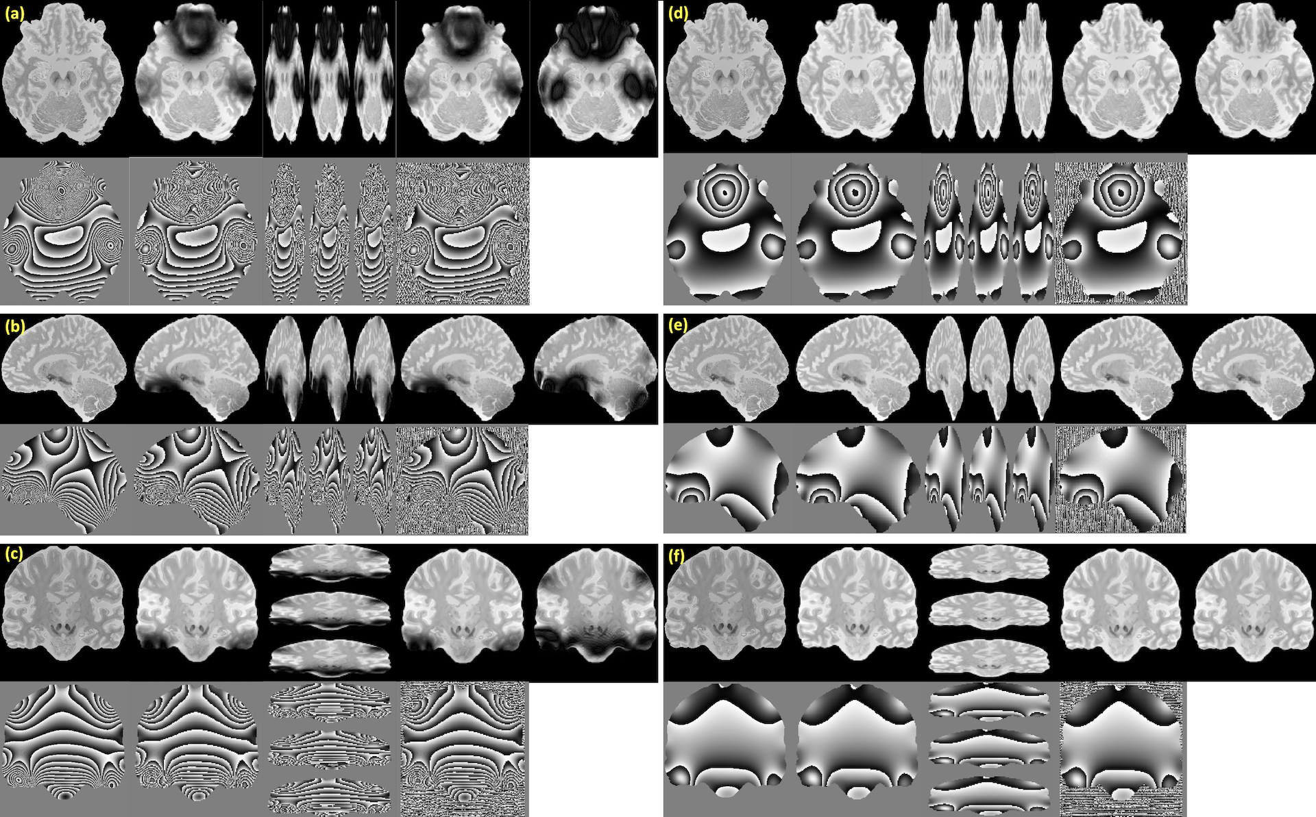
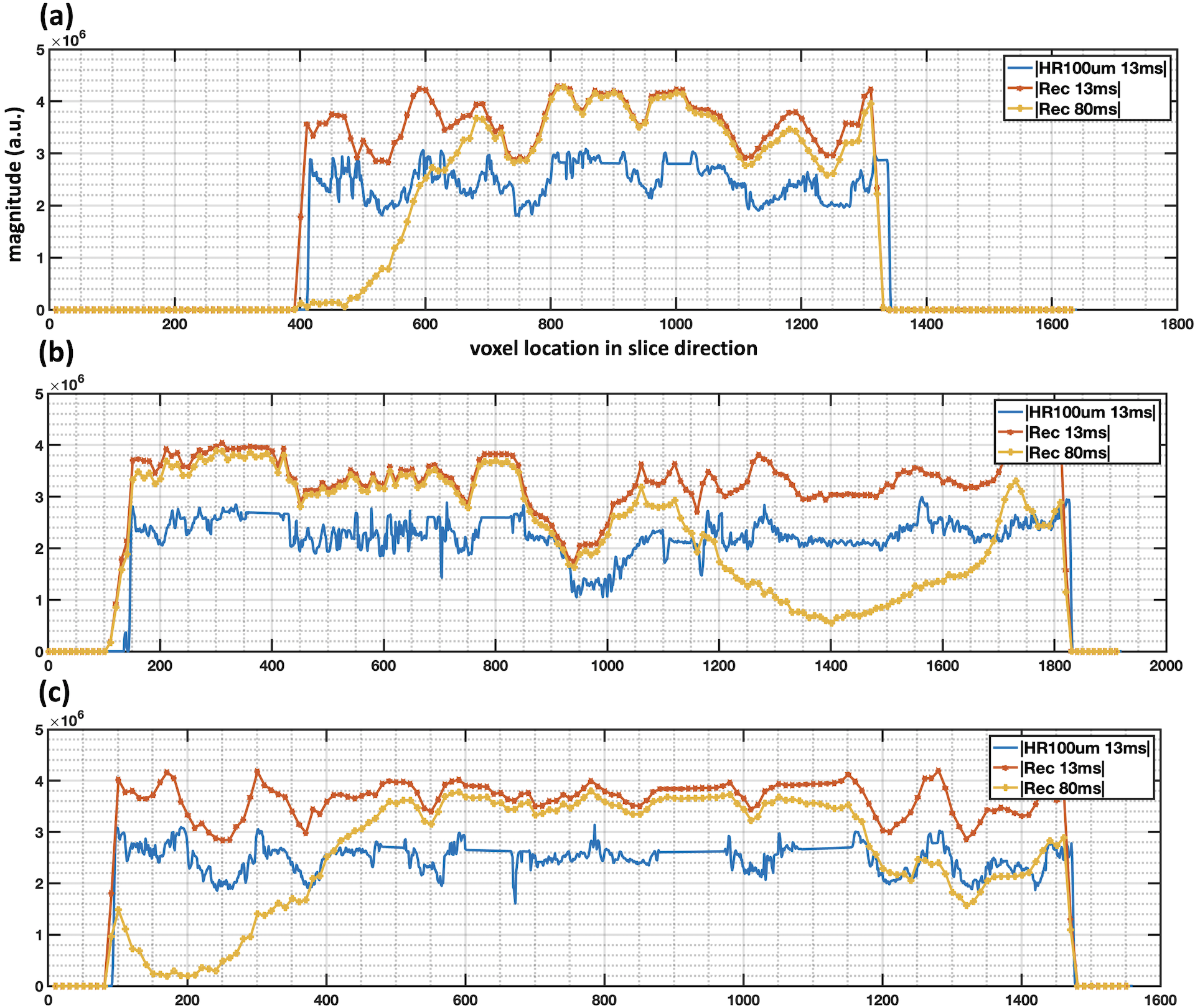
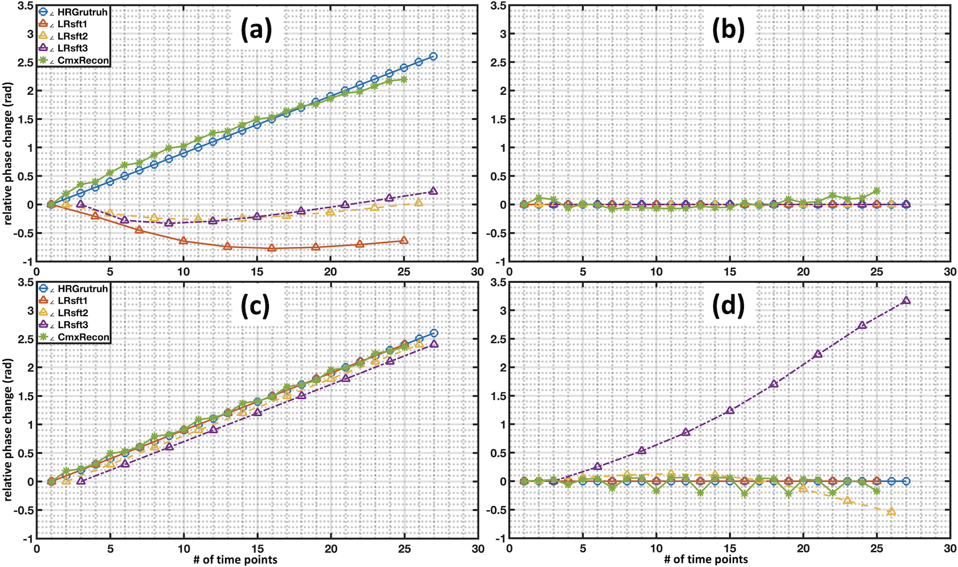
Fig. 1 shows a more pronounced artifact presented in low-resolution images with longer TE (80ms) for all three views, comparing to those with a shorter TE (13 ms). Images reconstructed using our method show reduced dephasing artifact comparing to those reconstructed with conventional magnitude-valued super-resolution. Fig. 2 shows corresponding 1D magnitude plots of BigBrain data (100 micron isotropic) and reconstructed data (1 mm isotropic) using our method, which emphasizes the effect of TE on dephasing artifact in different brain regions.Fig. 3 shows the phase-variations of simulated dynamic low-resolution image vector and reconstructed dynamic image vector. It can be noted that the reconstructed phase variations show improved consistency with the ground-truth phase-variation curves, with less signal leakage to neighboring voxels. Moreover, relying solely on low-resolution dynamic phase variation to predict the behavior of high-temporal-resolution phase variation is not reliable, particularly for boundary voxels with substantial differences in phase variations.
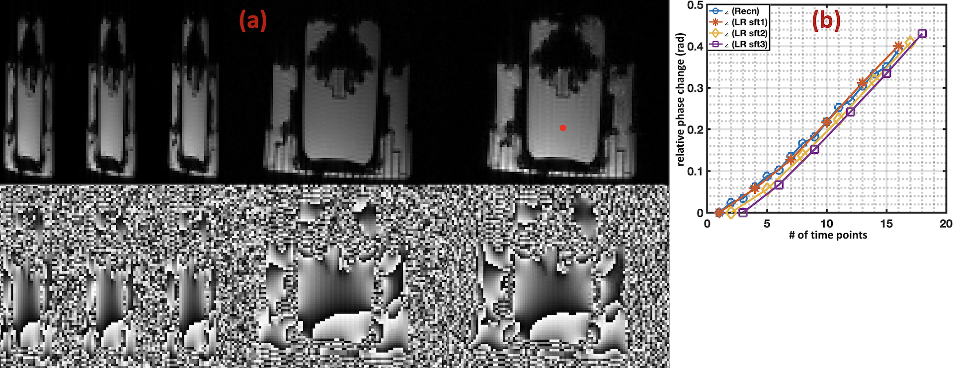
In Fig. 4, we can observe enhanced spatial image quality, including improved SNR and resolution, when comparing the reconstructed image to the initially acquired low-resolution images and the ground-truth image. Additionally, there is an improved temporal resolution of phase variations in selected voxels.
Conclusion:
Results from hybrid simulation and reconstruction of acquired images demonstrate a considerable potential of our proposed complex-valued spatial-temporal super-resolution for T2*-weighted dynamic MRI. Our approach shows promise in terms of improving image quality and providing precise depictions of dynamic phase variations. Phase information is highly valuable for reducing susceptibility artifact, benefiting applications that rely on phase mapping.Acknowledgements
No acknowledgement found.References
1 Tsai, R.Y.; Huang, T.S. Multiframe image restoration and registration. In Advances in Computer Vision and Image Processing; JAI Press: Greenwich, CT, USA, 1984; Volume 1, pp. 317–339.
2 Li L, Wang W, Luo H, Ying S. Super-Resolution Reconstruction of High-Resolution Satellite ZY-3 TLC Images. Sensors 2017; 17(5).
3 Van Reeth E, Tham IWK, Tan CH, Poh CL. Super-resolution in magnetic resonance imaging: A review. Concepts Magn. Reson. Part A 2012; 40A(6): 306–325.
4 Setsompop K, Fan Q, Stockmann J, et al. High-resolution in vivo diffusion imaging of the human brain with generalized slice dithered enhanced resolution: Simultaneous multislice (gSlider-SMS). Magn. Reson. Med. 2018; 79(1): 141–151.
5 Vu AT, Beckett A, Setsompop K, Feinberg DA. Evaluation of SLIce Dithered Enhanced Resolution Simultaneous MultiSlice (SLIDER-SMS) for human fMRI. NeuroImage 2018; 164: 164–171.
6 Sainz Martinez C, Bach Cuadra M, Jorge J. BigBrain-MR: a new digital phantom with anatomically-realistic magnetic resonance properties at 100-µm resolution for magnetic resonance methods development. Neuroimage. 2023 Jun;273:120074. doi: 10.1016/j.neuroimage.2023.120074. Epub 2023 Mar 31. PMID: 37004826.
Figures