1869
Dynamic imaging of the heart from scattering parameters using deep learning – an MR based feasibility study1Department of Radiology, University Medical Center Utrecht, Utrecht, Netherlands, 2Computational Imaging Group for MR diagnostics & therapy, Center for Image Sciences, University Medical Center Utrecht, Utrecht, Netherlands, 3Tesla Dynamic Coils BV, Zaltbommel, Netherlands, 4Department of Radiotherapy, University Medical Center Utrecht, Utrecht, Netherlands
Synopsis
Keywords: Image Reconstruction, Image Reconstruction, Cardiac motion, RF Arrays & Systems,Machine Learning/Artificial Intelligence
Motivation: To enable more accessible and less costly monitoring of cardiovascular mechanical function.
Goal(s): Perform a feasibility study into the potential of imaging the heart based on scattering parameters of an RF antenna array
Approach: An MRI inspired reconstruction network was trained based on 150 in silico simulations of MRI segmented heart models. The method predicts 2D-maps of dielectric property changes and was tested in silico and in vivo on a healthy control.
Results: In silico validation shows that it is feasible to reconstruct the shape and size of the heart, as well as left and right ventricular volumes, based on RF scattering measurements.
Impact: This work shows the feasibility of imaging the heart from differential scattering parameter measurements and by using MRI inspired reconstruction. Preliminary results warrant further investigation into acquiring paired MRI and RF scattering measurements in human subjects.
INTRODUCTION
Scattering parameters of RF antennas placed on the human body are modulated by the dielectric properties of the body. This principle can be used in MRI to detect motion1-4, and in microwave imaging to create images of dielectric objects5. In this work, we demonstrate the feasibility of making spatial images of the heart based on measurement of multi-frequency scattering parameters. Our method utilizes MRI inspired deep learning networks6,7 to transform scattering parameters into 2D images of the heart. The reconstruction problem is cast as reconstructing a change in dielectric properties compared to a reference phase. We trained and tested the method in silico using heart models from segmented MRI scans. The method was also tested vivo with S-parameters acquired on a healthy control. RF based imaging could provide a more accessible and less costly alternative to e.g. MRI and ultrasound for detection of changing mechanical function of the heart. We envision applications in monitoring chronic cardiovascular diseases such as heart failure.METHODS
Signal ModelDifferential scattering parameter $$$\Delta S_{ij,t}$$$ in a cardiac phase t compared to a reference cardiac phase (t=0) can be expressed by equation (1)5.
$$\Delta S_{ij,t}=S_{ij}(t)-S_{ij}(0)=\frac{j\omega}{a_ia_j}\int_{V}^{}(\epsilon(r,t)-\epsilon(r,0))E_i(r,t)\cdot E_j(r,0) dV$$ (1)
$$$E_i(r,t)$$$ is the E-field of element i at phase t and $$$E_j(r,0)$$$ is the E-Field of element j at the reference phase t=0. $$$a_i$$$ and $$$a_j$$$ are forward-power waves8, and $$$\Delta \epsilon(r,t)=(\epsilon(r,t)-\epsilon(r,0))$$$ is the complex permittivity variation. The proposed method for motion imaging is based on the inversion of equation (1). Spatial encoding arises from the distinct complex electric field patterns for all measurement frequencies and antenna combinations.
Reconstruction Method
We designed a neural network (Figure 1) inspired by AUTOMAP4 to reconstruct 2D images of in the dielectric anatomy based on S-parameters.
The method works in 2D: it predicts a dielectric property map that is integrated over the slice direction. In addition to predicting dielectric property maps, another network consisting of 3 fully connected layers is trained to predict hemodynamic parameters (volumes of the left and right ventricle).
In-Silico Training
The method was trained in silico, using electromagnetic simulations (Sim4Life, Zurich MedTech, Zurich, Switzerland). A subject-specific thorax model was simulated, derived from MRI images of the subject on which in-vivo data was also acquired (male, 23y, 1.83m, 63kg). To train with various cardiac anatomies, 140 heart models were placed in the thorax (MICCAI 2017)11. Output of the simulations was an 8x8 scattering matrix for diastole and systole. The architecture was trained by minimizing RMSE between predicted and ground-truth 2D $$$\Delta\epsilon$$$ images. The simulated antenna setup was an 8-channel dipole array9, (Figure 2). S-parameters were simulated at 20-frequencies from 55MHz to 1.3GHz
Inference
Inference was done in silico with 10 heart models from the MICCAI database. 2D $$$\Delta\epsilon$$$ images were estimated based on simulated scattering parameters and compared to the ground-truth.
Finally, inference was done with S-parameters measured on a volunteer after obtaining IRB approval and informed consent. S-parameters were measured (setup in Figure 2), and with a 2 channel network analyzer (Copper Mountain Technologies, Indianapolis, USA). Entries of the scattering matrix where measured subsequently. All data was acquired simultaneously with ECG for synchronization (ECG Shield, Olimex, Plovdiv, Bulgaria).
RESULTS AND DISCUSSION
Figure 3 shows the in silico validation. A qualitative spatial match between ground-truth and predicted 2DΔϵ images can be observed.The ground-truth 2DΔϵ image (based on the segmented MRI imaging of the volunteer) and the predicted 2DΔϵ image using the measured ΔS-parameters are shown in Figure 4. A qualitative match can be observed also in this case (the contrast lower than to the validation set is due to a very thin layer of fat around the heart).
There are artifacts at the edges of the image, likely due to noise (not present in the training set) and deviations between simulation and measurement setup.
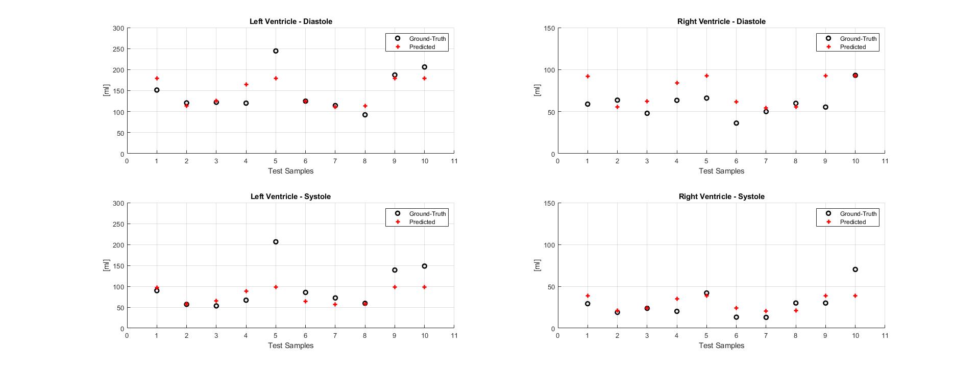
The predicted left and right ventricular volumes of an in silico validation on 10 test subjects are shown in Figure 5.
CONCLUSION
The potential of RF-based imaging for spatial imaging of heart motion was investigated. Preliminary results demonstrate an in silico proof of concept, where the dimension and shape of the heart can be reconstructed. Furthermore, accurate prediction of cardiac mechanical biomarkers such as ventricular volume appears feasible. These preliminary result provide confidence to warrant future investigation into a training workflow based on in vivo acquired S-parameters measurements and MR images. Furthermore, extension to 3D imaging will be explored, as well as using physics informed deep learning approaches that include estimated E-field distributions.Acknowledgements
This project was funded partially by Dutch Heart Foundation Dekker grant 03-006-2022-0024References
[1] D. Buikman, P. Helzel T Fau - Röschmann, and P. Röschmann, "The rf coil as a sensitive motion detector for magnetic resonance imaging," (in eng), no. 0730-725X (Print).
[2] J. Ludwig, P. Speier, F. Seifert, T. Schaeffter, and C. Kolbitsch, "Pilot tone–based motion correction for prospective respiratory compensated cardiac cine MRI," Magnetic Resonance in Medicine, vol. 85, no. 5, pp. 2403-2416, 2021/05/01 2021, doi: https://doi.org/10.1002/mrm.28580.
[3] R. J. M. Navest et al., "The noise navigator for MRI-guided radiotherapy: an independent method to detect physiological motion," Phys Med Biol, vol. 65, no. 12, p. 12NT01, Jun 18 2020, doi: 10.1088/1361-6560/ab8cd8.
[4] B. R. Steensma, C. A. Louka, A. J. E. Raaijmakers, and C. A. T. van den Berg, "Measuring stroke volume with wearable RF antennas: a validation study with EM simulations and MRI," in Conference of the International Society of Magnetic Resonance in Medicine, London, 2022, 31 ed., p. 3952.
[5] A. S. Beaverstone, D. S. Shumakov, and N. K. Nikolova, "Frequency-Domain Integral Equations of Scattering for Complex Scalar Responses," IEEE Transactions on Microwave Theory and Techniques, vol. 65, no. 4, pp. 1120-1132, 2017, doi: 10.1109/TMTT.2016.2638428.
[6] O. Ronneberger, P. Fischer, and T. Brox, "U-Net: Convolutional Networks for Biomedical Image Segmentation," in Medical Image Computing and Computer-Assisted Intervention – MICCAI 2015, Cham, N. Navab, J. Hornegger, W. M. Wells, and A. F. Frangi, Eds., 2015// 2015: Springer International Publishing, pp. 234-241.
[7] B. Zhu, J. Z. Liu, S. F. Cauley, B. R. Rosen, and M. S. Rosen, "Image reconstruction by domain-transform manifold learning," Nature, vol. 555, no. 7697, pp. 487-492, 2018/03/01 2018, doi: 10.1038/nature25988.
[8] K. Kurokawa, "Power Waves and the Scattering Matrix," IEEE Transactions on Microwave Theory and Techniques, vol. 13, no. 2, pp. 194-202, 1965, doi: 10.1109/TMTT.1965.1125964.
[9] A. J. Raaijmakers et al., "The fractionated dipole antenna: A new antenna for body imaging at 7 Tesla," Magn Reson Med, vol. 75, no. 3, pp. 1366-74, Mar 2016, doi: 10.1002/mrm.25596.
[10] A. Christ et al., "The Virtual Family—development of surface-based anatomical models of two adults and two children for dosimetric simulations," Physics in Medicine & Biology, vol. 55, no. 2, p. N23, 2009/12/17 2010, doi: 10.1088/0031-9155/55/2/N01.
[11] A. Bernard O Fau - Lalande et al., "Deep Learning Techniques for Automatic MRI Cardiac Multi-Structures Segmentation and Diagnosis: Is the Problem Solved?," (in eng), no. 1558-254X (Electronic).
Figures
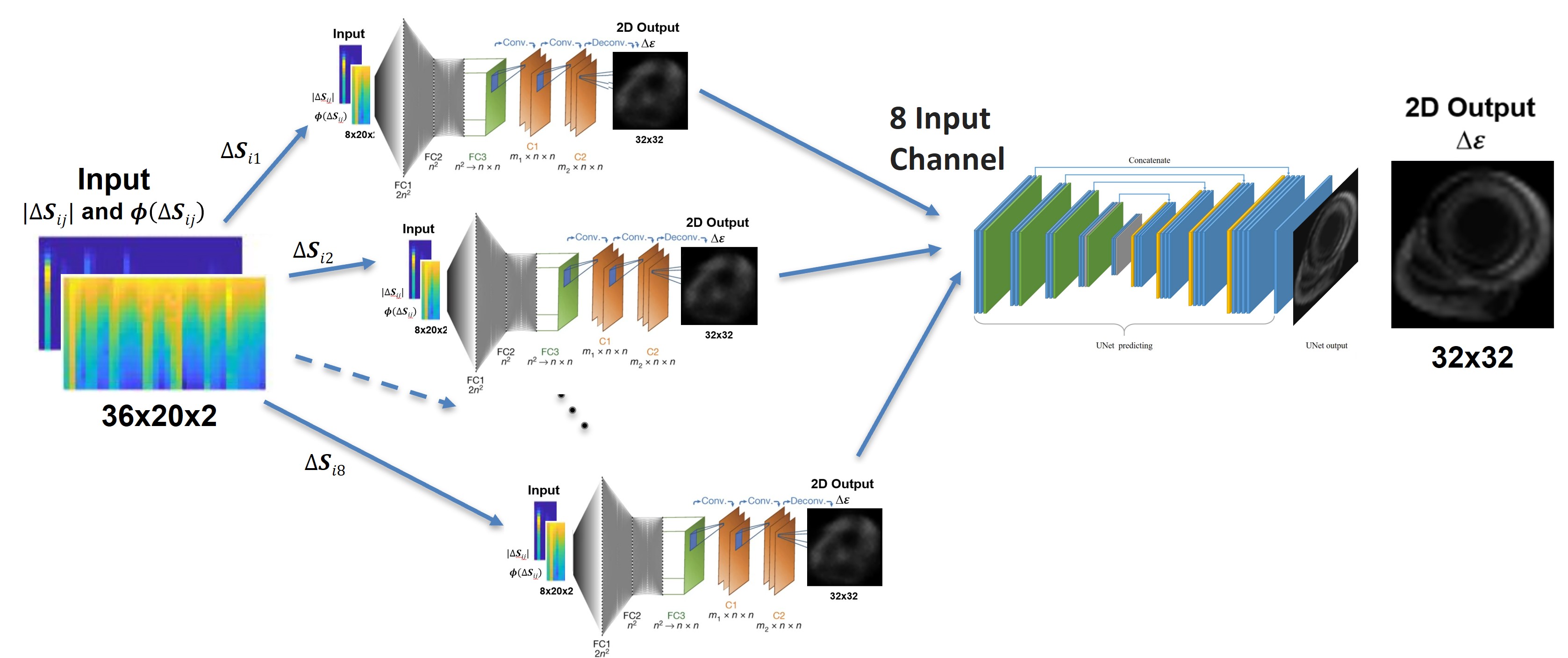
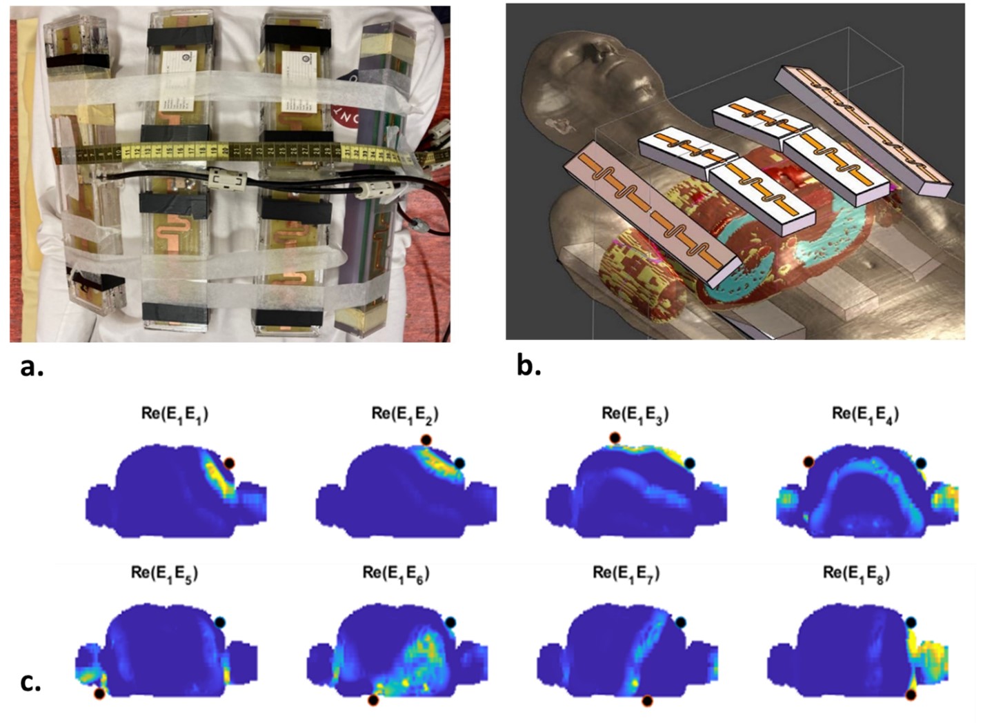
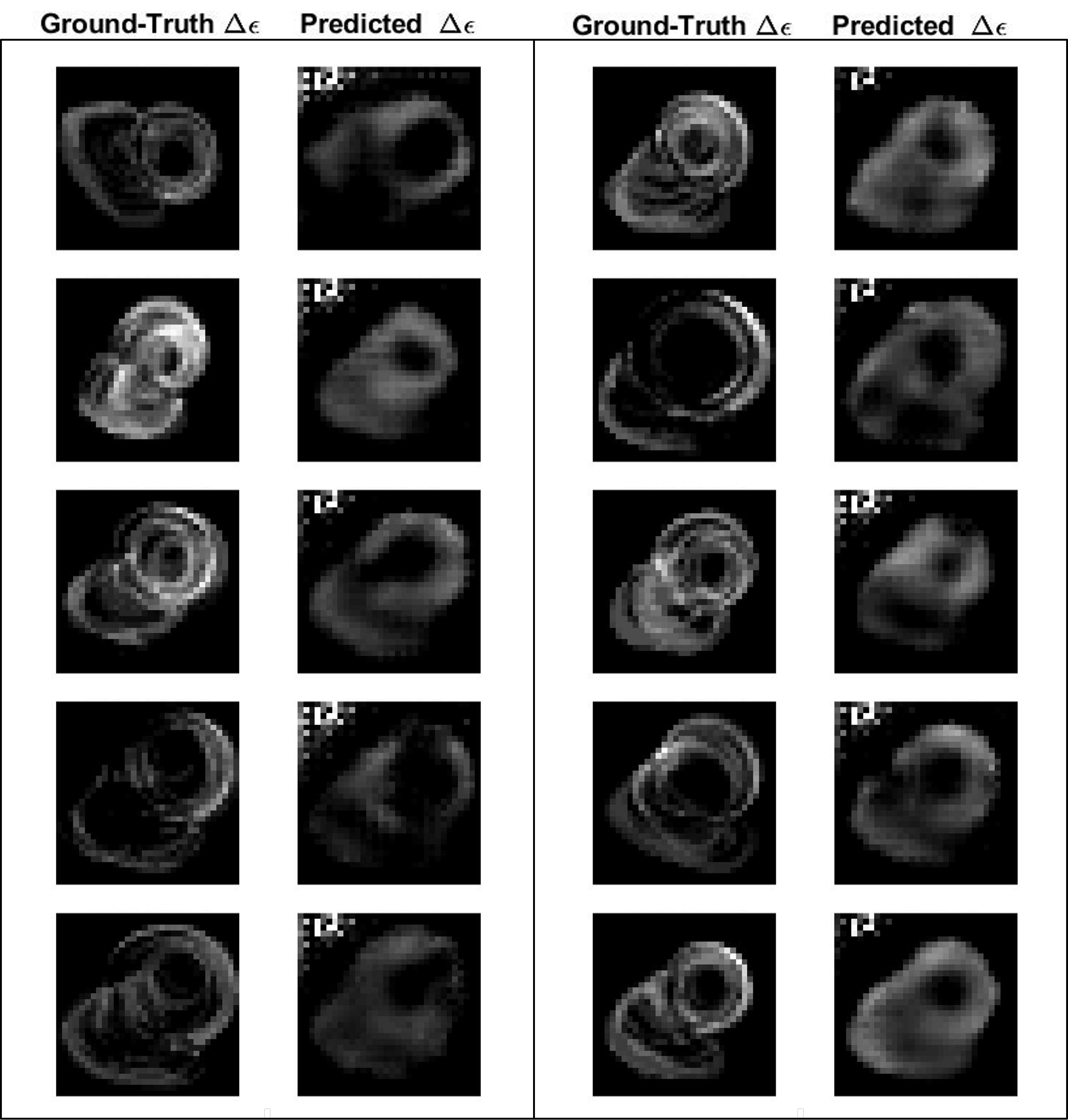
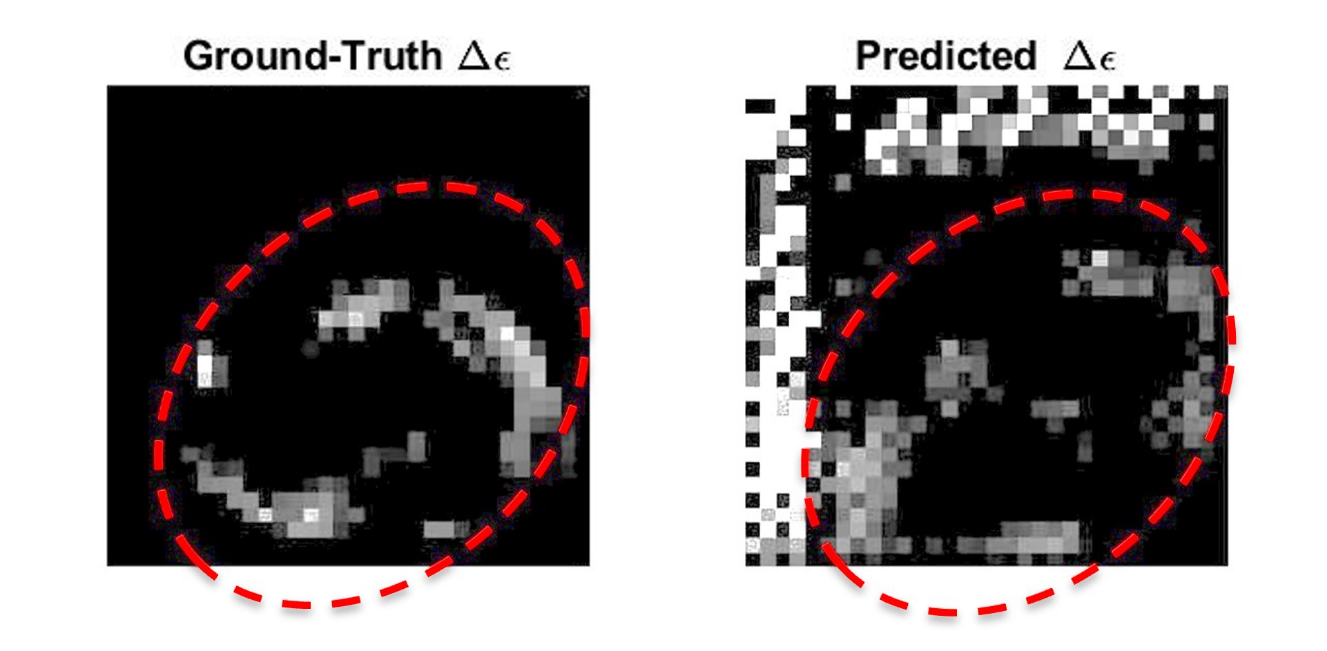
Figure 4: ground-truth and predicted 2D∆ϵ-maps on a healthy control. Compared to Figure 3, the color scale has been modified to compensate for the low contrast due to a very thin layer of fat around the heart.
Note that the in silico training was done with a subject specific torso model based on a segmented MRI scan of the healthy control. In the training, different heart models were incorporated (segmented hearts from the MICCAI database).