1867
Optimally-truncated SVD and PCA applied to spatio-temporal hyperpolarized 13C cardiac data1Radiology, University of California, San Francisco, San Francisco, CA, United States, 2University of California, Berkeley, Berkeley, CA, United States, 3University of California, San Francisco, San Francisco, CA, United States
Synopsis
Keywords: Hyperpolarized MR (Non-Gas), Cardiovascular
Motivation: Current hyperpolarized data analysis techniques, first validated for interrogating cancer, are now being utilized for HP 13C cardiac MRI which is metabolically diverse.
Goal(s): Our goal was to explore the viability of applying advanced data analysis techniques to previously acquired HP 13C cardiac data to derive novel and potentially impactful information.
Approach: We applied optimally-truncated SVD based PCA to fasted and fed state [1-13C] pyruvate images and k-means clustering on resulting data.
Results: Low-rank (r=3) PCA results captured sufficient data to represent the pyruvate images and identify plausible separable components of interest. Clustering spatially resolving data sufficiently for potential further constrained analyses.
Impact: Hyperpolarized 13C cardiac MRI can be analyzed using PCA to derive novel and potentially impactful voxel-wise analysis. As use of HP 13C MRI expands to new applications, advanced analysis techniques can be utilized to better characterize complex alterations in metabolism.
Introduction
Hyperpolarized (HP) 13C imaging can provide in vivo measurements of metabolic fluxes [1-3]. It has been translated into human studies of cardiac metabolism and recently expanded to investigate metabolic implications in Hypertrophic Cardiomyopathy (HCM) [4]. Existing HP imaging methods and analysis pipelines, currently performed for brain, abdomen and pelvis were adapted and validated with healthy volunteers (HV). Common-place HP analysis outputs such as area-under-the-curve (AUC) ratios and first order kinetic rate quantifications can also be used to characterize and stratify normal and abnormal substrate utilization in the heart. Additional adaptations to analysis techniques for cardiac focused exams may still improve both qualitative and quantitative results. Principal component analysis (PCA) has been utilized in MRI for denoising and image reconstruction and successfully applied in low signal-to-noise ratio (SNR) applications [5]. We applied PCA to HV HP 13C cardiac imaging data to explore the potential to gain additional novel information.Methods
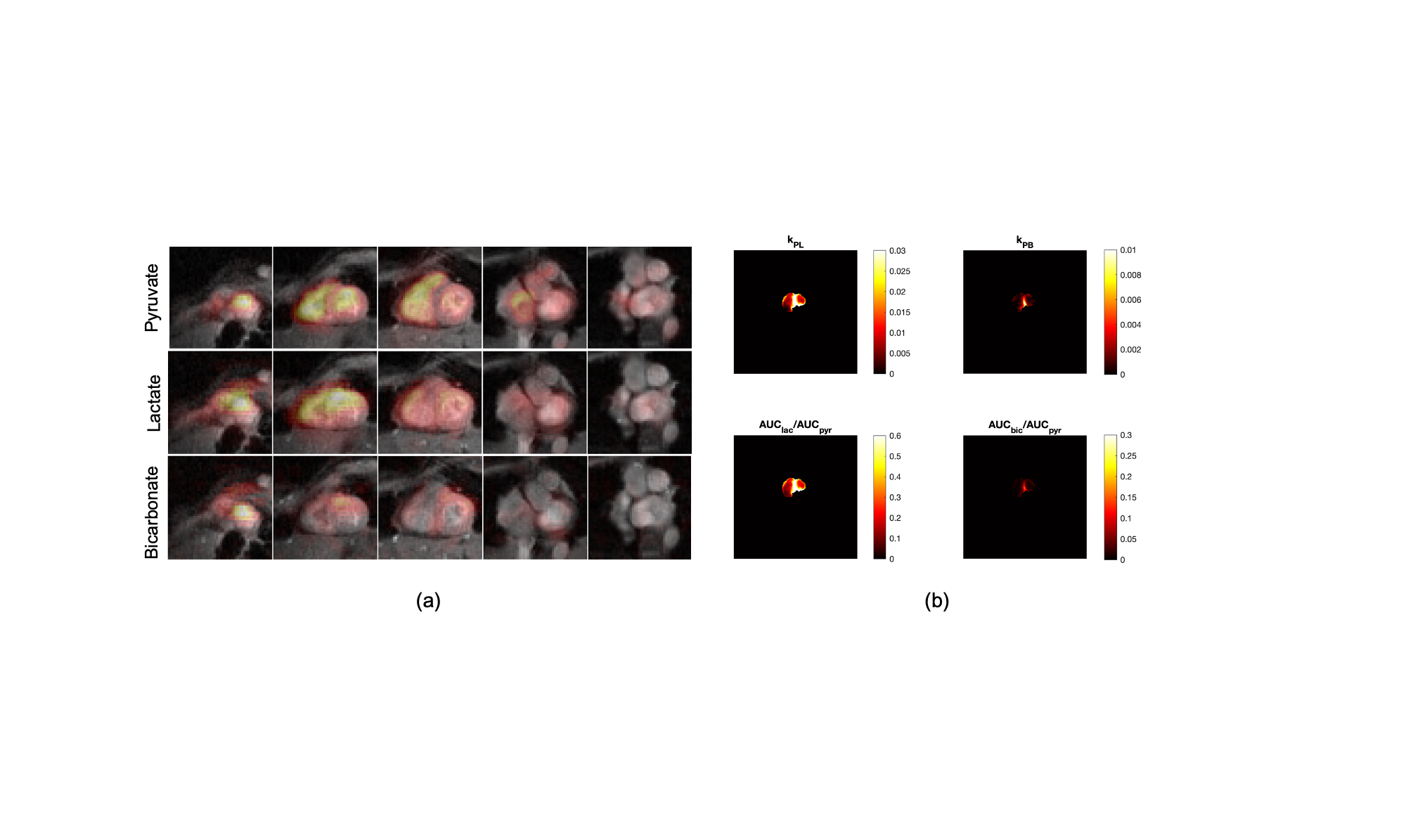
As a part of an ongoing study to investigate cardiac metabolism [4], HP 13C imaging was previously performed using a volumetric multi-slice (nSlice=5), multi-metabolite imaging sequence, resulting in short-axis images of [1-13C]pyruvate, [1-13C]lactate and 13C-bicarbonate. Two scans with identical imaging protocols were performed per study and subjects underwent an oral glucose challenge between scans. Typical data analysis consists of AUC calculations for all acquired metabolites and substrate-product AUC ratios. First-order kinetic rates are also fit based on a 3-site, 1-compartment pharmacokinetic model [5] (Figure 1).PCA was performed on the [1-13C]pyruvate images of a single HV dataset, including both fasted and fed state images. Singular value decomposition (SVD) was utilized as the basis for PCA and optimally truncated [6] to form a low-rank representation of metabolite dynamics. All voxels from both injections were projected into the truncated principal component space and further analyzed using k-means clustering. To compare and qualitatively assess cluster results, manual cardiac segmentations were also generated. Anatomical masks were created using a freehand ROI polygon tool based on CINE images spatially and temporally matched to the HP images.Results
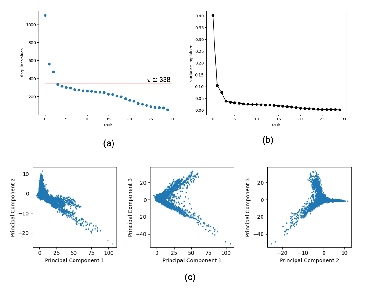
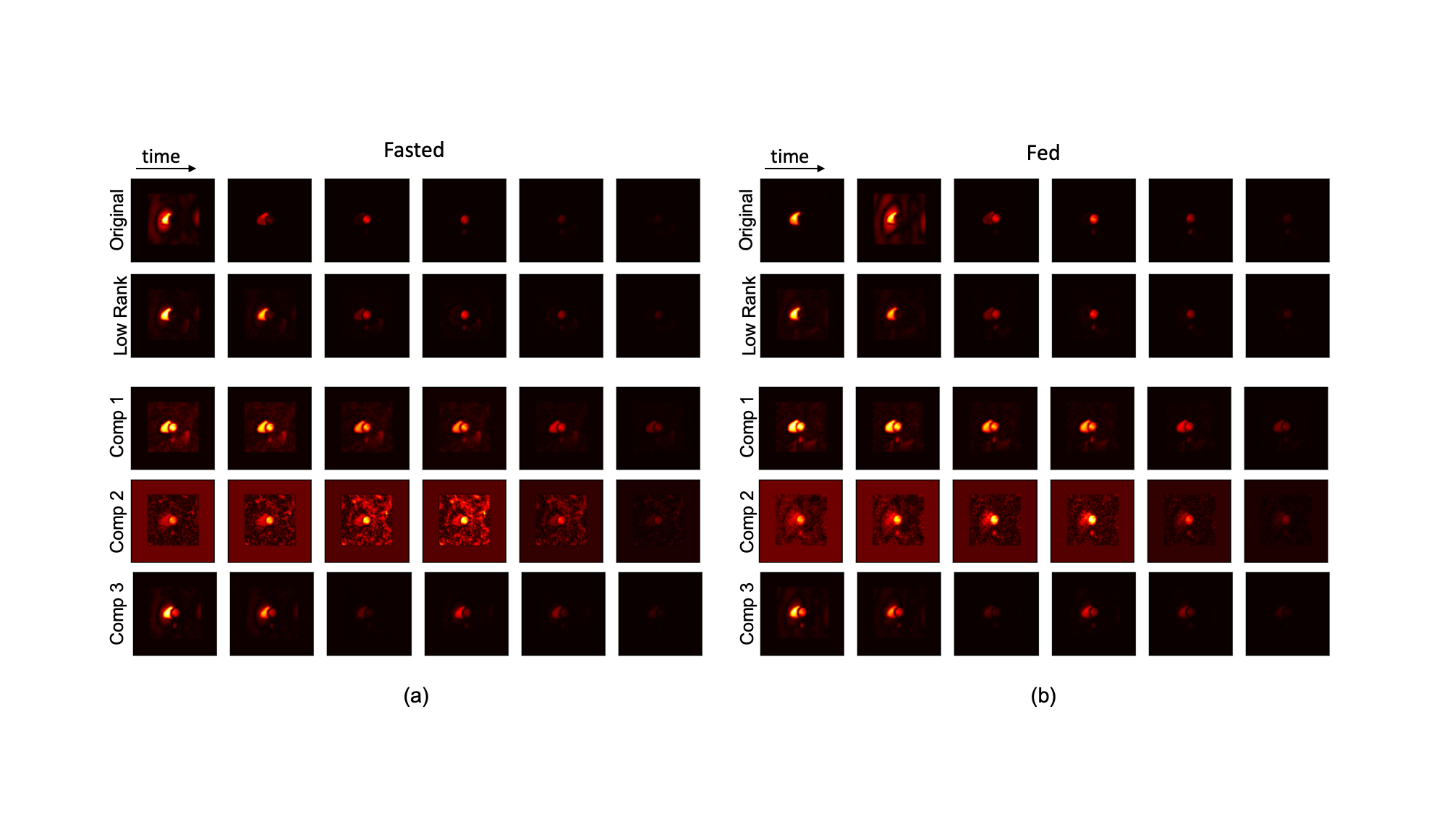
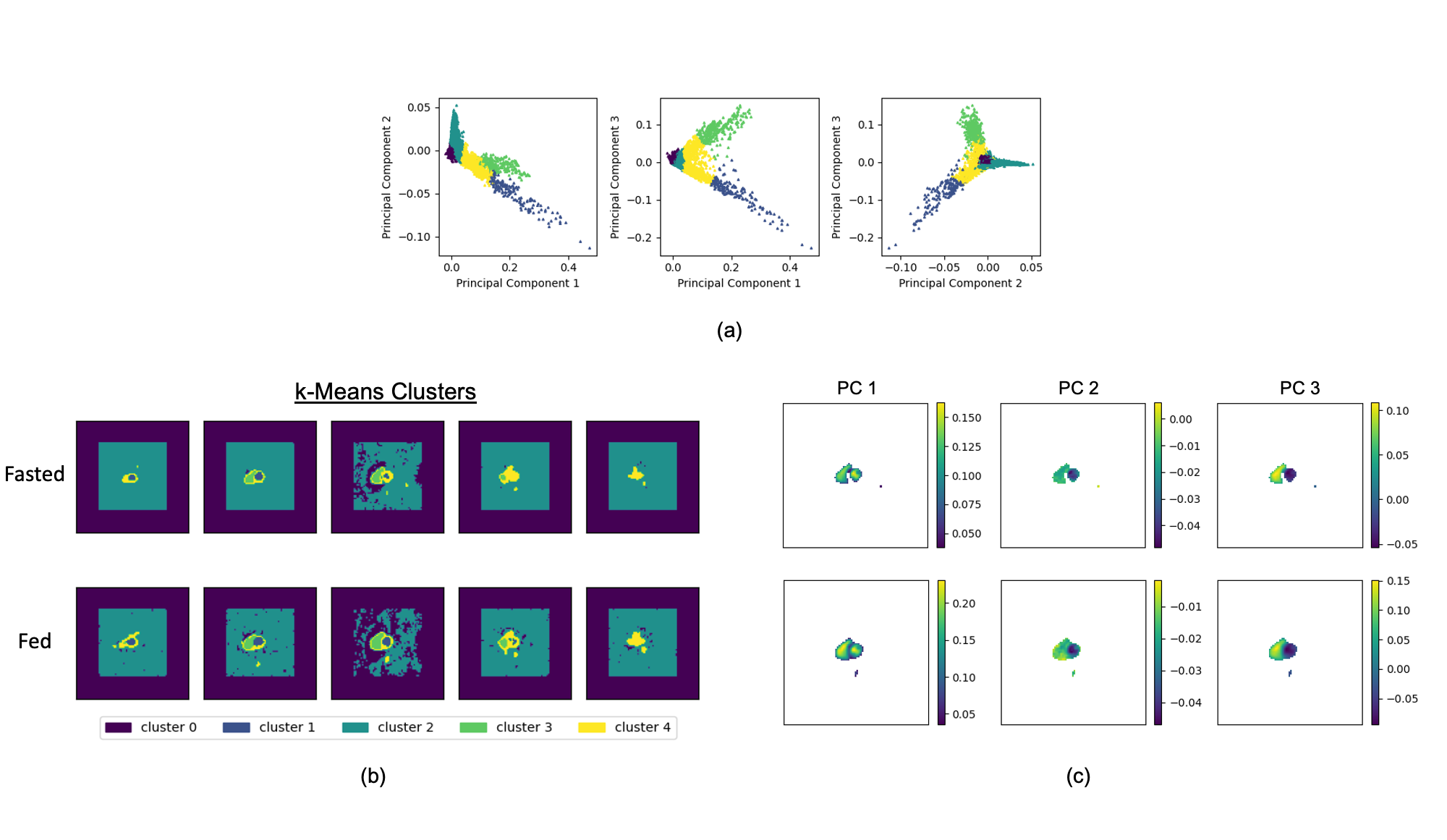
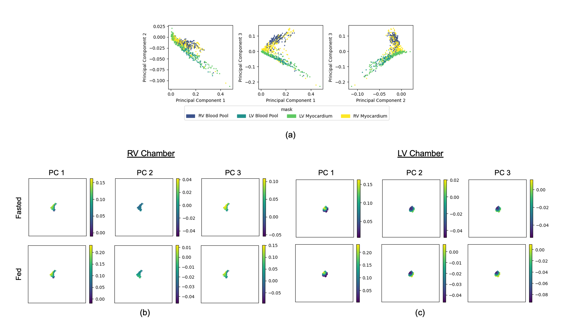
The optimal truncation point derived from the results of the SVD, resulted in rank 3 as the optimal low rank reconstruction (Figure 2a). The truncated singular values explained ~58% of the total variance (Figure 2b). All voxels, from both injections, were projected into the low rank principal component space (Figure 2c). The full dynamics were also visualized for the pyruvate acquisitions. Figure 3 shows a single, representative slice across the first 6 timepoints of the original dynamics, the low-rank representation, and each individual component image. The voxel-wise projections into the principal component space were also analyzed via k-means clustering (nCluster = 5)(Figure 4a), cluster results are depicted for each injection and each slice (Figure 4b), and also the PC-projected data for non-background clusters are shown (Figure 4c). Anatomical masks were applied to label the PC-projected voxels (Figure 5), background voxels were omitted for visual clarity. The right ventricle and left ventricle chamber masks were then isolated and visualized in image space for a single slice for each of the low-rank contributing components (Figure 5).Discussion
This work demonstrated that principal component analysis can be applied to formulate a low rank representation of hyperpolarized cardiac imaging data. The technique of optimally truncated SVD resulted in reduced dimensionality which reduced noise while maintaining an acceptable representation of the pyruvate dynamics which can be seen in Figure 3. Individual components of the low-rank data suggest that a temporally resolved separable model is a viable direction for further analysis. Component 3 seems to represent a cyclic element of the data which we hypothesize could relate to bolus circulation. A separable model and its depiction of the circulating metabolite signal has the potential to inform and improve accuracy of pharmacokinetic model-based fitting. Additionally, k-means clustering on the PC data projections can provide spatially meaningful clustering which could allow for further voxel-wise analysis of component dependence. The clustering of the background voxels is impacted by the zero-filling which is used to match the pyruvate data field-of-view (FOV) of these studies to lactate and bicarbonate image FOVs. In future, we intend to perform analysis on original FOV data. Clusters moderately agree with the anatomical-based masks which emphasizes that PC-projected data may inform spatially-constrained analyses or have the potential to identify spatial abnormalities. More investigations and simulation-based verifications are required to understand how the separable components relate to circulation, metabolic conversion, and other influences. Our intended future work includes learning on additional metabolites, additional subjects and HCM patients.Acknowledgements
No acknowledgement found.References
- Golman K, Petersson JS, Magnusson P, et al. Cardiac metabolism measured noninvasively by hyperpolarized 13 C MRI. Magnetic Resonance in Med. 2008;59(5):1005-1013. doi:10.1002/mrm.21460
- Miller JJ, Lau J, Tyler D. Hyperpolarized MR in cardiology: probing the heart of life. In: Advances in Magnetic Resonance Technology and Applications. Vol 3. Elsevier; 2021:217-256. doi:10.1016/B978-0-12-822269-0.00006-3
- Timm KN, Miller JJ, Henry JA, Tyler DJ. Cardiac applications of hyperpolarised magnetic resonance. Progress in Nuclear Magnetic Resonance Spectroscopy. 2018;106-107:66-87. doi:10.1016/j.pnmrs.2018.05.002
- Larson PEZ, Tang S, Liu X, et al. Regional Quantification of Cardiac Metabolism with Hyperpolarized [1 - 13 C]-Pyruvate MRI Evaluated in an Oral Glucose Challenge. Radiology and Imaging; 2023. doi:10.1101/2023.10.16.23297052
- Vaziri S, Autry AW, Lafontaine M, et al. Assessment of higher-order singular value decomposition denoising methods on dynamic hyperpolarized [1-13C]pyruvate MRI data from patients with glioma. NeuroImage: Clinical. 2022;36:103155. doi:10.1016/j.nicl.2022.103155
- Larson PEZ, Chen H, Gordon JW, et al. Investigation of analysis methods for hyperpolarized 13C‐pyruvate metabolic MRI in prostate cancer patients. NMR in Biomedicine. 2018;31(11). doi:10.1002/nbm.3997
- Gavish M, Donoho DL. The Optimal Hard Threshold for Singular Values is \(4/\sqrt {3}\). IEEE Trans Inform Theory. 2014;60(8):5040-5053. doi:10.1109/TIT.2014.2323359
Figures