1866
Universal parallel transmit pulses for pulse-acquire based whole-brain MRSI1German Center for Neurodegenerative Diseases (DZNE), Bonn, Germany, 2High-field MR Center, Department of Biomedical Imaging and Image-guided Therapy, Medical University of Vienna, Vienna, Austria, 3Department of Physics and Astronomy, University of Bonn, Bonn, Germany
Synopsis
Keywords: Spectroscopy, Spectroscopy
Motivation: Using single-channel transmit coils, the $$$B_1$$$ distribution is very inhomogenous in the brain. This leads to flipangle variations and consequently signal loss in a large portion of the brain. Using parallel transmit these variations might be mitigated.
Goal(s): The goal of this work is to develop universal slab-selective homogeneuos excitation pulses for whole-brain MRSI acquisitions.
Approach: Slab-selective $$$k_t$$$ points excitation have been calculated under consideration of multiple off-center frequencies. Flip angle homogeneity and excitation phase linearity was examined.
Results: Resulting spectra can be quantified using single-channel optimized basis functions. Clear gray matter white matter contrast is visible in metabolic maps
Impact: Universal excitation pulses, especially designed for whole-brain MRSI have been applied to pulse-acquire MRSI. Anatomical features could be found in the metabolic maps, even in the cerebellum. This might be a promising step towards reliable whole-brain MRSI.
Introduction
Ultra-high field scanners and fast MRSI readouts have shortened the acquisition time of whole-brain MRSI to feasible scan times1. The flip angle is set to the expected Ernst angle to be as SNR efficient as possible. However, the excitation field inhomogeneity at ultra-high fields leads to flipangle variations, and consequently reduced SNR in large portions of the brain, especially the cerebellum. Parallel-transmit (pTx) could offer a more homogenous excitation and increased SNR.Methods
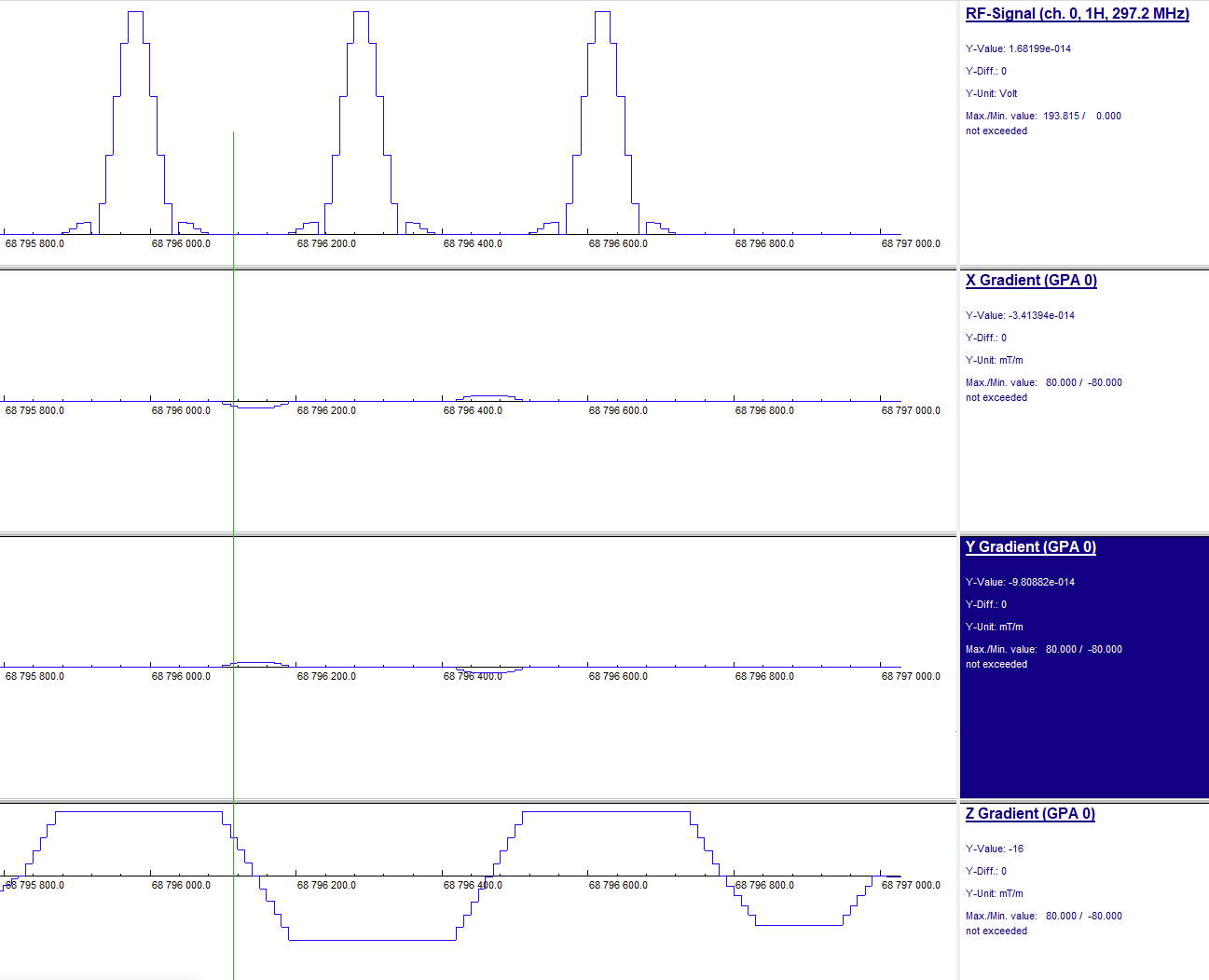
In a preceding database study, 30 subjects were measured at three 7 Tesla sites (1x Siemens 7T+, 2x Siemens Terra) using 32-channel receive, 8-channel transmit coils (Nova Medical). For each subject a $$$B_0$$$ maps, individual channel $$$B_1$$$ maps and a MPRAGE2 were acquired.Universal slab selective $$$k_t$$$-points pulses3 with three, sinc-shaped subpulses were calculated. Slab selection gradients are inverted after each subpulse. During the ramptimes, $$$k_t$$$ blips are used to navigate the excitation $$$k$$$-space. MRSI has additional requirements for excitation pulses, as the pulses need to perform well over a span of off-center frequencies. First, the flip-angle should be independent of the off-center frequency. The excitation phase should depend linearly on the off-center frequency. A single sinc-shaped pulse, as used in conventional excitation fulfills these requirements natively. Nine different off-center frequencies (0, ±60, ±120, ±260, ±400 Hz) were added to the $$$B_0$$$ map of each subject. Pulse calculation was performed using 270 virtual subjects (30 subjects * 9 frequencies). The duration of each sinc-shaped (TBP=4) subpulse is 220µs and a gradient ramptime of 50µs leads to a total pulse duration of 960µs. Figure 1 shows a screenshot of one excitation pulse in the sequence simulation (IDEA freamework).
The calculated pulses were used in a pulse-acquire based MRSI sequence with a concentric ring $$$k$$$-space trajectory4. Whole brain MRSI was performed in one subject (TE,TR=1.3,600ms, 42° excitation, 5mm isotropic resolution, FoV=(220x220x175)mm3). A 135mm slab was excited using pulses detuned to 2.4ppm. Acquisition time was 10:07min. Additionally, an MPRAGE5 was acquired.
Coil combination was performed using iMUSICAL6. Spectral quantification was performed with LCModel7, using basis functions calculated for single-transmit-channel measurements. This was performed for each voxel within a brain mask, extracted from the MPRAGE.
Results
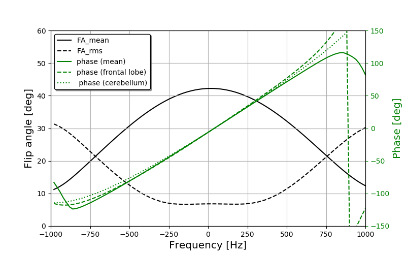
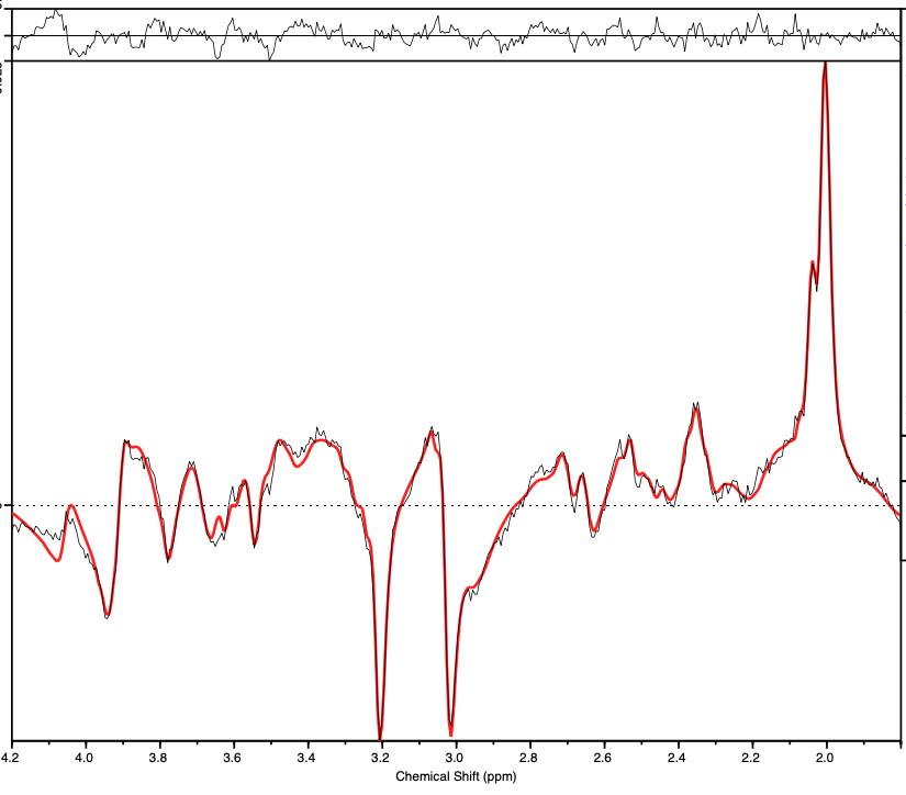
Figure 2 shows the flip angle and the excitation phase of the universal pulses as a function of the off-center frequency. The flipangle reduces for off-center frequencies. While the mean flip angle for on-resonant spins is the target of 42°, and the mean flip angle for spins with an off-center frequency of 500Hz is still above 30°. There is a slight asymmetry in the frequency dependence. The RMS of the flipangle is fairly stable at around 8° for low off-center frequencies and start to increase at around ±300Hz. The excitation phase is depicted for three different brain segments and shows linear behavior inside the optimized frequency.One example of the LCModel quantification can be seen in Figure 3. The fit matches the data well and very little structure can be seen in the fit residuum. First order phase was not corrected for, and the baseline was removed.
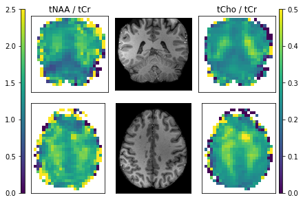
In Figure 4 a coronal slice and a transversal slice of the tNAA/tCr and the tCho/tCr ratios are depicted. Corresponding slices from the MPRAGE are also depicted. The gray matter to white matter contrast is clearly visible in both contrasts. Lower tNAA/tCr in the cerebellum is observable. Some extreme values are found at the edges of the brain.
Discussion
Universal excitation pulses can be applied to pulse-acquire MRSI. The excitation phase depends linearly on the off-center frequency. Consequently, single-channel optimized basis functions fit well to the data in the LCModel quantification.In principle, more subpulses might lead to an even more homogeneous excitation pulse. However, the linearity of the excitation phase was difficult to achieve with more than three subpulses. Also, the pulse duration becomes problematic, if more subpulses are used.
Good contrast metabolic maps could be obtained using these pulses. This indicates that it is feasible to use universal excitation pulses for whole-brain MRSI measurements. The extreme values at the edges of the brain can be explained by lipid contamination. Due to the high flip angle homogeneity, the metabolic maps show anatomical features even in the cerebellum.
In the future, the universal pulse homogenization properties will be quantified and results compared to single-channel-transmit MRSI.
Conclusion
Universal excitation pluses have been successfully applied to whole-brain MRSI. The resulting spectra can be fitted using single-channel basis functions due to the linearity of the excitation phase. Clear gray matter to white matter contrast can be seen in metabolic maps, even in the cerebellum.Acknowledgements
This work received financial through the German Federal Ministry of Education and Research (BMBF; funding code 01ED2109A) as part of the SCAIFIELD project under the aegis of the EU Joint Programme - Neurodegenerative Disease Research (JPND) (www.jpnd.eu) and was supported by the National Institutes of Health under award number R01EB031787.References
1. Hangel, G, Spurny-Dworak, B, Lazen, P, et al. Inter-subject stability and regional concentration estimates of 3D-FID-MRSI in the human brain at 7 T. NMR in Biomedicine. 2021; 34(12)
2. Mugler and Brookeman. Three-dimensional magnetization-prepared rapid gradient-echo imaging (3D MP RAGE). Magnetic Resonance in Medicine. 1990;15(1)
3. Jamil, R, Mauconduit, F, Gras, V, Boulant, N. General gradient delay correction method in bipolar multispoke RF pulses using trim blips. Magn Reson Med. 2020; 85: 1004–1012.
4. Furuyama JK, Wilson NE, Thomas MA. Spectroscopic imaging using concentrically circular echo-planar trajectories in vivo. Magn Reson Med 2012; 67
5. Mugler and Brookeman. Three-dimensional magnetization-prepared rapid gradient-echo imaging (3D MP RAGE). Magnetic Resonance in Medicine. 1990;15(1)
6. Moser, P.; Bogner, W.; Hingerl, L.; Heckova, E.; Hangel, G.; Motyka, S.; Trattnig, S.; Strasser, B. Non-Cartesian GRAPPA and coil combination using interleaved calibration data—Application to concentric-ring MRSI of the human brain at 7T. Magn. Reson. Med. 2019, 82
7. Stephen Provencher: Estimation of metabolite concentrations from localized in vivo proton NMR spectra. Magn Reson Med 30, 672 (1993)
Figures