1863
A 3/2-channel 1H/13C RF surface coil for localized 13C MRS in the human frontal lobe at 7 T1CIBM Center for Biomedical Imaging, Lausanne, Switzerland, 2Animal Imaging and Technology, Ecole Polytechnique Fédérale de Lausanne (EPFL), Lausanne, Switzerland, 3Laboratory for Functional and Metabolic Imaging (LIFMET), Ecole Polytechnique Fédérale de Lausanne (EPFL), Lausanne, Switzerland
Synopsis
Keywords: Spectroscopy, Spectroscopy, 13C, RF coil
Motivation: Studying cerebral metabolism in the human frontal lobe using 13C MR spectroscopy is of great interest but presents challenges due to the low sensitivity of 13C nuclei and SAR limitations at ultrahigh magnetic fields.
Goal(s): Our goal was designed a 3/2-channel 1H/13C RF coil specifically for conducting 13C MRS measurements in the human frontal lobe.
Approach: The feasibility and effectiveness of the coil design were demonstrated with bench measurements and the application of adiabatic carbon editing (ACE)-STEAM and ISIS-DEPT sequences.
Results: The coil enables the acquisition of naturally abundant 13C metabolite signals in both in vitro and in vivo with high sensitivity.
Impact: The 3/2-channel 1H/13C RF coil, designed and optimized for 13C MRS study in the human frontal lobe, benefits high transmit efficiency and provides large FOV in the forehead.
Introduction
Using 13C MRS to study metabolism in the frontal lobe is a powerful approach for investigating the pathophysiology of various neuropsychiatric disorders1. However, in vivo 13C MRS measurement faces the challenges of low sensitivity and the stringent SAR limitation at ultrahigh fields for human applications, particularly in the frontal lobe. Local TxRx RF coils, widely used for 13C MRS2, provide higher Tx efficiency and SNR while lower SAR compared to volume coils. They can be advantageous for applications focused on specific brain regions.In this study, we developed a 3/2-channel 1H/13C RF surface coil used for 13C MRS measurements in the human frontal lobe to benefit from a high Tx efficiency, yet to further increase FOV beyond what was achieved in previously published design3. We evaluated the coil performance on the bench and validated the feasibility and efficiency of this coil design by employing two sequences to acquire the in vitro and in vivo localized natural abundant 13C MR spectra: (1) ACE-STEAM4 to acquire indirect 1H-[13C] MR spectra from phantoms; (2) ISIS-DEPT5 to obtain direct 13C-[1H] MR spectra from phantoms and the human frontal lobe.
Method
Coil design and bench measurementThe 3/2-channel 1H/13C surface coil (Figure 1) consists of three 10 cm diameter circular 1H loops and two 8.5 cm diameter circular 13C loops made from 3 mm diameter copper tubes. The mutual coupling between the loops was minimized by overlapping. Bazooka cable traps were constructed for both the 1H and 13C frequencies, using Teflon tubes (1 cm in diameter, 5 cm in length for 1H, and 8 cm for 13C), in combination with capacitors (15 pF for 1H and 214 pF for 13C) to minimize common-mode signals on the coaxial cables. To ensure effective isolation at the 1H frequency, two lowpass filters (Trilithic Inc., Indianapolis, USA) were incorporated. Bench measurements were conducted using a network analyzer (Agilent, Santa Clara, California, USA).
Pulse sequences
Two pulse sequences were used to acquire spectra to demonstrate the performance of the coil: (a) ACE-STEAM: STEAM6 with broadband inversion pulse (HS8, 6ms, BW=10.04 kHz) on 13C channel during TM; (b) ISIS-DEPT: 3D-ISIS localization7 combined with DEPT8. No 13C and 1H decoupling was applied.
MR measurement
All MR measurements were performed on a 7T human MR scanner (Siemens, Erlangen, Germany). The 13C frequency and transmit voltage were fine-tuned using a small sphere containing 99% 1-13C formic acid, placed between the two 13C loops.
Two 1.5 L cylindrical phantoms were prepared: (1) 114 mM of myo-inositol, and (2) 100 mM of glutamate. Phantom spectra were acquired using the ACE-STEAM (TE/TM/TR=7.9/30/3500ms, 4096 datapoints, BW=4kHz, HS8 pulse centered at 34 ppm on 13C channel) and ISIS-DEPT (TR=3500ms, 4096 datapoints, θ=90° for myo-inositol and 45° for glutamate, BW=20kHz) within the voxel (3.6×2.0×3.6 cm3 for myo-inositol and 3.0×2.0×2.0 cm3 for glutamate) located near the coil surface.
In vivo measurements were conducted on a healthy volunteer (male, 32 years) with informed consent obtained in accordance with the Swiss cantonal ethics committee. Unlocalized DEPT (θ=90°, 4096 datapoints, BW=20kHz, TR=3.5s, 96 averages) and ISIS-DEPT (448 averages, VOI=7.2×2.7×4.7 cm³) were performed in the frontal lobe.
Result and Discussion
The isolation between the 1H and 13C loops was less than -25 dB at the 13C frequency. Introducing the RF filters further reduced the coupling at the 1H frequency, as illustrated in Figure 2.Within a 12-mL voxel using ACE-STEAM, significant signals of the 13C-coupled glutamate H2, H3, and H4 were observed on the difference spectra, as shown in Figure 3(b). Using ISIS-DEPT allows detecting signals of 1H-coupled myo-inositol C1-C6 (total amount ~30 µmol within the voxel) and glutamate C3 and C4 (~12 µmol) with an SNR exceeding 50 in 15 minutes.
The developed coil provided sufficient FOV coverage for the human forehead (Figure 4). In an in vivo natural abundance measurement, with a 5-minute acquisition without localization, strong signals of myo-inositol, glycerol, and lipids were observed. A 3.5s TR can be realized using ISIS-DEPT within the FDA guidelines. In a 91-mL voxel with 26-minute scanning, an SNR of 6 was achieved in the natural abundance 13C MR spectra of myo-inositol (anticipated total amount <9 µmol). Additionally, the absence of lipids indicates excellent localization performance.
Conclusion
The 3/2-channel 1H/13C surface coil combined with the ACE-STEAM and ISIS-DEPT sequences proves to be highly effective for conducting in vivo 13C MRS measurement in the human frontal lobe at 7 T. It offers sufficient spatial coverage and excellent spectral quality while complying with RF safety regulations, ensuring the feasibility of dynamic 13C MRS measurements within the frontal lobe following 13C labeled substrate administration.Acknowledgements
This work was supported by the Swiss National Science Foundation (grants n° 320030_189064). We acknowledge the CIBM Center for Biomedical Imaging for providing expertise and resources to conduct this study.References
1. Sailasuta N, Robertson LW, Harris KC, et al. Clinical NOE 13C MRS for neuropsychiatric disorders of the frontal lobe. Journal of magnetic resonance. 2008, 195(2): 219-225.
2. Adriany G, Gruetter R. A half-volume coil for efficient proton decoupling in humans at 4 tesla. Journal of Magnetic Resonance (1969). 1997;125(ARTICLE):178-84.
3. Roig ES, Magill AW, Donati G, et al. A double‐quadrature radiofrequency coil design for proton‐decoupled carbon‐13 magnetic resonance spectroscopy in humans at 7T. Magnetic resonance in medicine. 2015, 73(2): 894-900.
4. Pfeuffer J, Tkáč I, Choi I Y, et al. Localized in vivo 1H NMR detection of neurotransmitter labeling in rat brain during infusion of [1‐13C] D‐glucose. Magnetic Resonance in Medicine. 1999, 41(6): 1077-1083.
5. Aue WP, Mueller S, Seelig J. Localized 13C NMR spectra with enhanced sensitivity obtained by volume-selective excitation. Journal of Magnetic Resonance (1969). 1985 Feb 1;61(2):392-5.
6. Tkáč I, Starčuk Z, Choi I Y, et al. In vivo 1H NMR spectroscopy of rat brain at 1 ms echo time. Magnetic Resonance in Medicine. 1999, 41(4): 649-656.
7. Ordidge RJ, Connelly A, Lohman JA. Image-selected in vivo spectroscopy (ISIS). A new technique for spatially selective NMR spectroscopy. Journal of Magnetic Resonance (1969). 1986 Feb 1;66(2):283-94.
8. Doddrell DM, Pegg DT, Bendall MR. Distortionless enhancement of NMR signals by polarization transfer. Journal of Magnetic Resonance (1969). 1982 Jun 15;48(2):323-7.
Figures
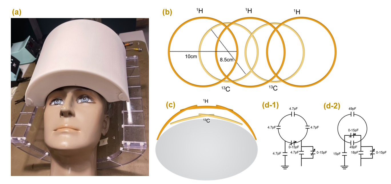
Figure 1: Configuration of the 3-channel 1H/2-channel 13C RF coil. (a) All loops are enclosed within the casing to cover the human frontal lobe; (b) schematic top view: the 1H loops with a diameter of 10 cm above the 13C loops with a diameter of 8.5 cm; (c) schematic side view: geometrical isolation between 13C and 1H loops; (d-1) Design and capacitor values for 1H loop; (d-2) Design and capacitor values for 13C loop.
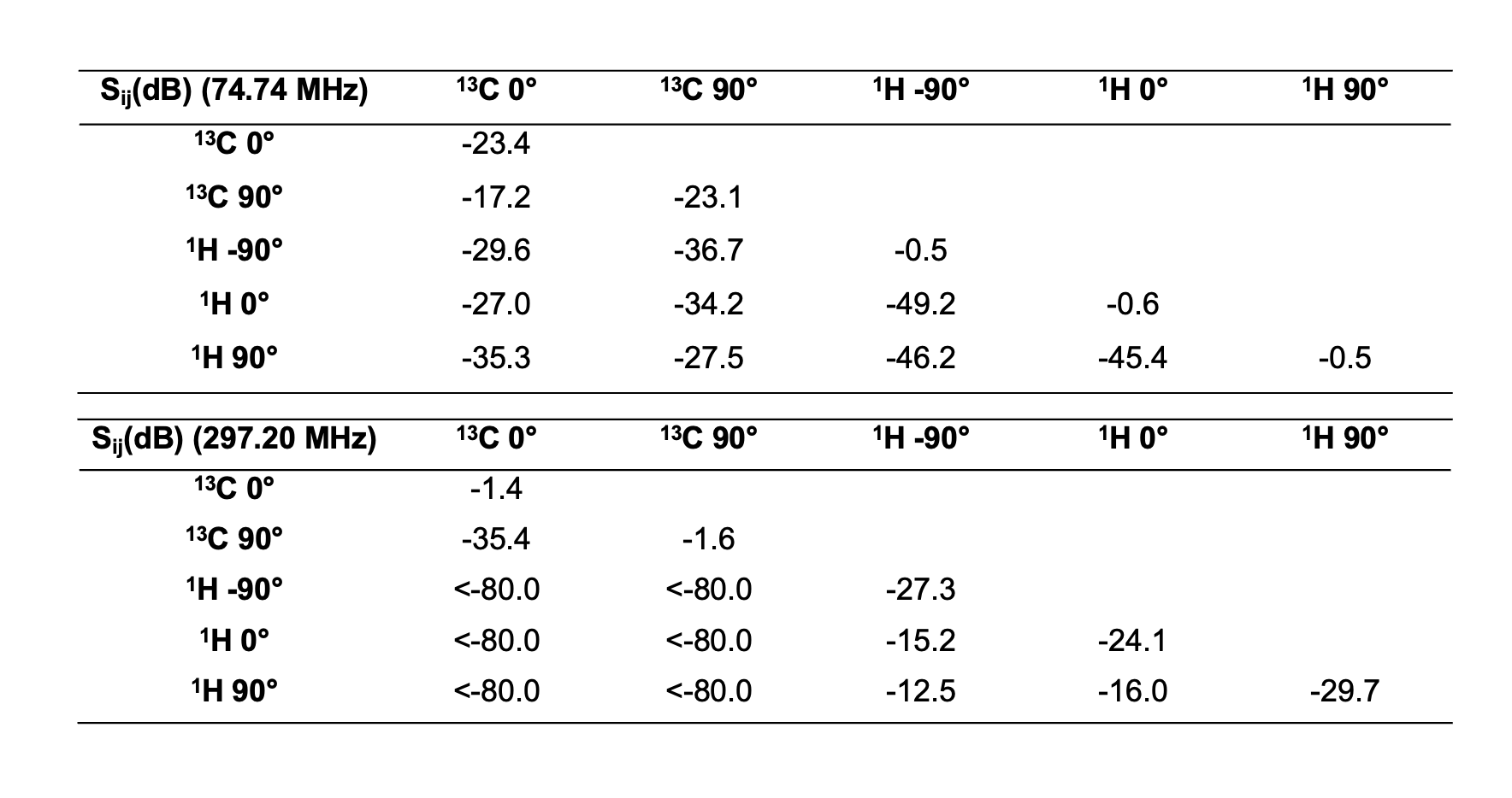
Figure 2: Measured mutual coil coupling (S-parameters) at 13C frequency (74.74 MHz) and at the 1H frequency (297.20 MHz). The coil was loaded with the self-built cylindrical phantom containing 114 mM myo-Inositol in 1.5 L PBS solution.
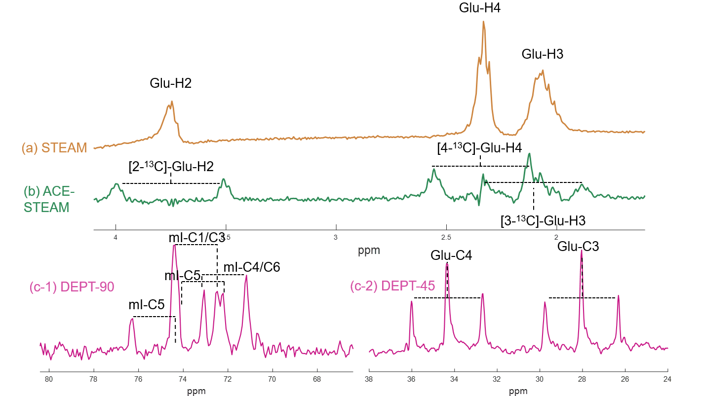
Figure 3: In vitro 1H and 13C MR spectra. (a) STEAM (TE/TM/TR=7.9/30/3500 ms, 4096 datapoints, BW=4kHz, 32 averages, VOI=3.0×2.0×2.0 cm3); (b) ACE-STEAM (TE/TM/TR=7.9/30/3500 ms, 4096 datapoints, BW=4kHz, 6 ms-HS8 pulse centered at 34 ppm in the 13C channel, 384 averages, VOI=3.0×2.0×2.0 cm3); (c-1) ISIS-DEPT (TR=3500 ms, 4096 datapoints, θ=90°, BW=20kHz, 256 averages, VOI=3.6×2.0×3.6 cm3); (c-2) ISIS-DEPT (TR=3500 ms, 4096 datapoints, θ=45°, BW=20kHz, 256 averages, VOI=3.0×2.0×2.0 cm3).
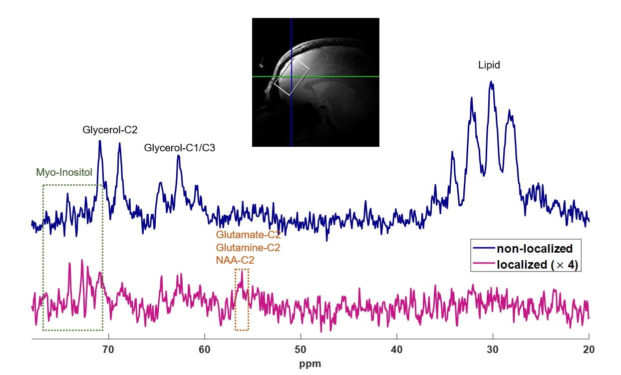
Figure 4: Voxel localization and spectra measured in the human frontal lobe with (1) non-localized DPET (top, 96 averages; TR=3.5 s, 4096 datapoints, θ=90°, BW=20kHz); (2) ISIS-DEPT (bottom, 448 averages, VOI=7.2×2.7×4.7 cm3). Within the voxel, the natural abundance 13C signal of the myo-inositol can be observed at 70-76 ppm, as well as those of glutamate, glutamine, and NAA at 55-57 ppm, with the exclusion of the lipid signal at 25-35 ppm, suggesting excellent localization performance.