1862
NAD+(H) Dynamics Using 31P Functional MRS in the Brain: Insights into Energy Metabolism Mechanisms during Visual Stimulation.1Animal imaging and technology core, CIBM Center for Biomedical Imaging, École Polytechnique Fédérale de Lausanne (EPFL), Lausanne, Switzerland, 2Laboratory for Functional and Metabolic Imaging, École Polytechnique Fédérale de Lausanne (EPFL), Lausanne, Switzerland, 3Department of Radiology, Lausanne University Hospital (CHUV), Lausanne, Switzerland, 4School of Biology and Medicine, University of Lausanne, Lausanne, Switzerland
Synopsis
Keywords: Spectroscopy, Spectroscopy, functional MRS, visual stimulation, energy metabolism, ultra-high field, 7T
Motivation: Alterations in brain nicotinamide adenine dinucleotide (NAD+) levels, as observed in aging, neurodegenerative conditions, and psychiatric disorders, necessitate an in-depth exploration of NAD's functional dynamics.
Goal(s): Investigating the feasibility of using 31P functional MRS (fMRS) at 7T to measure NAD+ dynamics during a visual stimulation task.
Approach: 32 volunteers were measured using 31P fMRS at 7T, during exposure to an established visual stimulation task.
Results: The present study provides evidence for the possibility of measuring NAD+ dynamics in the occipital lobe using 31P fMRS during a visual stimulation task. Furthermore, the use of denoising algorithms may boost sensitivity to detect functional changes.
Impact: Functional 31P MR Spectroscopy could contribute to complex cognitive and clinical studies, investigating energy metabolism deficits. Eventually, this knowledge could contribute to the development of novel therapeutic strategies targeting brain energy deficits associated with neurodegenerative diseases and cognitive impairments.
Introduction
The coenzymes nicotinamide adenine dinucleotide (NAD+) and its reduced form (NADH) play an important role in bioenergetics, and NAD+ acts as a cosubstrate for various enzymes in cellular signaling. Deficits in brain NAD concentration have been reported in aging, neurodegenerative and psychiatric disorders. Monitoring their functional dynamics could reveal how these coenzymes contribute to cerebral energy metabolism supporting neural communication, synaptic plasticity, and the modulation of neurotransmitter release, thereby unraveling the underpinnings of cognitive processes. Importantly, an intracellular redox imbalance and mitochondrial dysfunction have been implicated in the pathophysiology of schizophrenia, and one study already showed differences in NAD content and NAD+/NADH ratio between schizophrenia patients and healthy controls [6]. The non-invasive measurement of NAD content in vivo has been demonstrated using 31P MRS at high magnetic fields [7]. Dynamic measurements of NAD+(H) fluctuations in the human brain, especially during functional activation, have remained challenging due to low sensitivity. This study therefore aims to investigate the feasibility of using 31P functional MRS (fMRS) at 7T to measure NAD+(H) dynamics in the occipital lobe during a visual stimulation task.Methods
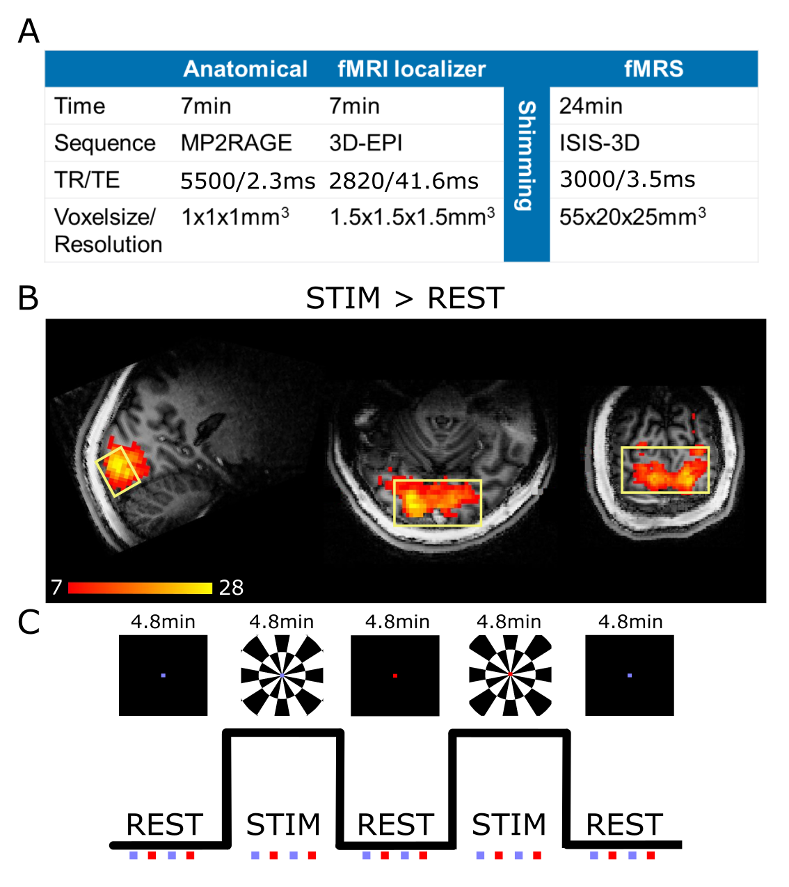
Thirty-two young, healthy participants (mean age: 23.3 ± 3.2 years, 15 female) were recruited for this study. Dynamic 31P MRS data were acquired using a 7T MRI scanner (Siemens Medical Solutions, Erlangen, Germany) and an ISIS 3D localization sequence (TR/TE=3000/3.5ms, voxel-size=55x20x25mm3, 16 averages/transient, Figure 1A). Before spectroscopy data acquisition, an anatomical image (M2PRAGE) and a short fMRI scan (3D-EPI) were obtained for accurate MRS voxel placement in the activated occipital area (Figure 1B). Participants were exposed to an established visual stimulation task (24 min) consisting of alternating blocks of rest (4.8 min) and flashing checkerboard stimuli (4.8 min) [3], while fMRS spectra were collected from the visual cortex (6 transients/block, in total 5 alternating blocks of rest and stimuli). In the rest condition, a black screen with a fixation dot was used. In both conditions, the fixation dot changed color four times a block (Figure 1C). The participants were asked to respond to the color change with a button press. FMRS data preprocessing (frequency and phase correction) was performed using in-house MATLAB scripts. Additional exploratory analyses on NAD+ and tNAD were performed using a Marchenko-Pastur principal component analysis (MP-PCA) denoising algorithm [4,5] on the complex-valued FIDs before quantification. A concentration quantification analysis was performed using LCModel software to estimate NAD+(H) and other 31P metabolites levels [1]. Statistics were performed using linear mixed effects models in R, using linewidth as a covariate.Results
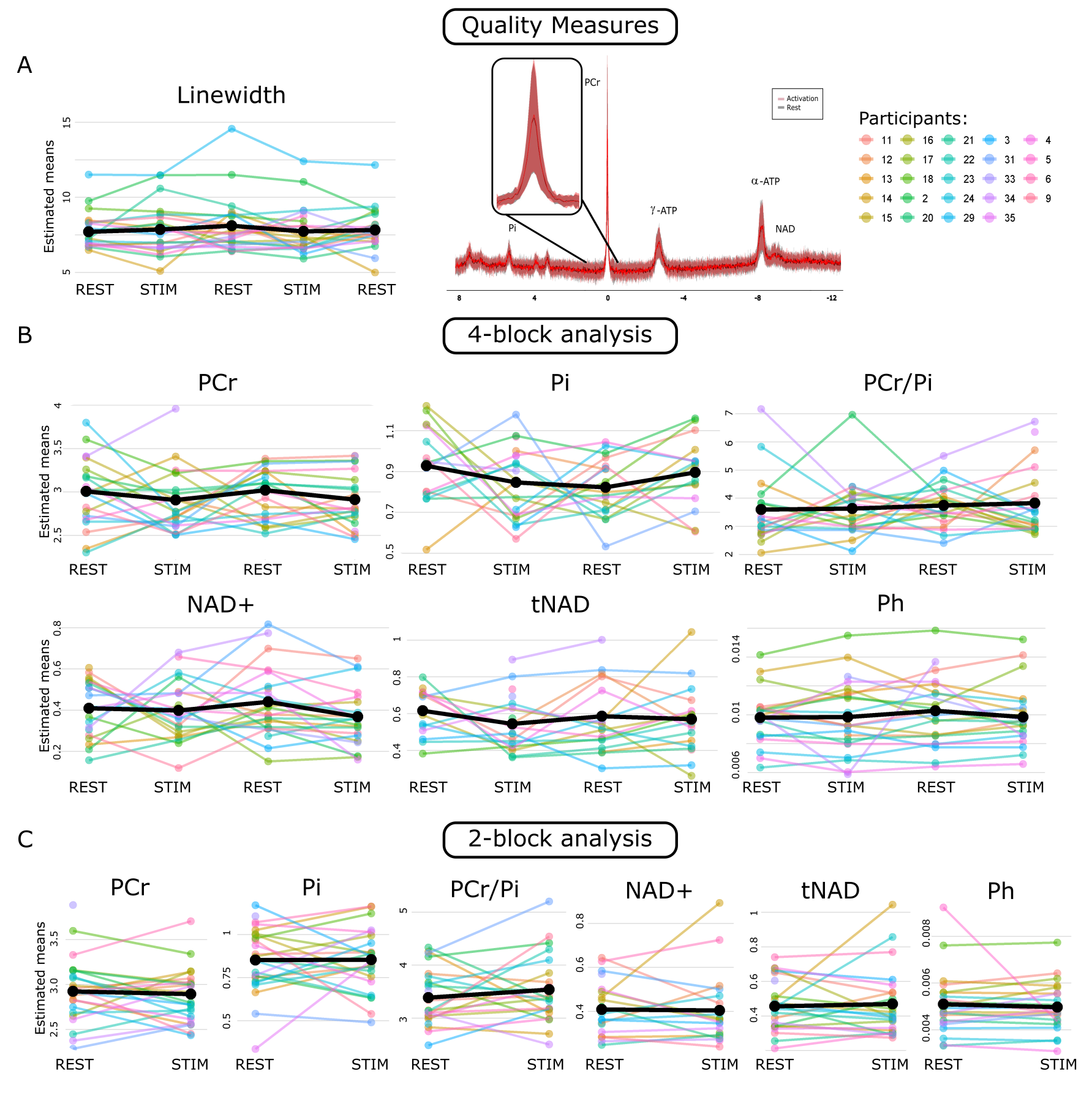
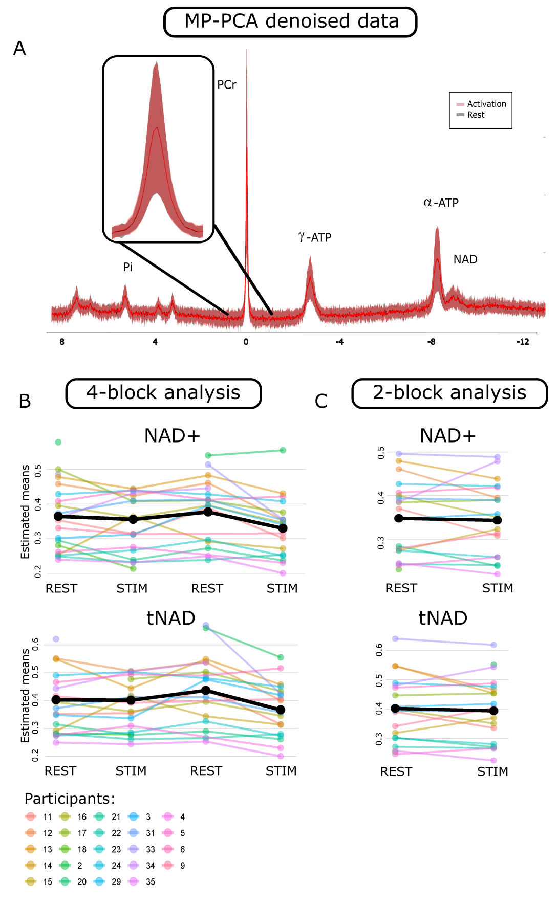
Four participants had to be excluded based on the button presses during the experiment (less than 18 out of 20), and five because of excessive motion, resulting in twenty-four datasets. The results indicate that the spectra were of good quality (mean FWHM=7.8Hz, SNR=7.3 for a measurement block of 6 transients). Results demonstrated no significant main effect of stimulation vs. rest blocks when averaging concentrations per block (4-block analysis) for PCr, Pi, PCr/Pi, NAD+, total NAD (tNAD) and pH (all p>0.05). Analysis of the average concentrations over all rest blocks and stimulation blocks (2-block analysis)also did not show significant differences for PCr, tNAD, NAD+, pH, Pi, and PCr/Pi (all p>0.05; Figure 2). For the exploratory analysis including an additional MP-PCA denoising, a significant effect of block (4-block analysis) was found for tNAD and NAD+ (both p<0.01; Figure 3), which was mainly driven by the second rest and activation block.Discussion/Conclusion
The present study provides evidence for the possibility of measuring NAD+ dynamics in the occipital lobe using 31P functional MRS during a visual stimulation task only when using an additional denoising algorithm before quantification. Further investigations involving simulation-based confirmation of the denoising steps on functional 31P MRS data are warranted to confirm the feasibility of this technique. After careful confirmation of the methods, changes in the investigated metabolite concentrations would indicate enhanced energy production and utilization in response to visual stimulation, supporting the notion of a task-related metabolic demand. If confirmed, this knowledge could contribute to more complex cognitive and clinical studies, investigating conditions in which energy metabolism plays a crucial role. Eventually, this knowledge could contribute to the development of novel therapeutic strategies targeting brain energy deficits associated with neurodegenerative diseases and cognitive impairments. Concluding, using functional 31P MRS to investigate energy metabolism changes during a (cognitive) task remains challenging due to low sensitivity, but denoising algorithms such as MP-PCA may provide a potential solution in this endeavor.Acknowledgements
No acknowledgement found.References
- Cuenoud, B., Ipek, Ö., Shevlyakova, M., Beaumont, M., Cunnane, S. C., Gruetter, R., & Xin, L. (2020). Brain NAD Is Associated With ATP Energy Production and Membrane Phospholipid Turnover in Humans. Frontiers in Aging Neuroscience, 12. https://www.frontiersin.org/articles/10.3389/fnagi.2020.609517
- Xin, L., Ipek, Ö., Beaumont, M., Shevlyakova, M., Christinat, N., Masoodi, M., Greenberg, N., Gruetter, R., & Cuenoud, B. (2018). Nutritional Ketosis Increases NAD+/NADH Ratio in Healthy Human Brain: An in Vivo Study by 31P-MRS. Frontiers in Nutrition, 5. https://www.frontiersin.org/articles/10.3389/fnut.2018.00062
- Barreto, F. R., Costa, T. B. S., Landim, R. C. G., Castellano, G., & Salmon, C. E. G. (2014). 31P-MRS Using Visual Stimulation Protocols with Different Durations in Healthy Young Adult Subjects. Neurochemical Research, 39(12), 2343–2350. https://doi.org/10.1007/s11064-014-1433-9
- Mosso, J., Simicic, D., Şimşek, K., Kreis, R., Cudalbu, C., & Jelescu, I. O. (2022). MP-PCA denoising for diffusion MRS data: Promises and pitfalls. NeuroImage, 263, 119634. https://doi.org/10.1016/j.neuroimage.2022.119634
- Veraart, J., Novikov, D. S., Christiaens, D., Ades-aron, B., Sijbers, J., & Fieremans, E. (2016). Denoising of diffusion MRI using random matrix theory. NeuroImage, 142, 394–406. https://doi.org/10.1016/j.neuroimage.2016.08.016
- Kim, S.-Y., Cohen, B. M., Chen, X., Lukas, S. E., Shinn, A. K., Yuksel, A. C., Li, T., Du, F., & Öngür, D. (2017). Redox Dysregulation in Schizophrenia Revealed by in vivo NAD+/NADH Measurement. Schizophrenia Bulletin, 43(1), 197–204. https://doi.org/10.1093/schbul/sbw129
- Zhu, X.-H., Lu, M., Lee, B.-Y., Ugurbil, K., & Chen, W. (2015). In vivo NAD assay reveals the intracellular NAD contents and redox state in healthy human brain and their age dependences. Proceedings of the National Academy of Sciences, 112(9), 2876–2881. https://doi.org/10.1073/pnas.1417921112
Figures