1855
Image navigators insensitive to B0 inhomogeneity for optimal prospective motion-corrected MRS1Radiology, University of Minnesota, Minneapolis, MN, United States
Synopsis
Keywords: Spectroscopy, Spectroscopy
Motivation: Navigator-based prospective motion-corrected MRS is often performed with 2nd order shims optimized in the whole brain to prevent degradation of navigator images, resulting in suboptimal linewidth.
Goal(s): Here, we report image-based prospective motion correction with 2nd order shims adjusted in the MRS voxel while maintaining good navigator image quality despite the strong B0 inhomogeneity outside the voxel.
Approach: This was achieved by segmenting multislice spiral navigator images to reduce blurring artifacts from inhomogeneous B0 fields.
Results: High-quality spectra with optimal linewidth were acquired in the presence of subject motion at 3T and 7T, demonstrating the feasibility of this new approach on clinical scanners.
Impact: Image-based prospective motion correction with optimal localized B0 shim (1st and 2nd order) in the MRS voxel is feasible in the human brain at 3T and 7T.
Introduction
Prospective motion and shim correction enables acquisition of good quality brain 1H MR spectra in the presence of subject motion. Among available techniques1, image-based navigators2-4 rely on acquiring navigator images without large artifacts to reliably estimate subject motion. Because dynamic update of 2nd-order shims is generally not possible, these shims must be adjusted either in the whole brain or in the localized VOI. Adjusting 2nd-order shims in the whole brain results in suboptimal linewidth5, especially in hard to shim regions such as the prefrontal cortex or pons6. Adjusting of 2nd-order shims in the localized VOI results in unacceptable degradation of navigator images due to the large inhomogeneous B0 field outside the shimmed voxel.Therefore, the aim of this study was to acquire good quality motion navigator images without artifacts in the presence of strong B0 field inhomogeneity with 1st and 2nd order shims optimized in an MRS voxel. Such artifacts can be minimized by shortening the echo-time by segmenting the readout into multiple shots. For this purpose, our recently proposed fast high-resolution motion navigator based on spiral in/out k-space trajectories was modified4 to handle segmented images.
Methods
Experiments were performed on a 3T Prismafit scanner with a 64-channel head coil and on a 7T Terra scanner with the Nova 1Tx/32Rx head coil. Study was approved by IRB and written informed consent was obtained from all participants.Initial reference segmented spiral navigator images were acquired in the whole brain (30 slices, matrix=64x64, slice thickness=3mm, gap=1.5mm, spatial interleaves=10 at 3T and 15 at 7T). MPRAGE was then acquired to position an 8mL VOI in prefrontal cortex (PFC). FAST(EST)MAP was used to adjust 1st and 2nd order shims. sLASER7 was modified to incorporate the motion navigator consisting of two segmented spiral navigator slices (97 ms per slice at 3T and 108 ms per slice at 7T) that did not overlap with the voxel. Motion was calculated at each TR using multislice-to-volume registration4.
Spectra were obtained in six subjects at both 3T and 7T. Spectra were acquired using three conditions: without subject motion and without correction (baseline), with subject motion and motion-correction disabled (NoCo), (iii) with subject motion and motion, shim and frequency navigators enabled (ShMoCo). During NoCo and ShMoCo measurements, the subjects were instructed to rotate the head slowly to the side (i.e. Z-rotation) four times when prompted by the operator.
Results
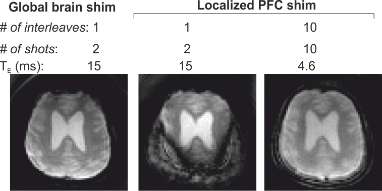
Figure 1 shows the spectral linewidth measured in PFC with three B0 settings. With global shim (shims optimized in the whole brain), a broad spectrum was measured (water linewidth=13.4Hz). When the linear shims were adjusted inside the voxel after global shim, the linewidth was improved (9.8 Hz). The linewidth was further improved (6.7Hz) when all 1st and 2nd order shims were optimized inside PFC. Likewise, the gain is SNR of NAA was almost 10% with voxel-based shim compared to global shim with linear shims adjusted in the voxel.Figure 2 shows spiral navigator images acquired from the whole brain using global and localized B0 shim settings. With two spiral-in/out acquisitions and global shim, acceptable images were measured throughout the brain. However, when the B0 shim was optimized in PFC, large signal blurring and loss of signal was apparent in regions outside PFC due to off-resonance effects. These artifacts were greatly reduced when increasing the spatial interleafs to 10.
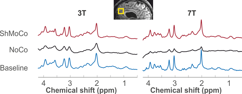
Figure 3 shows sLASER spectra acquired with NoCo and ShMoCo from two subjects. At baseline (no motion), high-quality spectra were measured in the PFC region; mean linewidth was 6±1Hz at 3T and 12±1Hz at 7T. With head rotation but motion correction disabled (NoCo), the spectral quality was dramatically reduced compared to baseline. With ShMoCo, high-quality data was measured consistent with baseline. No significant differences in linewidth or SNR were observed between baseline and ShMoCo (p=0.71).
Discussion and Conclusion
The current study demonstrates the feasibility of acquiring high-quality motion corrected MRS spectra with optimal spectral width (1st- and 2nd-order shims adjusted on the voxel) on both clinical 3T and 7T scanners. A single shim setting (localized 1st and 2nd order shims adjusted in the voxel) can be used to acquire both the navigator images and MR spectra. Another recently reported approach uses numerical optimization to find a compromise between optimal linewidth in the voxel and acceptable B0 inhomogeneity in the whole brain8. The method proposed here does not require any numerical B0 optimization.In conclusion, our new approach enables acquisition of good quality image navigators even in the presence of strongly inhomogeneous B0 field such as that obtained after shimming on a localized voxel.
Acknowledgements
This work was supported by funding from the National Institutes of Health (NIH) R01 EB030000, P41 EB027061, 1S10 OD017974-01, and S10 OD025256.References
- Maclaren J, Herbst M, Speck O, Zaitsev M. Prospective motion correction in brain imaging: a review. Magn Reson Med 2013;69(3):621-636
- Keating B, Deng W, Roddey JC, et al. Prospective motion correction for single-voxel 1H MR spectroscopy. Magn Reson Med. 2010; 64(3):672-679
- Hess AT, Tisdall MD, Andronesi OC, Meintjes EM, van der Kouwe AJW, Real-Time Motion and B0 Corrected Single Voxel Spectroscopy Using Volumetric Navigators, Magn Reson Med 2011; 66:314–323
- Adanyeguh I, Henry PG, Deelchand DK, Fast prospective motion correction for spectroscopic acquisitions. MRS Workshop 2022, Lausanne, Switzerland
- Juchem C, Cudalbu C, de Graaf RA, Gruetter R, Henning A, Hetherington HP, Boer VO. B0 shimming for in vivo magnetic resonance spectroscopy: Experts' consensus recommendations. NMR in Biomedicine 2021;34(5):e4350.
- Jayadev NB, Henry PG, Deelchand DK, Prospective Motion Correction for MR spectroscopy in the human brain using multi-slice Spiral Navigator, Proc. Intl. Soc. Mag. Reson. Med. 2022; 31: 4994
- Deelchand DK, Berrington A, Noeske R, Joers JM, Arani A, Gillen J, Schär M, Nielsen J-F, Peltier S, Seraji-Bozorgzad N, Landheer K, Juchem C, Soher BJ, Noll DC, Kantarci K, Ratai EM, Mareci TH, Barker PB, Öz G. Across-vendor standardization of semi-LASER for single-voxel MRS at 3T. NMR in Biomedicine 2021;34(5):e4218.
- Boer VO, Andersen M, Lind A, Lee NG, Marsman A, Petersen ET. MR spectroscopy using static higher order shimming with dynamic linear terms (HOS-DLT) for improved water suppression, interleaved MRS-fMRI, and navigator-based motion correction at 7T. Magnetic Resonance in Medicine 2020;84(3):1101-1112.
Figures