1849
B1+ Shimming in 1H RS-COKE Spectroscopic Imaging in the Human Brain at 7T1Imaging Centre of Excellence, University of Glasgow, Glasgow, Scotland, 2Siemens Healthcare Ltd, Frimley, United Kingdom, 3Life Sciences Core Facilities, Weizmann Institute of Science, Rehovot, Israel, 4Department of Brain Sciences, Weizmann Institute of Science, Rehovot, Israel, 5MR CoilTech Limited, Glasgow, Scotland, 6Dept. of Neuroradiology, NHS Greater Glasgow and Clyde, Glasgow, Scotland
Synopsis
Keywords: Spectroscopy, High-Field MRI, Metabolism, Neuro
Motivation: 7T MRI is adversely affected by inhomogeneity in the B1 transmit field. In MRSI applications, this can manifest as spatial variability in water-suppression and signal excitation, which may adversely affect quantification.
Goal(s): To use B1+ shimming to decrease the inhomogeneity of the transmit field and improve water suppression and metabolite quantification in RS-COKE MRSI.
Approach: B1+ shim weights for all RF pulses were optimised using magnitude least-squares and data compared with a circularly polarised mode acquisition.
Results: Metabolite quantification showed greater consistency, lower error estimates and improved water-suppression efficiency, in some subjects, when B1+ shimming was applied to the RS-COKE MRSI sequence.
Impact: The use of B1+ shimming in RS-COKE MRSI improves the quantification of metabolite concentrations in some subjects. This increased robustness will allow for its application to patient populations in future clinical research.
Background
7T offers such advantages for MRSI as higher SNR and greater spectral resolution1, but suffers from inhomogeneity in the RF transmit field (B1+). This leads to a range of actual flip-angles across the imaging volume, resulting in spatial variability in water-suppression (WS) and signal excitation.A previously presented Echo-Planar Spectroscopic Imaging (EPSI)2,3 variant, Readout-Segmented COnsistent K-t space EPSI (RS-COKE)4, employs readout-segmentation to decouple the echo spacing from the spatial resolution5, resulting in greater spectral bandwidth, necessary for 7T EPSI. This is combined with COKE6, which introduces alternating phase-encoding blips between readout lobes to move Nyquist ghosts from the spectral to image domain, where they are more easily corrected.
Here we present RS-COKE with B1+ shimming7, a parallel-transmit (pTx) technique, which varies RF pulse phase and amplitudes to achieve a more homogenous B1+ at 7T.
Methods
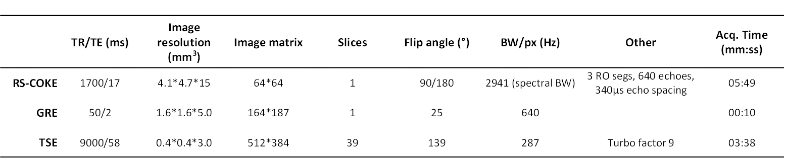
Healthy subjects were scanned, with local ethical approval, on a 7T scanner (MAGNETOM Terra pTx, Siemens Healthcare, Erlangen, Germany), using a custom-built, 8-channel transmit, 64-channel receive head coil8,9.A T2-weighted-TSE was acquired for RS-COKE slice placement, followed by a GRE image used to estimate receive channel weights for combination10. Parameters for all sequences are reported in Table 1. RS-COKE was run four times, in circularly-polarised (CP) mode (i) with and (ii) without VAPOR WS11 and with B1+ shimming applied to WS, excitation and refocussing pulses (iii) with and (iv) without WS. Per-channel B1+ maps were collected for pulse design. The slice-selective B1+ shim-weights for each pulse were optimised by magnitude least-squares12 with constraints on peak RF amplitude (B1+max = 175V) and standard deviation of flip angle (15% of nominal).
A bespoke processing pipeline13 was employed for frequency-dependent phase correction, k-space trajectory correction and smoothing at readout-segment transitions, which suppresses strong ghosting artifacts from subcutaneous lipids. No further lipid suppression was used. Automatic zero- and first-order phase correction were performed, as were residual eddy-current correction and water-scaling. The processed data were fitted voxel-wise in LCModel14. Metabolite concentrations are reported in arbitrary units as no a priori knowledge of relaxation times or tissue composition were included. Finally, WS efficiency (%), was calculated as:
$$WS_{eff} = \frac{I_{un} - I_{su}}{I_{un}} x 100\%$$
where Iun is the intensity of the unsuppressed water peak and Isu of the suppressed water peak15.
Results
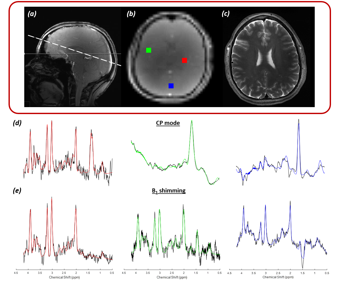
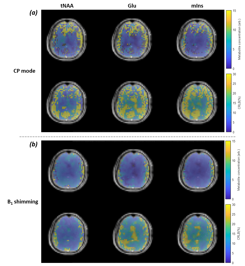
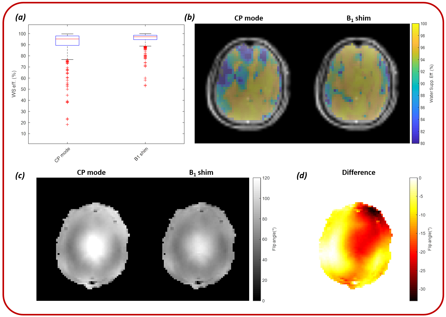
All data presented are for one volunteer. Figure 1 shows the RS-COKE slice position, the GRE image, the T2-weighted-TSE at the RS-COKE slice and spectra for three voxels. Concentrations and Cramér–Rao Lower Bounds (CRLB) for total n-acetyl aspartate (tNAA), glutamate (Glu) and myo-inositol (mIns) for CP mode, and with B1+ shimming are reported in Table 2. Metabolite and CRLB maps are shown in Figure 2 for tNAA, Glu and mIns for both modes. WS efficiency boxplots are shown in Figure 3 along with voxel-wise WS efficiency maps, simulated flip-angle maps for CP mode and B1+ shimming, and a flip-angle difference map.Discussion
Data presented for CP mode show that a high-quality spectrum was acquired for the red voxel, but those in the green and blue voxels are of poor quality with fitting of metabolite peaks largely not possible, as evidenced by high CRLB values. With B1+ shimming, the spectrum from the red voxel is comparable with CP mode, with similar values of concentration and CRLB, but the fitting is improved for the green and blue voxels. This can be seen in both the quality of the spectra where clear, resolvable metabolite peaks are evident, and in, the CRLB error estimates, which are all below 20%.The metabolite and CRLB maps show that B1+ shimming significantly improves the uniformity of concentration and error estimates for each of the metabolites, particularly in lateral regions.B1+ shimming also shows improvements in the WS efficiency across the slice, particularly evident in the right frontal lobe, even though the WS pulses are not slice selective.
In volunteers with smaller heads, high-quality results were achieved in CP mode, with B1+ shimming results comparable. In two volunteers with larger heads, significant lipid contamination was observed in CP mode, but not with B1+ shimming. Upon further investigation, this proved not to be related to the segment transitions, which was a possible source of ringing artefact in the readout direction. The source of the lipid signal in CP mode remains unclear and further investigation is underway.
Conclusion
This study shows the capacity of B1+ shimming in RS-COKE MRSI to improve spectral quality and quantification in some subjects. In particular, the use of B1+ shimming improves the capability of the RS-COKE technique to perform robust mapping of metabolites in the human brain in vivo.Acknowledgements
No acknowledgement found.References
1. Henning A. Proton and multinuclear magnetic resonance spectroscopy in the human brain at ultra-high field strength: A review. Neuroimage. 2018;168:181-98.
2. Mansfield P. Spatial mapping of the chemical shift in NMR. Magnetic Resonance in Medicine. 1984;1(3):370-86.
3. Posse S, DeCarli C, Le Bihan D. Three-dimensional echo-planar MR spectroscopic imaging at short echo times in the human brain. Radiology. 1994;192(3):733-8.
4. Keith GA, Seginer A, Porter DA, Schmidt R. Readout-segmented COKE: a technique for echo-planar spectroscopic imaging at 7 tesla with high spectral bandwidth and suppression of Nyquist errors in the spectral domain. ESMRMB; Virtual2020.
5. Keith GA, Vicari M, Woodward RA, Porter DA, editors. In vivo echo-planar spectroscopic imaging (EPSI) at 7 Tesla with readout segmentation for improved spectral bandwidth. ISMRM; 2019; Montreal.
6. Schmidt R, Seginer A, Tal A. Combining multiband slice selection with consistent k-t-space EPSI for accelerated spectral imaging. Magn Reson Med. 2019;82(3):867-76.
7. Boer VO, Klomp DWJ, Juchem C, Luijten PR, de Graaf RA. Multislice 1H MRSI of the human brain at 7 T using dynamic B0 and B1 shimming. Magnet Reson Med. 2012;68(3):662-70.
8. Williams SN, Allwood-Spiers S, McElhinney P, Paterson G, Herrler J, Liebig P, et al. A Nested Eight-Channel Transmit Array With Open-Face Concept for Human Brain Imaging at 7 Tesla. Frontiers in Physics. 2021;9.
9. Gunamony S, Feinberg D. An 8-channel transmit 64-channel receive compact head coil for Next Gen 7T scanner with head gradient insert. ISMRM; 2022; London.
10. Hall EL, Stephenson MC, Price D, Morris PG. Methodology for improved detection of low concentration metabolites in MRS: optimised combination of signals from multi-element coil arrays. Neuroimage. 2014;86:35-42.
11. Tkac I, Starcuk Z, Choi IY, Gruetter R. In vivo 1H NMR spectroscopy of rat brain at 1 ms echo time. Magn Reson Med. 1999;41(4):649-56.
12. Setsompop K, Wald LL, Alagappan V, Gagoski BA, Adalsteinsson E. Magnitude least squares optimization for parallel radio frequency excitation design demonstrated at 7 Tesla with eight channels. Magn Reson Med. 2008;59(4):908-15.
13. Seginer A, Keith GA, Porter DA, Schmidt R. Artifact suppression in readout-segmented consistent K-t space EPSI (RS-COKE) for fast (1) H spectroscopic imaging at 7 T. Magn Reson Med. 2022;88(6):2339-57.
14. Provencher SW. Estimation of metabolite concentrations from localized in vivo proton NMR spectra. Magn Reson Med. 1993;30(6):672-9.
15. Keith G, Porter D. Improved whole brain water suppression efficiency with four-pulse WET in echo-planar spectroscopic imaging (EPSI) at 7 tesla. ISMRM; Montreal2019. p. 2482.
Figures