1847
1H-MRS of NAD+ and tryptophan in human brain at 3T in under 5 minutes using a spectrally-selective single-slice pulse sequence1CAMIPM, Radiology, University of Pennsylvania, Philadelphia, PA, United States
Synopsis
Keywords: Spectroscopy, Spectroscopy
Motivation: NAD+ and tryptophan are important in energy metabolism, DNA repair, mitochondrial function, and aging. Both have recently been observed in brain at 7T, but observation at 3T is more challenging and has not been shown previously.
Goal(s): To detect NAD+ and tryptophan at 3T in brain in a clinically feasible scan time less than 5 minutes.
Approach: A single slice spectroscopy sequence with spectrally selective excitation was developed and optimized, allowing high sensitivity acquisition.
Results: The H2 (9.3 ppm) and H6 (9.1 ppm) peaks of NAD+ and the indole (10.1 ppm) peak of tryptophan are both unambiguously observed in four healthy subjects.
Impact: The ability to identify NAD+ and tryptophan at 3T in less than 5 minutes has the potential to significantly enhance the adoption of this method as part of existing neuroimaging protocols.
Introduction
Historically, 1H spectroscopy has mostly been confined to the spectral region upfield from water. However, recent interest in detection of nicotinamide adenine dinucleotide (NAD+) has sparked a renewed curiosity in the downfield side of the spectrum (>4.7 ppm). NAD+ is a vital coenzyme in energy metabolism, DNA repair, mitochondrial function, and aging, and it is currently being investigated as a potential disease biomarker and therapeutic target. Our group has recently observed another downfield peak in human brain which has been attributed to the indole protons of tryptophan (Trp)1, a precursor for production of NAD+ through the de novo synthesis pathway. Both of these metabolites are challenging to detect in vivo because of their low concentrations (< 1 mM) and polarization transfer effects with water and, therefore, require specialized pulse sequences.Previous work has shown that NAD+ can be detected in brain at UHF (≥ 7T) using pulse sequences that rely on spectrally selective excitation to avoid saturating downfield signals.2-4 A similar sequence was used by Nanga et. al. to detect Trp at 7T.1 Dziadosz et. al. has recently shown detectability of NAD+ at 3T using both a metabolite cycling technique to avoid water presaturation and spectrally selective excitation that took about 30 minutes interleaved to acquire.5 Here, we show detectability of both the NAD+ and Trp metabolites at 3T for the first time using an optimized single slice pulse sequence with spectrally selective excitation in an acquisition time of less than 5 minutes.
Methods
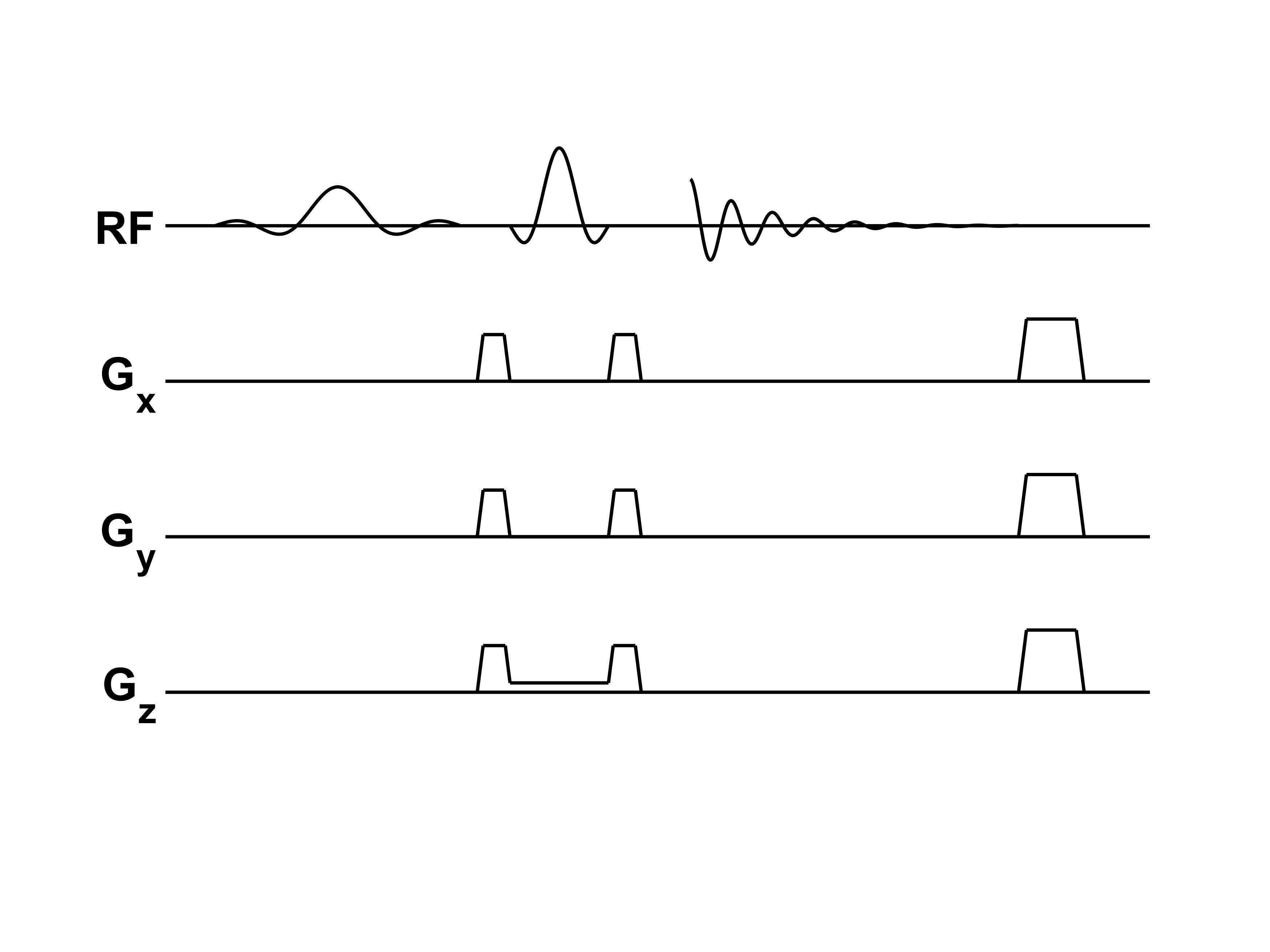
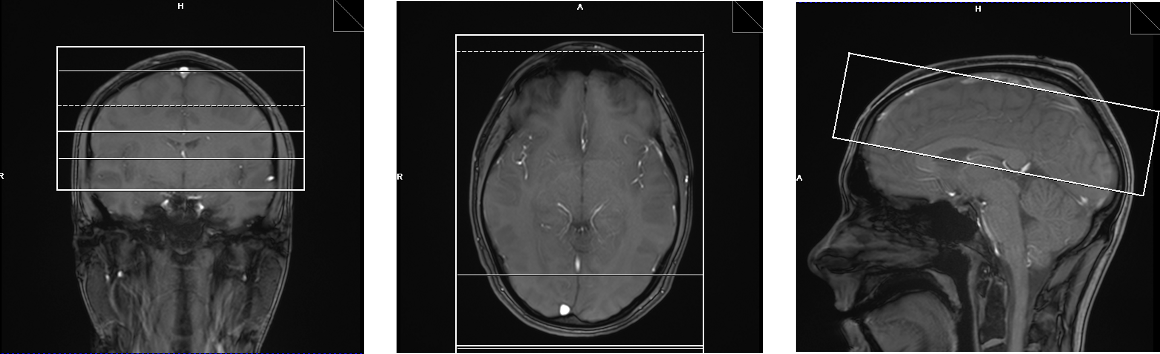
Four healthy subjects were scanned at 3T (MAGNETOM Prisma, Siemens Healthcare, Erlangen, Germany) using a 64 channel Rx head coil (Nova Medical) under an IRB-approved protocol. The pulse sequence is shown in Figure 1 and consists of a spectrally selective 90-degree Hamming-filtered sinc pulse followed by a low bandwidth, slice selective 180-degree SLR pulse. Excitation was centered at 9.7 ppm with a bandwidth of 2 ppm to fully excite resonances of NAD+ and Trp while only partially exciting the adenosine moieties around 8.5 ppm. TE/TR were 33/1000 ms, and 256 averages (with a 4-step RF phase cycling and 2-step gradient polarity cycling) were acquired in a scan time of 4 min 20 sec. An additional 36 sec water reference scan was acquired with the excitation frequency placed at 4.7 ppm. A 60 mm thick slice covering the superior brain region above the ventricles was obliquely placed as illustrated in Figure 2 to maximize brain coverage while avoiding regions that negatively affect the shim quality. The water signal from the reference scan was used to determine the receive coil coefficients applied in the downfield scan.Results
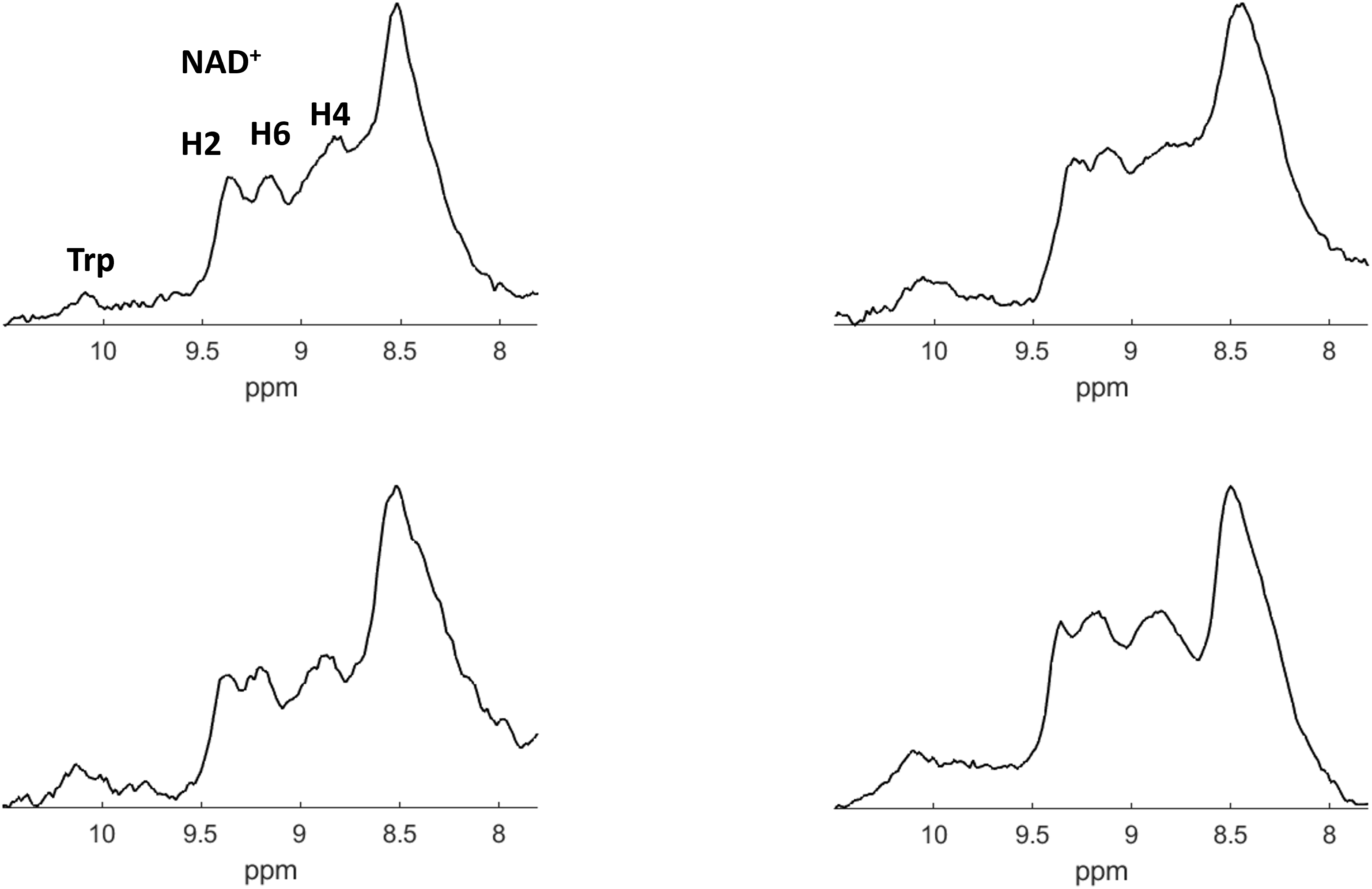
Downfield spectra from the four subjects are shown in Figure 3. In each spectrum, the H2 (9.3 ppm) and H6 (9.1 ppm) peaks of NAD+ are clearly visible along with the indole peak of Trp (10.1 ppm). Though present, the H4 (8.9 ppm) peak of NAD+ is less resolvable, as it is somewhat in the transition band of the excitation pulse along with the adenosine peak around 8.5 ppm.Discussion
In order to observe such low concentration resonances in a reasonably short scan time, the proposed pulse sequence attempted to maximize signal sensitivity in several ways. First, an entire slice was localized, allowing for a larger scan volume and a shorter TE than sequences that localize to a voxel. The reduction in TE was especially important here, as the spectrally selective excitation pulse was made longer than in our previous work at 7T in order to remain narrow band (2 ppm/256 Hz) with a sharper transition band (5 lobe). Second, a short TR was used to avoid prolonged dead time between excitations. In addition, the bandwidth of the localization pulse was kept low in order to take advantage of the chemical shift-induced slice displacement, thereby reducing water excitation. Quantitation is ongoing and will be compared with results obtained at 7T.While the benefit of UHF has been shown for the detection of these low sensitivity downfield resonances, clinical 7T scanners still have limited availability. To make NAD+/Trp spectroscopy more accessible, reliable detection at 3T is crucial. And the ability to do this in under 5 minutes (under 10 minutes, including imaging localization and shimming) allows for this protocol to be appended to existing neuroimaging protocols without requiring a dedicated session.
Conclusion
We have shown that the downfield resonances of NAD+ and Trp can be detected in human brain at 3T in under 5 minutes using a high-sensitivity spectrally selective, single slice sequence, potentially allowing more widespread usage.Acknowledgements
This work was supported by the National Institute of Biomedical Imaging and Bioengineering of the National Institutes of Health under award Number P41EB029460, by the National Institute of Aging of the National Institutes of Health under award numbers R01AG071725 and R01AG063869, and by the National Heart, Lung, and Blood Institute of the National Institutes of Health under award Numbers R01HL137984, R01HL169378, and F31HL158217.References
1. Nanga RP, Elliott MA, Swain A, Wilson N, Swago S, Soni ND, Witschey WR, Reddy R. Identification of l‐Tryptophan by down‐field 1H MRS: A precursor for brain NAD+ and serotonin syntheses. Magnetic Resonance in Medicine. 2022 Dec;88(6):2371-7.
2. de Graaf RA, De Feyter HM, Brown PB, Nixon TW, Rothman DL, Behar KL. Detection of cerebral NAD+ in humans at 7T. Magnetic resonance in medicine. 2017 Sep;78(3):828-35.
3. Gonçalves SI, Ligneul C, Shemesh N. Short echo time relaxation‐enhanced MR spectroscopy reveals broad downfield resonances. Magnetic resonance in medicine. 2019 Oct;82(4):1266-77.
4. Bagga P, Hariharan H, Wilson NE, Beer JC, Shinohara RT, Elliott MA, Baur JA, Marincola FM, Witschey WR, Haris M, Detre JA. Single‐Voxel 1H MR spectroscopy of cerebral nicotinamide adenine dinucleotide (NAD+) in humans at 7T using a 32‐channel volume coil. Magnetic resonance in medicine. 2020 Mar;83(3):806-14.
5. Dziadosz M, Hoefemann M, Döring A, Marjańska M, Auerbach EJ, Kreis R. Quantification of NAD+ in human brain with 1H MR spectroscopy at 3 T: Comparison of three localization techniques with different handling of water magnetization. Magnetic resonance in medicine. 2022 Sep;88(3):1027-38.
Figures