1845
Constrained Optimized Water Suppression (COWS) for Macromolecule Measurements with 1H Magnetic Resonance Spectroscopy1Biomedical Engineering, Columbia University, New York, NY, United States, 2Radiology, Columbia University, New York, NY, United States
Synopsis
Keywords: Spectroscopy, New Signal Preparation Schemes, Magnetic Resonance Spectroscopy, Macromolecules
Motivation: A new water suppression module optimized for both macromolecule detection and short repetition time sequences such as magnetic resonance spectroscopic imaging.
Goal(s): To have an efficient, flexible, water suppression algorithm where macromolecules can be measured.
Approach: Single voxel localization by adiabatic selective refocusing (semi-LASER) measurement of macromolecules were conducted in the prefrontal cortex, posterior frontal (PFL) and occipital lobes. Both VAPOR and the customizable water suppression (COWS) algorithm were performed for each experiment.
Results: Both WS methods performed well with residual water signals below the 2.01-ppm NAA singlet signals. COWS demonstrated better water suppression than VAPOR overall, especially in the PFL.
Impact: A new customizable water suppression algorithm, COWS, was tested in vivo for improved efficiency and speed when compared to the gold standard (VAPOR).
Introduction
Proton magnetic resonance spectroscopy (MRS) is a non-invasive technique that can be used to measure biochemical compounds in living tissue. Macromolecules (MM) are large chemical species made up of proteins1,2 that exhibit broad linewidths, and typically overlap with smaller chemical species, referred to as metabolites. Although macromolecules may bias metabolite spectra at short-TE, they themselves may be clinically relevant. Fluctuations in macromolecule concentrations have been detected in pathologies such as Multiple Sclerosis3 and Normal Pressure Hydrocephalus4, and have been shown to help differentiate solitary metastasis from glioblastomas1,5.The concentration of water is 5,000-10,000 times higher than that of metabolites and macromolecules6,7, and excellent water suppression (WS) is needed to accurately quantify these chemicals. Currently, the gold standard for WS is Variable Power Radio Frequency Pulses with Optimized Relaxation Delays8 (VAPOR). Macromolecule measurements typically employ a double inversion recovery preparation1, which interferes with the standard VAPOR WS timings. Moreover, VAPOR cannot be used for longer spectroscopy imaging techniques such as MRSI9.
Previously, our lab presented the development of a flexible WS algorithm, Constrained Optimization Water Suppression (COWS), that allows for WS schemes tailored to application-specific sequence timings. COWS enables the creation of WS schemes for regular MRS with shorter duration, as opposed to an optimized 7-pulse VAPOR WS scheme, without compromising WS efficiency. In this study, we assess the WS performance of a newly designed COWS scheme that is dedicated to MM measurement, employing double-inversion preparation for metabolite nulling. We demonstrate similar or improved performance of COWS compared to VAPOR despite its significantly reduced time requirement.
Material and Methods
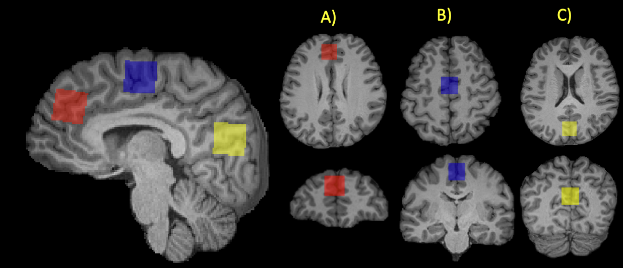
The COWS algorithm was used to design a WS scheme comprising 7 pulses, with a minimum duration of 28 ms between all pulse, and 21 ms between the seventh WS and the single-voxel MRS excitation. All water coherence pathways are automatically dephased by the crusher scheme generated by COWS via DOTCOPS10,11.To validate the efficiency of the COWS and VAPOR WS modules, 1H MR spectra were collected from a sample of 10 healthy participants (female=5, age=30 +/- 9 years) on a 3-Tesla MAGNETOM Prisma full-body (60-cm bore) magnetic resonance scanner using built-in transmission with the vendor’s 32-receive-channel head and neck coil (Siemens Healthineers, Erlangen, Germany). T1 weighted (repetition time (TR) = 2300 ms, echo time (TE) = 2.26 ms, field of view (FOV) = 256 x 256 x 192 mm3, with 1 mm slice thickness) images were acquired sagittally. Single voxel MRS using a semi-localization through adiabatic selective refocusing (semi-LASER) 12 sequence (TR = 2000 ms, TE = 26 ms, number of repetitions (NR) = 64, number of spectral points (SP) = 2048, voxel size = 20 mm3) were conducted in three brain regions: the prefrontal cortex, the posterior frontal (PFL), and the occipital lobes (OL) (Figure 1). Macromolecule spectra were acquired using double inversion recovery, for both VAPOR (number of pulses (np) = 7, duration (t) = 626 ms), and COWS (np = 7, t = 236 ms).
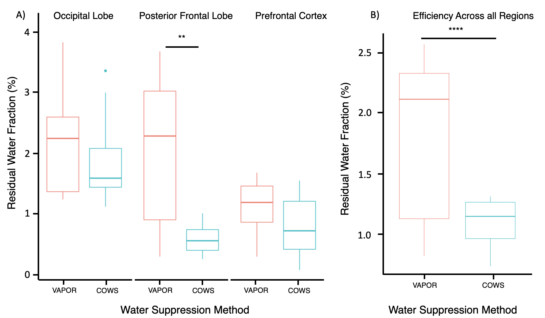
To determine the accuracy of each WS scheme, residual water fraction was calculated from the magnitude spectrum, where the amplitude of water in the water-suppressed spectrum is divided by the amplitude of water in the water-unsuppressed spectrum, then multiplied by 100%. A lower residual water fraction indicates better WS. Next, t-tests with Shapiro-Wilks were performed to investigate differences between COWS and VAPOR.
Results and Discussion
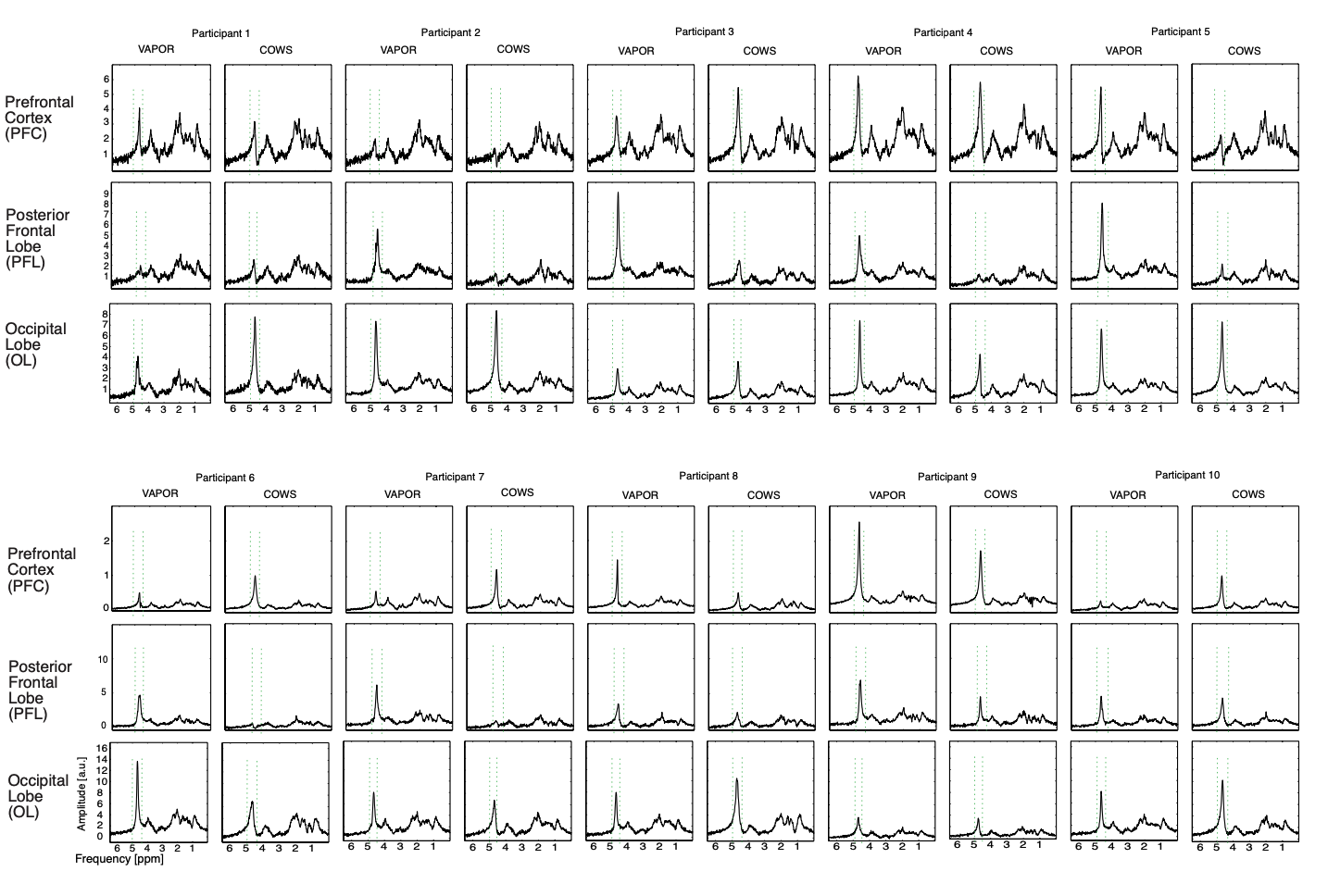
All macromolecule spectra with residual water can be seen across all three brain regions in Figure 2. Both WS methods performed very well with residual water signals well below the 2.01-ppm NAA singlet signals. The average residual water fraction for macromolecule spectra were 1.11% for COWS and 1.82% for VAPOR. Regionally, COWS had a better residual water fraction than VAPOR in the PFC (0.801% / 1.18%), PFL (0.612% / 2.06%), and OL (1.90% / 2.23%). A significant difference was observed in the PFL (p = 0.0048, CI = [0.56 2.34]). There were no significant differences observed in the PFC (p = 0.089, CI = [-0.06 0.81]) nor the OL (p = 0.38, CI = [-0.44 1.096]). The COWS (236 ms) WS module was faster than VAPOR (636 ms). Overall, COWS demonstrated better water suppression than VAPOR, (p-value (p) = 10-6, 95% confidence interval (CI) = [1.25 2.23]) (Figure 3).Conclusion
An in-vivo experiment to test the efficiency of COWS WS against the gold standard, VAPOR, was performed. COWS performed better than VAPOR when suppressing water in acquisitions designed to measure macromolecules. The COWS module also is substantially shorter than VAPOR, making it more suitable for short TR experiments such as MRSI.Acknowledgements
No acknowledgement found.References
1. Landheer K, Gajdošík M, Treacy M, Juchem C. Concentration and effective T 2 relaxation times of macromolecules at 3T. Magn Reson Med 2020;84:2327–2337.
2. Cudalbu C, Behar KL, Bhattacharyya PK, et al. Contribution of macromolecules to brain 1 H MR spectra: Experts’ consensus recommendations. NMR Biomed 2021;34:e4393.
3. Mader I. Proton MR spectroscopy with metabolite-nulling reveals elevated macromolecules in acute multiple sclerosis. Brain 2001;124:953–961.
4. Akiyama Y, Yokoyama R, Takashima H, et al. Accumulation of Macromolecules in Idiopathic Normal Pressure Hydrocephalus. Neurol Med Chir (Tokyo) 2021;61:211–218.
5. Crisi G, Orsingher L, Filice S. Lipid and Macromolecules Quantitation in Differentiating Glioblastoma From Solitary Metastasis: A Short–Echo Time Single-Voxel Magnetic Resonance Spectroscopy Study at 3 T. J Comput Assist Tomogr 2013;37:265–271.
6. Tkáč I, Deelchand D, Dreher W, et al. Water and lipid suppression techniques for advanced 1 H MRS and MRSI of the human brain: Experts’ consensus recommendations. NMR Biomed 2021;34(5):e4459.
7. Zhu H, Barker PB. MR Spectroscopy and Spectroscopic Imaging of the Brain. In: Modo M, Bulte JWM, editors. Magnetic Resonance Neuroimaging. Vol 711. Methods in Molecular Biology. Totowa, NJ: Humana Press; 2011:203–226.
8. Tkáč I, Staruk Z, Choi I-Y, Gruetter R. In vivo1H NMR spectroscopy of rat brain at 1 ms echo time. Magn Reson Med 1999;41:649–656.
9. Tkáč I, Deelchand D, Dreher W, et al. Water and lipid suppression techniques for advanced 1H MRS and MRSI of the human brain: Experts’ consensus recommendations. NMR Biomed 2021;34:e4459.
10. Landheer K, Juchem C. Dephasing optimization through coherence order pathway selection (DOTCOPS) for improved crusher schemes in MR spectroscopy. Magn Reson Med 2019;81:2209–2222.
11. Landheer K, Juchem C. Simultaneous optimization of crusher and phase cycling schemes for magnetic resonance spectroscopy: an extension of dephasing optimization through coherence order pathway selection. Magn Reson Med 2020;83:391–402.
12. Landheer K, Gajdošík M, Juchem C. A semi-LASER, single-voxel spectroscopic sequence with a minimal echo time of 20.1 ms in the human brain at 3 T. NMR Biomed 2020;33:e4324.
Figures