1836
The Impact of Myo-Inositol on Cognitive Performance: A 7T Magnetic Resonance Spectroscopy Study1Psychiatry, Psychotherapy and Psychosomatics, RWTH Aachen University, Aachen, Germany, 2Institute of Neuroscience and Medicine, INM-4, Forschungszentrum Jülich GmbH, Jülich, Germany, 3Center for Computational Life Sciences, RWTH, Aachen, Germany, 4JARA – BRAIN – Translational Medicine, Aachen, Germany, 5Neurology, RWTH Aachen University, Aachen, Germany, 6Institute of Neuroscience and Medicine, INM-11, Forschungszentrum Jülich GmbH, Jülich, Germany
Synopsis
Keywords: Spectroscopy, Spectroscopy, 7T ultra high field MRS, Cognition
Motivation: This study aims to contribute novel insights into the potential cognitive role of myo-inositol and its implications for brain metabolism using 7 Tesla magnetic resonance spectroscopy (7T-MRS).
Goal(s): Our primary goal was to determine if myo-inositol concentrations within the posterior cingulate cortex (PCC) influence cognitive performance.
Approach: Ultra-high field 7T-MRS was used to measure myo-inositol concentrations in relation to cognitive performance, as assessed with the Trail-Making-Test (TMT-A/B). Data analysis involved multiple linear regressions with covariate control and bootstrapping for robustness.
Results: Our study reveals a significant (p<0.05) positive association between higher myo-inositol levels in the PCC and enhanced cognitive performance in TMT-A/B.
Impact: This study suggests the potential of myo-inositol as a biomarker for cognitive functioning. Furthermore, the findings contribute to our understanding of brain neurochemistry, supporting the advancement of personalised medicine for cognitive impairments.
Introduction
Myo-inositol (Ins), a multifaceted metabolite, has captured scientific interest for its diverse roles, including marking glial activity and involvement in inflammatory processes1. Notably, its association with cognitive-related conditions underscores the importance of understanding and regulating Ins. Its potential as a prophylactic agent against cognitive decline in disorders such as Alzheimer's disease and mild cognitive impairment is particularly compelling2. Additionally, the posterior cingulate cortex (PCC) is known for its structural and functional alterations in several cognitive-impacting conditions, including Alzheimer’s disease, attention deficit hyperactivity disorder (ADHD), autism, and aging 3-5. The enhanced signal-to-noise ratio of 7 Tesla magnetic resonance spectroscopy (7T MRS) markedly improves the differentiation, detection and quantification of neurometabolites such as Ins 6,7. Exploiting this cutting-edge technology, this study explores the influence of Ins concentrations on cognitive tasks.Methods
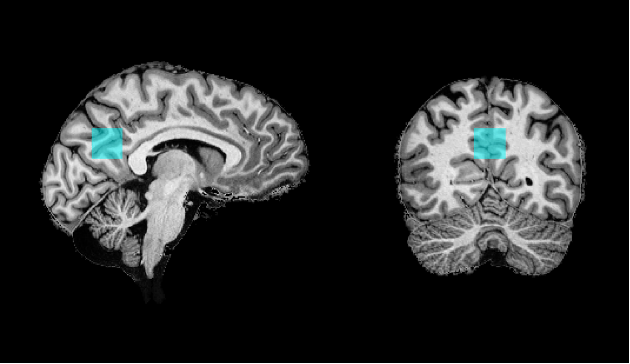
MR data were collected from 43 healthy individuals (26 males, 17 females; age range: 18-38 years) using a 7T MAGNETOM Terra scanner (Siemens Healthineers). MRS was used to quantify the absolute concentration of Ins within the PCC, as depicted in Figure 1.Structural MRI: MP2RAGE sequence - TR/TE 4500ms/1.99ms, voxel-size 0.75 mm3 isotropic resolution. Single-voxel MRS: STEAM sequence with ultra-short echo-time: TE = 4.6ms; TM = 28 ms; TR = 8200ms; 64 averages; voxel-size 20×20×20mm3 8-10. MR-spectra were pre-processed (motion, frequency and phase drift corrections), fitted and quantified using the FID-A package and LCModel (6.3-0I)11-14.The cognitive impairment sensitive Trail Making Test (TMT A/B) was performed 15,16 to rate the speed of visual search, processing, and execution. Data analysis was conducted using SPSS, and multiple linear regression was employed to explore the predictive value of Ins concentrations on TMT performance, controlling for age and gender as covariates. Bootstrapping was performed to enhance the reliability and robustness of our results.Results
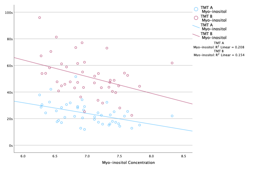
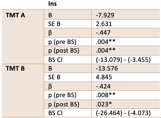
The impact of Ins on TMT parts A and B was statistically significant. These results persisted even after robust bootstrapping procedures were applied, further reinforcing the validity of our findings. For TMT-A, the bootstrapped p-value was .004, while for TMT-B, it was .023, indicating a reliable and noteworthy effect of Ins concentration on these cognitive tasks. For additional details, refer to the covariates analysis presented in Table 1 and Figure 2 for scatterplot visualisation.Discussion
In this study, a significant association between Ins levels and performance TMT A and B was observed, suggesting that elevated Ins concentrations in the PCC are predictive of enhanced visual search capabilities, processing speed, and mental flexibility. The results further implicate Ins in the executive and orienting components of attention 17. This is in agreement with nutrition-based research demonstrating cognitive improvements with inositol-stabilised arginine silicate (ASI) supplementation, which has been linked to better TMT outcomes 18. The cognitive-enhancing effects associated with Ins may stem from its role in arginine metabolism, where it reduces arginase activity, thereby increasing arginine availability 19. Arginine, a semi-essential amino acid, is involved in numerous critical functions, including cell proliferation, immune response, stress regulation, and gene expression1,20-24. Its significance in brain metabolism is partially attributed to its role as a precursor for nitric oxide, which induces vasodilation and, therefore could be what improves cerebral blood flow and potentially enhances PCC function. Contrasting findings in a separate MRS study on individuals with Down Syndrome showed a negative correlation between Ins levels and cognitive performance, indicating region-specific and possibly gene expression-related variations in Ins function 25. These findings underscore the potential of Ins as a cognitive enhancer and warrant further exploration of its neurological roles.Conclusion
In summary, our study highlights a significant link between elevated Ins levels in the PCC and improved cognitive performance in tasks involving visual search, processing speed, executive function and mental flexibility. These findings suggest the potential cognitive-enhancing role of Ins. Further investigation is warranted, particularly regarding region-specific variations and gene expression-related factors. This research advances our understanding of brain metabolism and its impact on fundamental cognitive processes.Acknowledgements
The authors thank Petra Engels, Elke Bechholz, and Anita Köth for their help with the scans, and Claire Rick for her careful proofreading of the abstract. We would like to acknowledge E.J. Auerbach and M. Marjanska (Center for Magnetic Resonance Research and Department of Radiology, University of Minnesota, USA) for the development of the STEAM sequence for the Siemens platform which were provided by the University of Minnesota under a C2P agreement.References
1 Fisher, S. K., Novak, J. E. & Agranoff, B. W. Inositol and higher inositol phosphates in neural tissues: homeostasis, metabolism and functional significance. J Neurochem 82, 736-754, doi:10.1046/j.1471-4159.2002.01041.x (2002).
2 López-Gambero, A. J., Sanjuan, C., Serrano-Castro, P. J., Suárez, J. & Rodríguez de Fonseca, F. The Biomedical Uses of Inositols: A Nutraceutical Approach to Metabolic Dysfunction in Aging and Neurodegenerative Diseases. Biomedicines 8, doi:10.3390/biomedicines8090295 (2020).
3 Johnson, K. A. et al. Preclinical prediction of Alzheimer's disease using SPECT. Neurology 50, 1563-1571, doi:10.1212/wnl.50.6.1563 (1998).
4 Buckner, R. L., Andrews-Hanna, J. R. & Schacter, D. L. The brain's default network: anatomy, function, and relevance to disease. Ann N Y Acad Sci 1124, 1-38, doi:10.1196/annals.1440.011 (2008).
5 Greicius, M. Resting-state functional connectivity in neuropsychiatric disorders. Curr Opin Neurol 21, 424-430, doi:10.1097/WCO.0b013e328306f2c5 (2008).
6 Otazo, R., Mueller, B., Ugurbil, K., Wald, L. & Posse, S. Signal‐to‐noise ratio and spectral linewidth improvements between 1.5 and 7 Tesla in proton echo‐planar spectroscopic imaging. Magnetic Resonance in Medicine: An Official Journal of the International Society for Magnetic Resonance in Medicine 56, 1200-1210 (2006).
7 Motyka, S. et al. The influence of spatial resolution on the spectral quality and quantification accuracy of whole‐brain MRSI at 1.5 T, 3T, 7T, and 9.4 T. Magnetic Resonance in Medicine 82, 551-565 (2019).
8 Tkác, I., Oz, G., Adriany, G., Uğurbil, K. & Gruetter, R. In vivo 1H NMR spectroscopy of the human brain at high magnetic fields: metabolite quantification at 4T vs. 7T. Magn Reson Med 62, 868-879, doi:10.1002/mrm.22086 (2009).
9 Gruetter, R. & Tkác, I. Field mapping without reference scan using asymmetric echo-planar techniques. Magn Reson Med 43, 319-323, doi:10.1002/(sici)1522-2594(200002)43:2<319::aid-mrm22>3.0.co;2-1 (2000).
10 Tkác, I., Starcuk, Z., Choi, I. Y. & Gruetter, R. In vivo 1H NMR spectroscopy of rat brain at 1 ms echo time. Magn Reson Med 41, 649-656, doi:10.1002/(sici)1522-2594(199904)41:4<649::aid-mrm2>3.0.co;2-g (1999).
11 Simpson, R., Devenyi, G. A., Jezzard, P., Hennessy, T. J. & Near, J. Advanced processing and simulation of MRS data using the FID appliance (FID-A)-An open source, MATLAB-based toolkit. Magn Reson Med 77, 23-33, doi:10.1002/mrm.26091 (2017).
12 Soher, B. J., Semanchuk, P., Todd, D., Steinberg, J. & Young, K. in Proc Int Soc Magn Reson Med. 1410.
13 Govindaraju, V., Young, K. & Maudsley, A. A. Proton NMR chemical shifts and coupling constants for brain metabolites. NMR Biomed 13, 129-153, doi:10.1002/1099-1492(200005)13:3<129::aid-nbm619>3.0.co;2-v (2000).
14 Govind, V., Young, K. & Maudsley, A. A. Corrigendum: proton NMR chemical shifts and coupling constants for brain metabolites. Govindaraju V, Young K, Maudsley AA, NMR Biomed. 2000;13: 129-153. NMR Biomed 28, 923-924, doi:10.1002/nbm.3336 (2015).
15 Reitan, R. TMT, trail making test a & B. South Tucson, AR: Reitan Neuropsychology Laboratory (1992).
16 Arnett, J. A. & Labovitz, S. S. Effect of physical layout in performance of the Trail Making Test. Psychological Assessment 7, 220 (1995).
17 Petersen, S. E. & Posner, M. I. The Attention System of the Human Brain: 20 Years After. Annual Review of Neuroscience 35, 73-89, doi:10.1146/annurev-neuro-062111-150525 (2012).
18 Gills, J. L. et al. Acute Inositol-Stabilized Arginine Silicate Improves Cognitive Outcomes in Healthy Adults. Nutrients 13, 4272 (2021).
19 Emerson, K., Perez-Ojalvo, S., Komorowski, J. & Greenberg, D. Arginase Enzyme Activity in Human Serum as a Marker of Cognitive Function: The Role of Inositol in Combination with Arginine Silicate. International Journal of Biomedical and Biological Engineering 17, 17-22 (2023).
20 Fujiwara, T. et al. L-arginine stimulates fibroblast proliferation through the GPRC6A-ERK1/2 and PI3K/Akt pathway. Plos one 9, e92168 (2014).
21 Kim, S.-H., Roszik, J., Grimm, E. A. & Ekmekcioglu, S. Impact of l-arginine metabolism on immune response and anticancer immunotherapy. Frontiers in oncology 8, 67 (2018).
22 Pervin, M., Unno, K., Konishi, T. & Nakamura, Y. L-arginine exerts excellent anti-stress effects on stress-induced shortened lifespan, cognitive decline and depression. International journal of molecular sciences 22, 508 (2021).
23 Morris, S. M., Jr. Arginine: beyond protein. Am J Clin Nutr 83, 508s-512s, doi:10.1093/ajcn/83.2.508S (2006).
24 Morris, S. M., Jr. Recent advances in arginine metabolism: roles and regulation of the arginases. Br J Pharmacol 157, 922-930, doi:10.1111/j.1476-5381.2009.00278.x (2009).
25 Beacher, F. et al. Hippocampal myo-inositol and cognitive ability in adults with Down syndrome: an in vivo proton magnetic resonance spectroscopy study. Archives of general psychiatry 62, 1360-1365 (2005).
Figures