1830
Evidence of impaired dACC glutamate modulation under task-specific motor control in obsessive compulsive disorder (OCD) using 1H fMRS1Psychiatry and Behavioral Neurosciences, Wayne State University, Detroit, MI, United States, 2Brain Imaging Research Division, Wayne State University, Detroit, MI, United States, 3Translational Neuroscience Program, Wayne State University, Detroit, MI, United States
Synopsis
Keywords: Spectroscopy, Spectroscopy, 1H MRS, fMRS, OCD
Motivation: The neurobiological mechanisms of poor top-down motor control in OCD are not well understood.
Goal(s): Our goal was to investigate neurobiological differences in glutamate modulation across different motor/inhibitory control responses in OCD compared to healthy controls.
Approach: 1H fMRS was conducted in adolescents with OCD and healthy controls.
Results: Task-specific impairments to the dACC glutamate modulation were observed in OCD compared to healthy controls.
Impact: These results providing compelling evidence towards characterizing neurosignaling changes in OCD pathophysiology.
Introduction
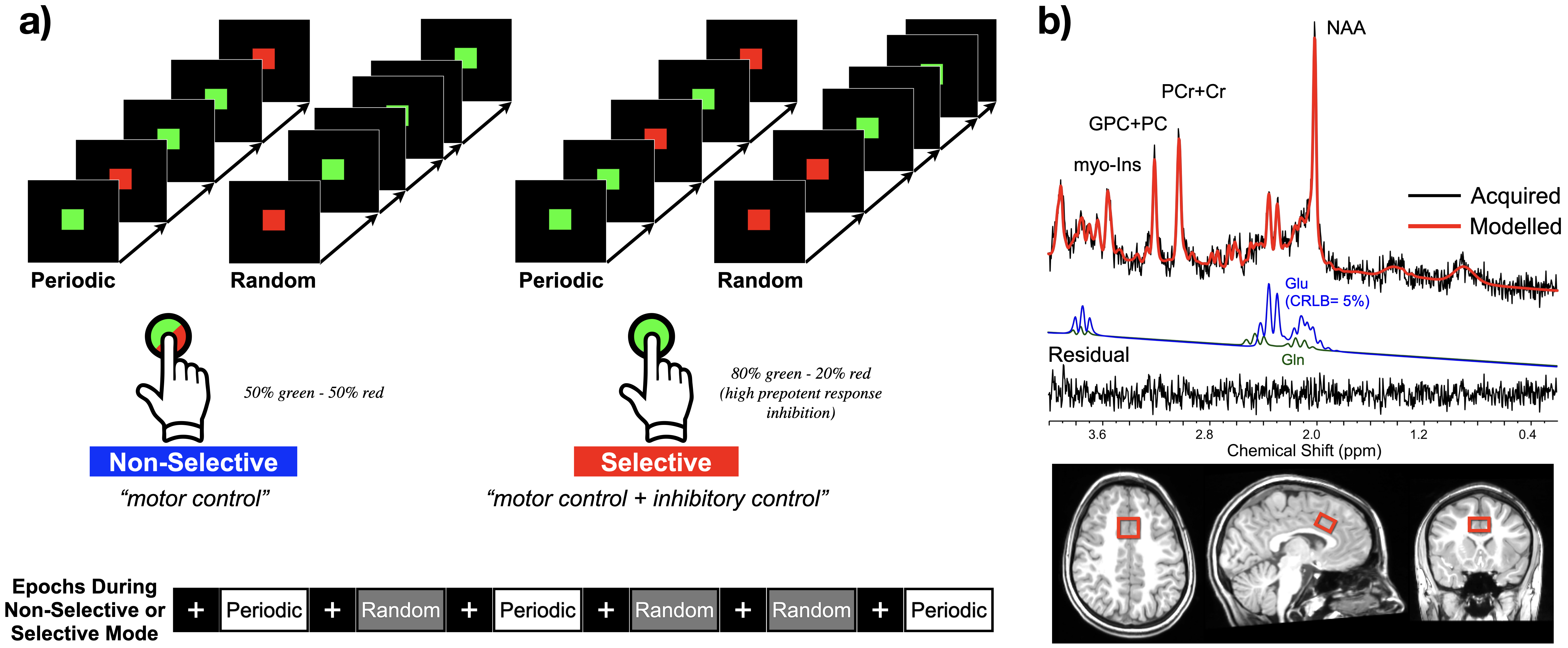
Brain networks rely heavily on the engagement of integrated actions of glutamatergic and GABAergic neurons that shifts the brain’s excitatory and inhibitory (E/I) synaptic drive. The E/I balance is particularly crucial for top-down control exerted by regions like the dorsal anterior cingulate cortex (dACC)2, 3. This control is impaired in several psychiatric conditions, most notably in obsessive compulsive disorder (OCD), where recurrent intrusive, obsessive thoughts and/or repetitive, compulsive behaviors interfere with the dACC’s functional role. fMRI studies4, 5 have demonstrated a dACC impairment during top-down motor control in OCD6, 7. However, the hemodynamic filtering of the fMRI signal does not permit assessment of the contributions of excitatory and/or inhibitory processes. Because shifts in the E/I balance are driven by variations in excitatory glutamatergic neurotransmission, they can reliably be detected with ¹H fMRS8, 9, a functional imaging technique not constrained by hemodynamics. Here, we used ¹H fMRS to investigate functional imbalances in the E/I synaptic drive of the dACC in OCD youth, using a specifically developed motor control task that used excitatory (Non-Selective) or inhibitory (Selective) response modes.Methods
A total of 34 OCD (13 males and 21 females; mean age, 17.8+2.2 years; age range 12.0 to 20.0 years) and 48 healthy individuals (HC; 20 males and 28 females; mean age, 17.1+2.4; age range 12.3 to 20.7 years) participated. The ¹H fMRS data were collected on a 3T Siemens Verio system using a 32-channel volume head-coil. Following the MPRAGE T1-weighted structural images, a single-voxel was prescribed midline in the dACC (2.0x1.7x1.2cm3 or 4.1cm3; Figure 1) using the AVP approach8, followed by B0-field shimming using FASTESTMAP. The two distinct task modes were: a) the “Non-Selective” (excitatory) mode in which participants always tapped their right forefinger to flashing green or red probes (50/50% distribution, 100% responses), and b) a “Selective” (inhibitory) mode in which participants inhibited responses to red probes (20% of responses). Each task mode included a .1s probe stimulus duration and six 32s task epochs interspersed with 16s rest epochs. Nineteen consecutive ¹H MRS measurements were acquired during each task mode (PRESS with OVS and VAPOR, TE=23ms, TR=2.67s, 6 averages/measurements, 2048 data points; scan time/mode 5:04min). A control condition (participants fixated on a crosshair, 3:28min) preceded the motor tasks forming a baseline for quantifying task-induced changes. The two ¹H MRS spectra from each task epoch and three consecutive pairs from the control condition, were phased (0th and 1st order) and frequency shift corrected prior to averaging and quantification using LCModel (Figure 1). A repeated measure GEE approach for each task mode was used to test significant glutamate level differences with the following terms in the model: Group (OCD, HC), Sex, Age, mean baseline condition glutamate level and interaction terms (Group*Sex and Group* Age).Results
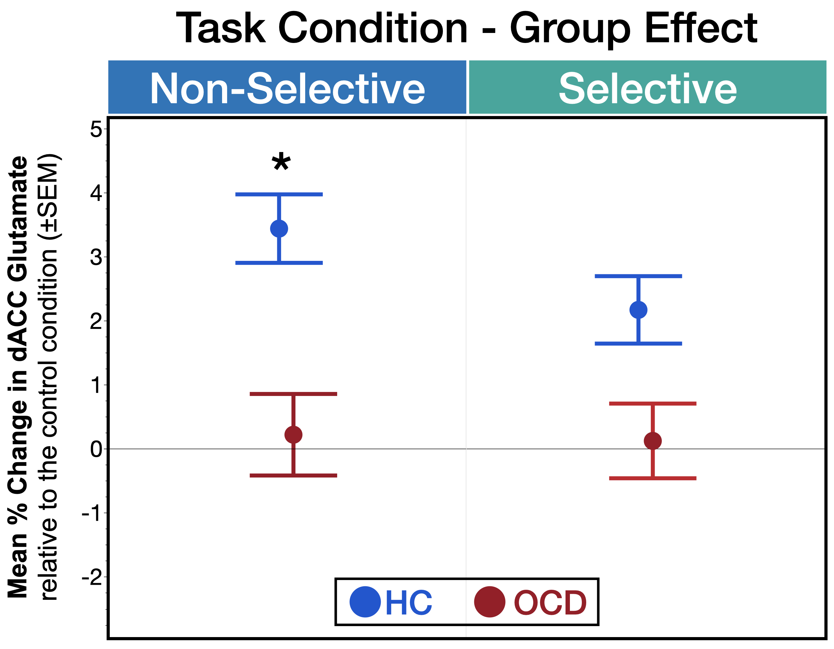
The Non-Selective mode drove a significant main effect of Group (χ2=4.19, p=.041; Figure 2), and Group*Sex (χ2=7.88, p=.0050) and Group*Age (χ2=5.87, p=.015) interactions. Post-hoc analyses demonstrated a significant decrease in dACC glutamate modulation in OCD females compared HC females (p<.0001).The Selective mode did not drive any significant group effects other than a Group*Sex (χ2=0.03, p=.15) trend, with post-hoc analyses demonstrating significantly lower dACC glutamate in OCD females compared to HC females (p=.021).
GLM analysis demonstrated no significant group differences in the glutamate baseline level or in the GM tissue fraction (or differences in Group*Sex or Group*Age interactions; all p>.05). Finally, response times on the task (available for 62% and 48% of the OCD and HC sample, respectively), demonstrated no significant Group or Group*Sex effect for both responding task modes (all p>.05).
Discussion
These results are the first to assess and demonstrated impaired task-specific functional neurochemistry of the dACC in OCD. Significantly lower mean glutamate levels (below baseline condition) during Non-Selective responding suggests a E/I shift towards relatively lower glutamatergic neurotransmission (and/or potentially greater inhibitory neurotransmission), with the task-dependent sensitivity of ¹H fMRS is emphasized by the lack of inter-group differences for the baseline condition. Our results provide a compelling approach toward in vivo characterization of the relative contributions of excitatory vs. inhibitory signaling in the pathophysiology of OCD.Acknowledgements
This work was supported by the NIMH under award number R01MH59299 (DRR, JAS and VAD), the Children’s Hospital of Michigan Foundation, Miriam Hamburger Endowed Chair, Paul and Anita Strauss Endowment, and by the Lycaki-Young Funds from the State of Michigan.
References
- Bari A, Robbins TW. Inhibition and impulsivity: behavioral and neural basis of response control. Prog Neurobiol. 2013;108:44-79. Epub 2013/07/17. doi: 10.1016/j.pneurobio.2013.06.005. PubMed PMID: 23856628.
- Paus T. Primate anterior cingulate cortex: where motor control, drive and cognition interface. Nat Rev Neurosci. 2001;2(6):417-24. Epub 2001/06/05. doi: 10.1038/3507750035077500 [pii]. PubMed PMID: 11389475.
- Haber SN, Calzavara R. The cortico-basal ganglia integrative network: the role of the thalamus. Brain Res Bull. 2009;78(2-3):69-74. doi: 10.1016/j.brainresbull.2008.09.013. PubMed PMID: 18950692.
- Asemi A, Ramaseshan K, Burgess A, Diwadkar VA, Bressler SL. Dorsal anterior cingulate cortex modulates supplementary motor area in coordinated unimanual motor behavior. Front Hum Neurosci. 2015;9:309. doi: 10.3389/fnhum.2015.00309. PubMed PMID: 26089783; PMCID: PMC4454840.
- Diwadkar VA, Asemi A, Burgess A, Chowdury A, Bressler SL. Potentiation of motor sub-networks for motor control but not working memory: Interaction of dACC and SMA revealed by resting-state directed functional connectivity. PloS one. 2017;12(3):e0172531. doi: 10.1371/journal.pone.0172531. PubMed PMID: 28278267; PMCID: PMC5344349.
- Diwadkar VA, Burgess A, Hong E, Rix C, Arnold PD, Hanna GL, Rosenberg DR. Dysfunctional Activation and Brain Network Profiles in Youth with Obsessive-Compulsive Disorder: A Focus on the Dorsal Anterior Cingulate during Working Memory. Front Hum Neurosci. 2015;9:149. Epub 2015/04/09. doi: 10.3389/fnhum.2015.00149. PubMed PMID: 25852529; PMCID: PMC4362304.
- Friedman A, Burgess A, Ramaseshan K, Easter P, Khatib D, Chowdury A, Arnold PD, Hanna GL, Rosenberg DR, Diwadkar VA. Brain network dysfunction in obsessive-compulsive disorder induced by simple uni-manual behavior: The role of the dorsal anterior cingulate cortex. Psychiatry Res Neuroimaging. 2017;260:6-15.
- Lauritzen M, Mathiesen C, Schaefer K, Thomsen KJ. Neuronal inhibition and excitation, and the dichotomic control of brain hemodynamic and oxygen responses. NeuroImage. 2012;62(2):1040-50. doi: 10.1016/j.neuroimage.2012.01.040. PubMed PMID: 22261372.
- Tatti R, Haley MS, Swanson OK, Tselha T, Maffei A. Neurophysiology and Regulation of the Balance Between Excitation and Inhibition in Neocortical Circuits. Biological psychiatry. 2017;81(10):821-31. doi: 10.1016/j.biopsych.2016.09.017. PubMed PMID: 27865453; PMCID: PMC5374043.
- Stanley JA, Raz N. Functional Magnetic Resonance Spectroscopy: The "New" MRS for Cognitive Neuroscience and Psychiatry Research. Frontiers in psychiatry. 2018;9:76. Epub 2018/03/30. doi: 10.3389/fpsyt.2018.00076. PubMed PMID: 29593585; PMCID: PMC5857528.
Figures