1824
Correlations between 5-ALA fluorescence and 7T MRSI in gliomas: Preliminary observations1High Field MR Centre, Medical University of Vienna, Vienna, Austria, 2Department of Neurosurgery, Medical University of Vienna, Vienna, Austria, 3Division of Neuropathology and Neurochemistry, Medical University of Vienna, Vienna, Austria, 4Division of Neuroradiology and Musculoskeletal Radiology, Medical University of Vienna, Vienna, Austria, 5Division of Oncology, Medical University of Vienna, Vienna, Austria, 6Christian Doppler Laboratory for MR Imaging Biomarkers, Vienna, Austria
Synopsis
Keywords: Spectroscopy, Tumor, Glioma, 5-ALA
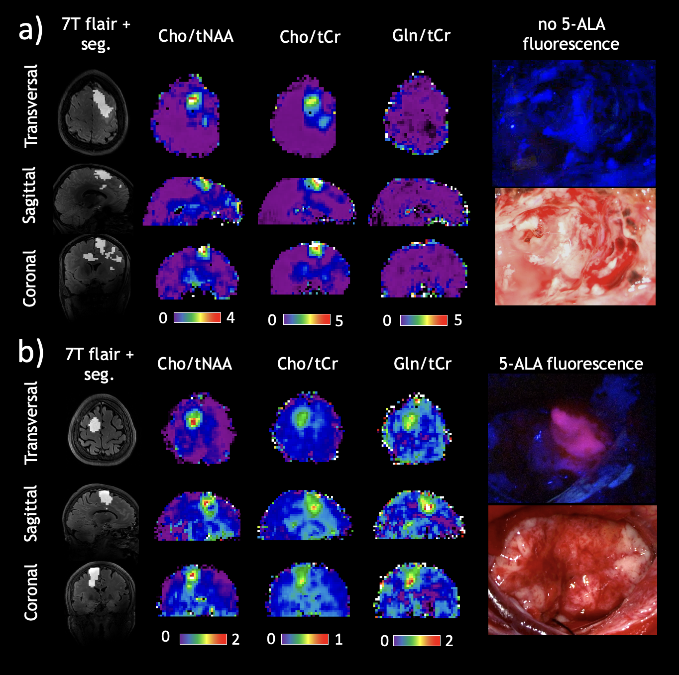
Motivation: Heterogeneity in gliomas represents a clinical and diagnostic issue. 5-ALA fluorescence used for intraoperative tumor delineation is different depending on the tumor type. Understanding the dysregulations in the heme pathway using 7T MRSI could help understanding metabolic heterogeneity in tumors.
Goal(s): Identifying 7T MRSI metabolites useful for investigating mechanisms of 5-ALA fluorescence and preoperative tumor characterization.
Approach: We retrospectively correlated 5-ALA fluorescence from clinical reports to metabolic ratios from 7T MRSI scans in a cohort of 23 patients.
Results: 5-ALA correlated with the Gln/tCr ratio (r2=0.597), which also correlated with the tumor grade.
Impact: Using 7T MRSI and 5-ALA, we improve guidance for neurosurgeons to resect aggressive tumor hotpots. Furthermore, 7T MRSI could be useful for in vivo studies of metabolic disturbances involved in 5-ALA activation.
Introduction
Gliomas present a group of primary brain tumors originating from glial cells and despite being relatively rare, they contribute largely to cancer mortality1. Heterogeneity or the presence of malignant hotpots, meaning that parts of the tumor show a higher malignancy than the rest, presents a diagnostic and clinical issue. Surgery with maximal safe resection (MSR) is the most efficient therapeutic method in increasing the overall survival and progression-free survival rate of glioma patients2. However, in partial resections of gliomas, parts of the tumor containing anaplastic foci could be overlooked, potentially leading to inadequate diagnosis and treatment, as well as a sooner recurrence3. Standard imaging techniques used for surgical planning such as structural MRI is usually not sufficient for determining these regions. Non-invasive imaging of certain metabolites using magnetic spectroscopy gives insight into altered metabolism in tumors and their microenvironments, which differ in different types4,5. MRSI levels of Cho, Cr, NAA as well as their ratios have been used to aid glioma diagnosis6. Compared to the standard 3T, high field 7T MRSI imaging allows for mapping of more biochemicals, leading to expectations of a more precise diagnostic tool7. A photosensitizing agent 5-aminolevulinic acid fluorescence (5-ALA) is used to aid surgical resections and improve MSR. 5-ALA is metabolized into protoporphyrin IX (PpIX) through the heme biosynthesis pathway, which accumulates in tumorous tissues and presents with fluorescence under blue light8. However, fluorescence is mostly present in high grade gliomas, the nature of which is thought to be linked to dysregulations in the heme synthesis pathway9. Understanding this could help us clarify metabolic heterogeneity in tumors10.The aim of our study was to correlate 5-ALA fluorescence in gliomas to the tumor grade and various metabolic ratios from 7T MRSI in order to explore new MRSI markers for determining malignant glioma hotspots as well as a better understanding of metabolic heterogeneity.Methods
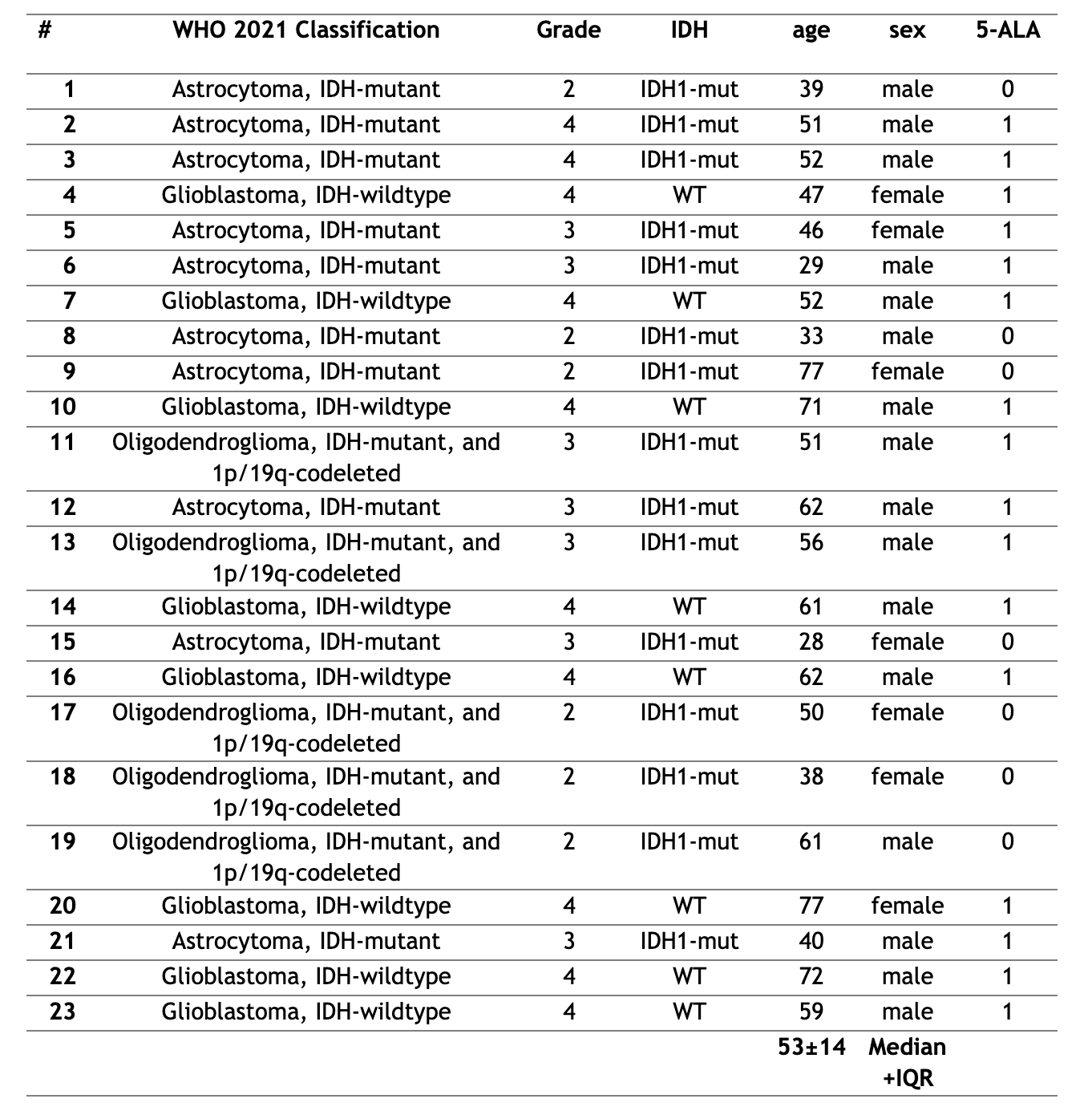
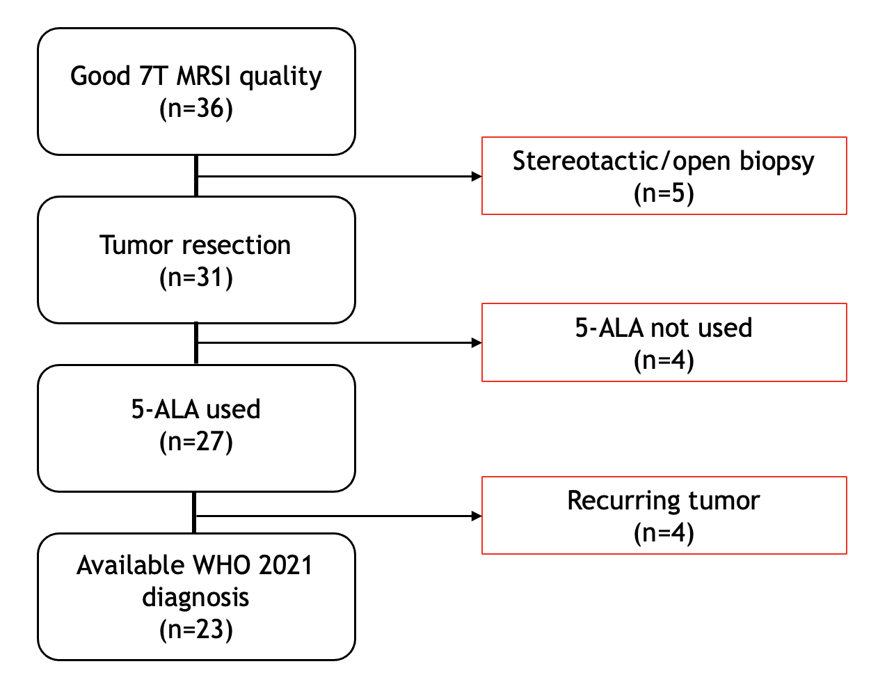
A cohort of 23 patients with clinical diagnoses of astrocytoma, oligodendroglioma or glioblastoma was retrospectively evaluated (Fig. 1). Exclusion criteria were: biopsy, recurring tumors and poor MRSI quality. Scans were conducted using a 7T scanner (Siemens Healthineers) with a 1Tx/32Rx-coil (Nova Medical). We acquired MRSI with a matrix of 64×64×39 and 3.4 mm nominal isotropic resolution, processed with an in-house pipeline including LCmodel11. The metabolic ratios evaluated for this study using python were tCho/tNAA, Glu/tNAA, Gln/tNAA, Ser/tNAA, Tau/tNAA, Gly/tNAA, Ins/tNAA, GSH/tNAA, tCho/tCr, Glu/tCr, Gln/tCr, Ser/tCr, Tau/tCr, Gly/tCr, Ins/tCr, and GSH/tCr. Reference tumor segmentation was performed by a neuroradiologist using clinical 3T MRI.5-ALA was administered orally (20 mg/kg body weight) three hours before surgery. A modified neurosurgical microscope (NC4 and Pentero, Carl Zeiss Surgical GmbH) was used for intraoperative visualization of PpIX fluorescence. Based on surgical reports we classified each patient into either “0 - no fluorescence”; or “1 - fluorescence”. Final statistical analysis explored differences between sex, age, tumor grade, IDH-mutation status, metabolic ratios, and 5-ALA fluorescence. Significant correlation coefficients were set to <-0.5 or >0.5.Results
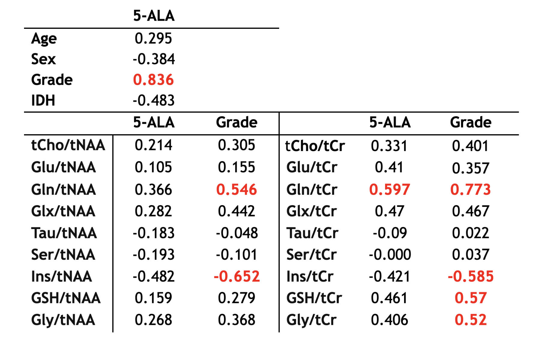
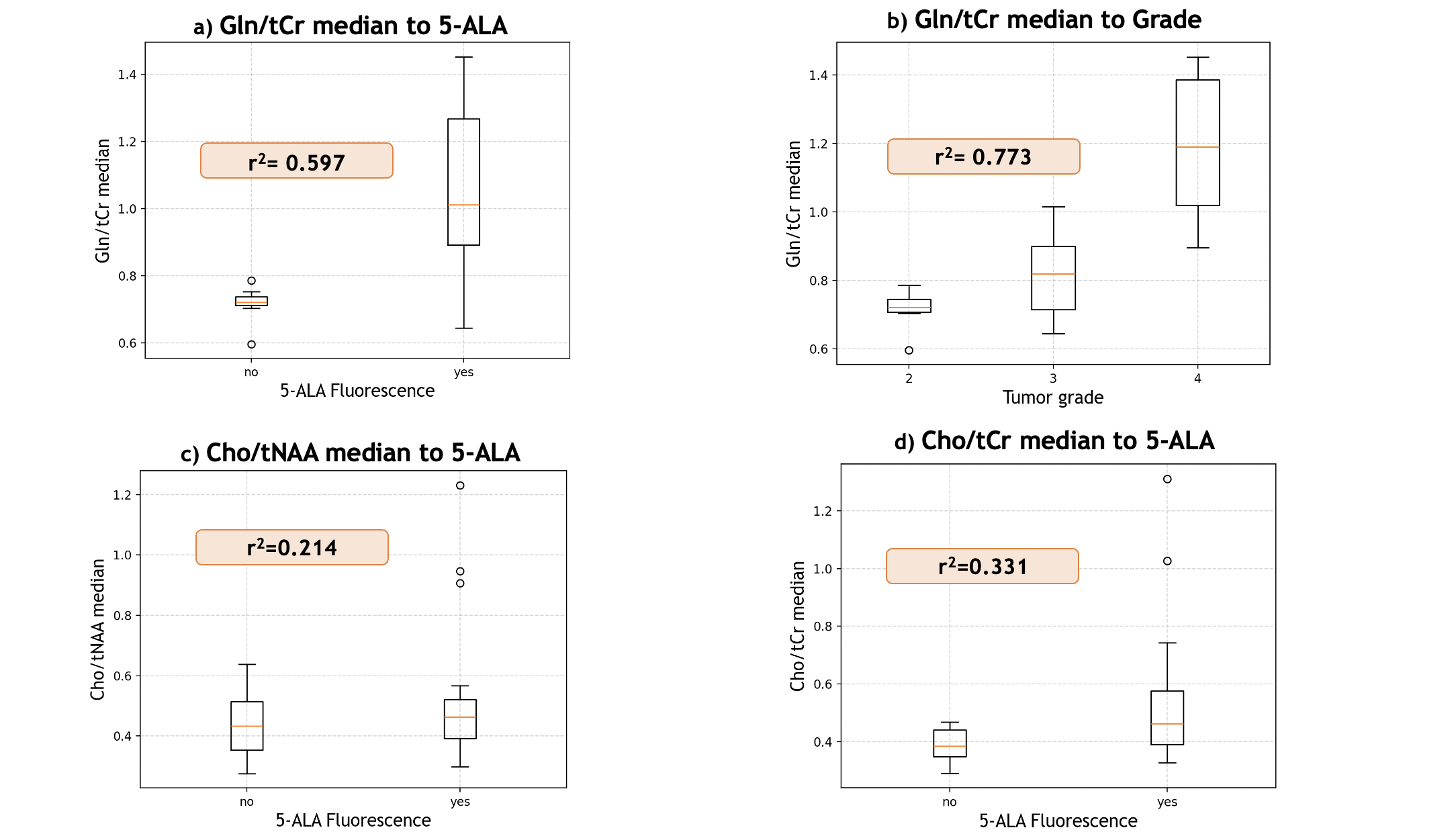
Of all, Gln/tCr ratios correlated with 5-ALA fluorescence over the threshold of 0.5. (r2=0.597). Figure 3 summarizes our results. Furthermore, ratios of Gln/tNAA (r2=0.546), Ins/tNAA (r2=-0.652), Gln/tCr (r2=0.773), Ins/tCr (r2=-0.585), GSH/tCr (r2=0.57) and Gly/tCr (r2=0.52) correlate well with tumor grade. Moreover, we found a correlation between 5-ALA fluorescence and tumor grade (r2=0.836).Discussion/Conclusion
To our knowledge, these are the first results that correlate 5-ALA fluorescence to 7T MRSI metabolic ratios in gliomas. Due to an increase in energy demand, glutamine metabolism is crucial in gliomas and seems to affect 5-ALA heterogeneity involving glutaminase 2 (GLS2) gene12. Our findings showing an increase in Gln/tCr in patients with 5-ALA fluorescence point to aberrations in the glutamine pathway, and show the potential of using 7T MRSI to study tumor metabolism in vivo. These results indicate that 5-ALA fluorescence could be a promising tool in exploring relevant hotspots for 7T MRSI tumor marker development, which could be useful in hotspot validation and more comprehensive surgical planning. Prospective studies with a larger cohort and a more extensive 5-ALA analysis could better define the correlations between 5-ALA and glutamine, as well as other metabolic ratios related to 5-ALA activation pathways. If the combination of 7T-MRSI with 5-ALA could better define tumor margins, this could optimize the extent of tumor resection and thus improve patient prognosis.Acknowledgements
This study was supported by the Austrian Science Fund (FWF) project KLI 1089 as well as a 2021 Comprehensive Cancer Center grant of the Medical University of Vienna. The financial support by the Austrian Federal Ministry for Digital and Economic Affairs, the National Foundation for Research, Technology and Development and the Christian Doppler Research Association is gratefully acknowledged.References
- Rasmussen BK, Hansen S, Laursen RJ, et al. Epidemiology of glioma: clinical characteristics, symptoms, and predictors of glioma patients grade I-IV in the the Danish Neuro-Oncology Registry. J Neurooncol. 2017;135(3):571-579. doi:10.1007/s11060-017-2607-5
- Bonosi L, Marrone S, Benigno UE, et al. Maximal Safe Resection in Glioblastoma Surgery: A Systematic Review of Advanced Intraoperative Image-Guided Techniques. Brain Sci. 2023;13(2):216. Published 2023 Jan 28. doi:10.3390/brainsci13020216
- Widhalm G, Kiesel B, Woehrer A, et al. 5-Aminolevulinic acid induced fluorescence is a powerful intraoperative marker for precise histopathological grading of gliomas with non-significant contrast-enhancement. PLoS One. 2013;8(10):e76988. Published 2013 Oct 18. doi:10.1371/journal.pone.0076988
- Korzowski A, Weckesser N, Franke VL, et al. Mapping an Extended Metabolic Profile of Gliomas Using High-Resolution 31P MRSI at 7T. Front Neurol. 2021;12:735071. Published 2021 Dec 23. doi:10.3389/fneur.2021.735071
- Di Nunno V, Franceschi E, Tosoni A, Gatto L, Bartolini S, Brandes AA. Tumor-Associated Microenvironment of Adult Gliomas: A Review. Front Oncol. 2022;12:891543. Published 2022 Jul 7. doi:10.3389/fonc.2022.891543
- Hangel G, Schmitz-Abecassis B, Sollmann N, et al. Advanced MR Techniques for Preoperative Glioma Characterization: Part 2 [published correction appears in J Magn Reson Imaging. 2023 Aug 11;:]. J Magn Reson Imaging. 2023;57(6):1676-1695. doi:10.1002/jmri.28663
- Bogner W, Gruber S, Trattnig S, Chmelik M. High-resolution mapping of human brain metabolites by free induction decay (1)H MRSI at 7 T. NMR Biomed. 2012;25(6):873-882. doi:10.1002/nbm.1805
- Widhalm G, Olson J, Weller J, et al. The value of visible 5-ALA fluorescence and quantitative protoporphyrin IX analysis for improved surgery of suspected low-grade gliomas [published online ahead of print, 2019 May 10]. J Neurosurg. 2019;1-10. doi:10.3171/2019.1.JNS182614
- Mischkulnig M, Roetzer-Pejrimovsky T, Lötsch-Gojo D, et al. Heme Biosynthesis Factors and 5-ALA Induced Fluorescence: Analysis of mRNA and Protein Expression in Fluorescing and Non-fluorescing Gliomas. Front Med (Lausanne). 2022;9:907442. Published 2022 May 18. doi:10.3389/fmed.2022.907442
- Widhalm G, Olson J, Weller J, et al. The value of visible 5-ALA fluorescence and quantitative protoporphyrin IX analysis for improved surgery of suspected low-grade gliomas [published online ahead of print, 2019 May 10]. J Neurosurg. 2019;1-10. doi:10.3171/2019.1.JNS182614
- Hingerl L, Strasser B, Moser P, et al. Clinical High-Resolution 3D-MR Spectroscopic Imaging of the Human Brain at 7 T. Invest Radiol. 2020;55(4):239-248. doi:10.1097/RLI.0000000000000626
- Kim S, Kim JE, Kim YH, et al. Glutaminase 2 expression is associated with regional heterogeneity of 5-aminolevulinic acid fluorescence in glioblastoma. Sci Rep. 2017;7(1):12221. Published 2017 Sep 22. doi:10.1038/s41598-017-12557-3
Figures