1814
Myocardial Tissue Composition Quantification at Systole, Diastole, Expiration and Inspiration1University of California San Francisco, San Francisco, CA, United States
Synopsis
Keywords: Myocardium, Quantitative Imaging
Motivation: We aim to develop a new method based on multi-compartment modeling to quantify myocardial tissue composition using MOLLI T1 mapping sequence.
Goal(s): In this pilot study, we evaluated how the cardiac function, respiration, and age may impact the quantification.
Approach: Data were acquired at end-systole/diastole, and end-expiration/inspiration. A series of maps were derived, including T1/T2 relaxation times, and fractions of three compartments: macromolecular proton, bound water and free water pools.
Results: The changes found in bound water and free water quantification matched human physiology. The developed method could provide a new non-invasive imaging tool for mapping tissue composition.
Impact: Quantifying myocardial tissue composition is valuable but very challenging. This pioneering work quantifies myocardial bound water and free water in a single breath-hold scan. It reveals the underlying tissue microstructure, which may open a new door for many applications.
INTRODUCTION
Quantifying myocardial tissue composition is valuable for diagnosing various cardiac diseases. We can utilize the widely available 2D breath-hold T1 mapping sequence to quantify myocardial tissue composition and assess myocardial fibrosis without contrast1,2 by developing a multi-compartment modeling with magnetization transfer (MT) effect for mapping and quantifying the macromolecular proton, bound water, and free water pools in myocardium. It's important to note that the physiology states such as cardiac motion, respiratory motion and aging may influence the quantification as their underlying microstructure varies. In this pilot study, we investigated whether this complex tissue composition quantification could serve as a new tool for gaining a deeper understanding and assessing various aspects of myocardial function, respiratory phase, and aging, by monitoring changes in tissue composition, going beyond reliance on a single relaxation time.METHODS
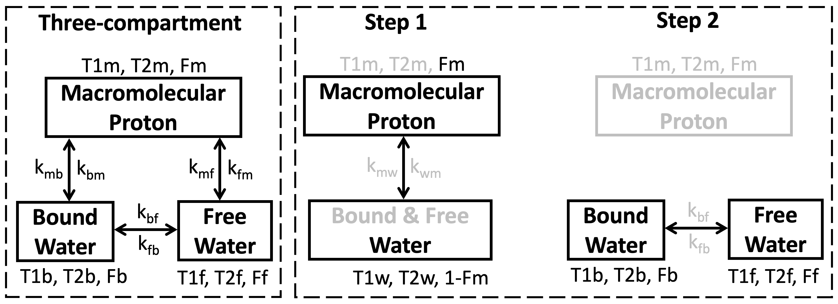
Three-compartment model used for tissue composition quantification is shown in Figure 1, where two steps were applied to derive the maps, including T1 relaxation times (T1w, T1b and T1f), T2 relaxation times (T2w, T2b, and T2f), and fractions (Fm, Fb and Ff) of the three compartments: macromolecular proton (MP), bound water and free water pools.Cardiac MRI were performed in 6 healthy subjects (5 females; age 48.3±22.5 (23-79) years) on 3T Vida MRI (Siemens, Germany). 2D cardiac T1 and T2 mapping were acquired at two cardiac phases (end-systole, end-diastole) and two respiratory phases (end-expiration, end-inspiration), respectively. MOLLI 5(3)3 T1 mapping was used and the data was processed using our multi-compartment modeling to derive a series of maps for tissue composition quantification1,2.The conventional T1 and T2 maps were obtained for comparison.
RESULTS
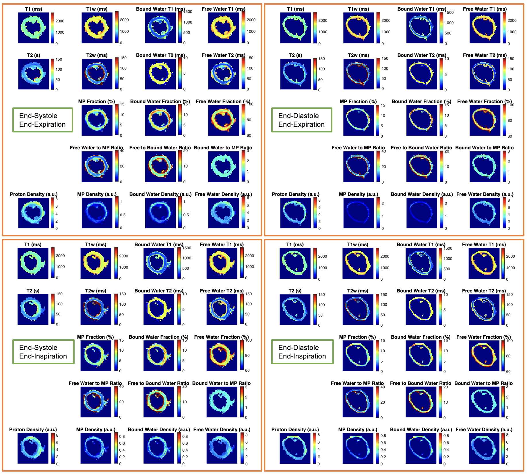
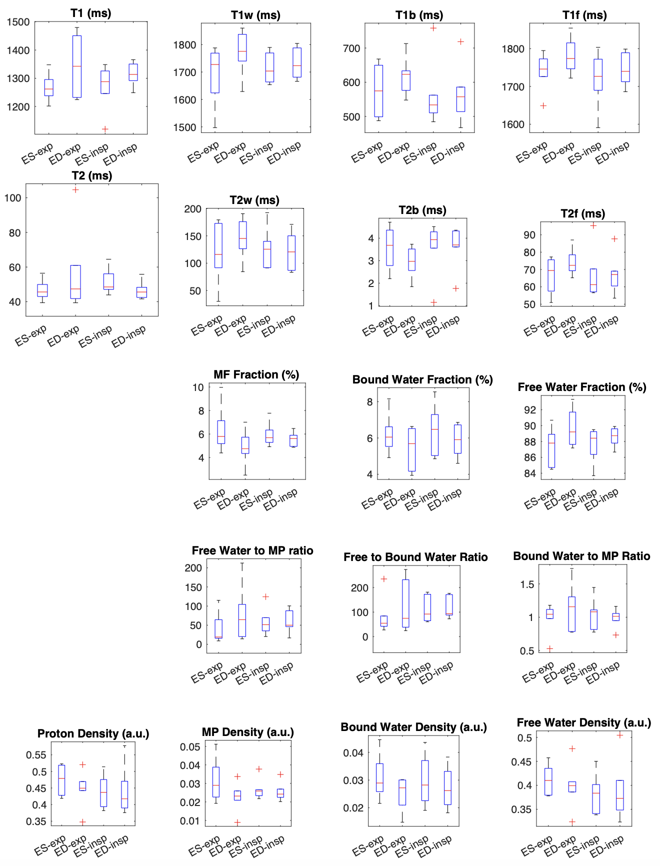
The quantitative tissue composition maps were successfully derived from the images acquired at four different states: end-systole & end-expiration (ES-exp), end-diastole & end-expiration (ED-exp), end-systole & end-inspiration (ES-insp), and end-diastole & end-inspiration (ED-insp). Figure 2 shows the four sets of maps from a representative subject.The map measurements in the septum of the 6 subjects are plotted in Figure 3 and listed in Table 1. Given a small sample size, no significant differences were found between two cardiac phases or respiratory phases. Some trends were observed in the measurements (Table 1). For both respiratory phases, T1 values (the reference one and those based on multi-compartment modeling) and free water fraction increased at ED, while the fractions of MP and bound water decreased at ED. Between two respiratory phases, free water T1 and proton density were found slightly lower at inspiration for both cardiac phases, while the bound water fraction was higher at inspiration.
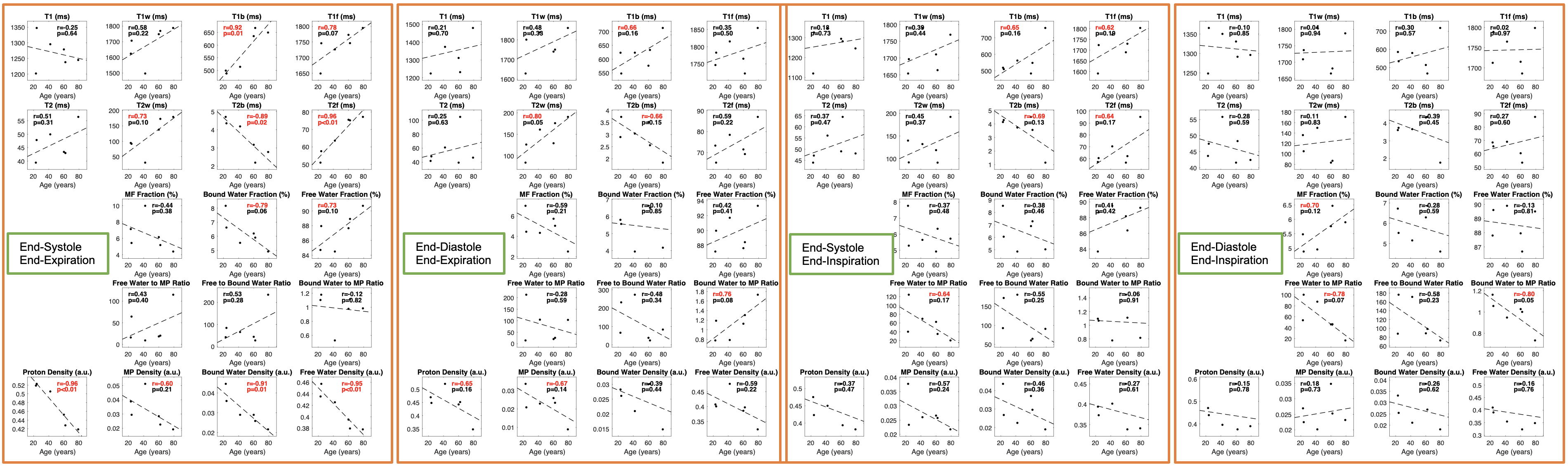
Linear Regression of the maps and age are shown in Figure 4. Some multi-compartment model-based maps at end-systole & end-expiration were highly correlated with age (p<0.05) while the conventional T1 and T2 mapping did not reach significance. Especially the maps of bound water showed strong correlations with age, such as T1 (r=0.92, p=0.01), T2 (r=-0.89, p=0.02), fraction (r=-0.79, p=0.06) and proton density (r=-0.91, p=0.01); similarly maps of free water, T1 (r=0.78, p=0.07), T2 (r=0.96, p<0.01), fraction (r=0.73, p=0.10) and density (r=-0.95, p<0.01). Some maps of the other three map sets achieved strong correlations with age (|r|>0.6) but did not reach significance (p<0.05).
DISCUSSION
Since coronary perfusion occurs during diastole rather than systole, more myocardial blood flow happens at diastole compared to systole, which could explain the elevated T1, and free water fraction (more blood flow) found at diastole in our results.It is known that inspiration drops intrathoracic pressure, dilates the thoracic vena cava and then decreases atrial filling. Our findings of reduced free water T1 and proton density (lower blood flow) at inspiration match the theory.
As known, aging cells show an increase in intracellular water volume, hydrogen bonds surrounding the contractile proteins (bound water) become free water as aging. Our findings of lower fraction of bound water and higher fraction of free water as aging well reflect this theory.
CONCLUSIONS
Tissue composition quantification was evaluated at different cardiac and respiratory phases and age in this pilot study. No significant differences in the map values were found among the phases given the small cohort, but some trends were observed and found to match human physiology. The elevated T1 and free water fraction at diastole reflect more blood flow in the myocardium, the reduced free water T1 and proton density at inspiration reflect atrial filling decrease with intrathoracic pressure. Findings of lower bound water and higher free water as aging (significant correlations) may demonstrate a new non-invasive imaging tool for assessing aging by mapping the tissue composition. Further investigations require a larger cohort, and it's important to evaluate gender differences.Acknowledgements
No acknowledgement found.References
1. J Liu, et al. Myocardial Tissue Composition Quantification with MOLLI Acquisition and Multi-Compartment Modeling. SCMR Annual Meeting 2023, 1350908.
2. J Liu, et al. MultI-Compartment Model based Parametric Mapping of the Heart with a Single MOLLI Acquisition (MIC-MOLLI), ISMRM Annual Meeting 2022, p1012.
Figures