1809
The diagnostic value of myocardial diffusion combined with T1mapping、ECV in the evaluation of myocardial salvage in acute myocardial infarction1Ningxia Hui Autonomous Region People's Hospital, Yinchuan, China, 2Philips Healthcare, Beijing, China
Synopsis
Keywords: Myocardium, Cardiomyopathy
Motivation: Cardiac MRI with diffusion-weighted imaging (DWI) non-invasively characterizes myocardial infarction, potentially identifying salvageable 'gray zone' areas without contrast agents.
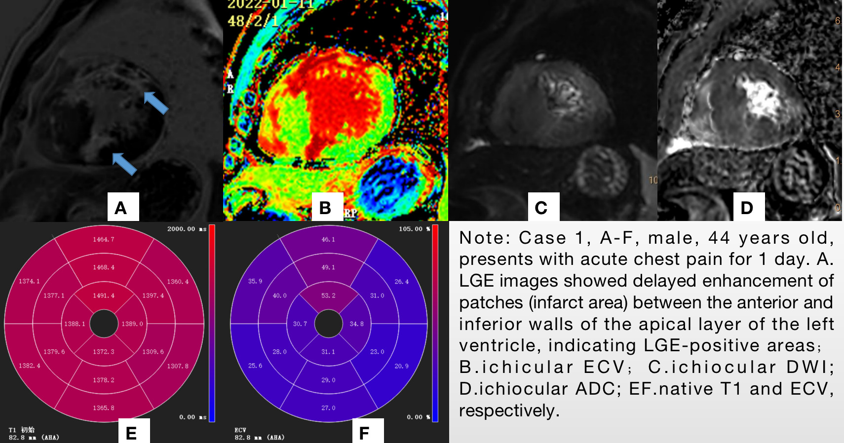
Goal(s): This study aimed to evaluate the diagnostic value of DWI combined with T1 mapping, ECV, and LGE in assessing viable myocardium in acute MI patients.
Approach: By comparing ADC values across infarcted, peri-infarct, and healthy myocardium, significant ADC discrepancies highlighted the viability of the 'gray zone.'
Results: These findings could influence treatment strategies to recover endangered myocardium, enhancing clinical outcomes in acute myocardial infarction scenarios.
Impact: Investigating the 'gray zone' through CMR DWI sequences and ADC value changes, this study advances our understanding of myocardial viability in acute MI, potentially guiding interventions to salvage myocardium and improve patient outcomes in acute ischemic events.
Introduction
The incidence of ischemic cardiomyopathy in China is rising annually, posing a significant health threat. Cardiac MRI has become a crucial non-invasive tool for the anatomical and functional assessment of cardiovascular diseases1. Late Gadolinium Enhancement (LGE) is considered the gold standard for myocardial infarction (MI) assessment, yet alternatives are needed when contrast agents are contraindicated due to renal impairment or allergies2. Diffusion-weighted imaging (DWI) has shown promise in acute stroke and is gaining traction for ischemic and non-ischemic cardiac diseases3-5. This study retrospectively evaluates the diagnostic performance of ADC values in acute MI, especially in identifying the 'gray zone'—the area of myocardium that is damaged but potentially salvageable, which is crucial for clinical treatment strategies and improving patient outcomes.Method Summary
In this study, 15 patients with suspected myocardial infarction (MI) (11 males, 4 females), all exhibiting delayed enhancement on LGE MRI images, were included. Cardiac MRI scans were performed using a Philips 3T scanner (Ingenia CX, Best, Netherlands), acquiring myocardial T1 mapping and DWI sequences. Images were analyzed using cardiovascular post-processing software (CVI42) to measure T1 values pre- and post-contrast, and the extracellular volume fraction (ECV) of the myocardium. Regions of interest (ROIs) were manually delineated within the enhanced segments on T1 mapping images, comparing the infarcted border zone ('gray zone') and remote unenhanced myocardium ('normal myocardium'). The ECV for these regions was calculated.DWI images were then processed using cardiovascular post-processing workstation (ISP), where ROIs were outlined based on the extent of delayed enhancement observed on LGE images. ADC values for the infarcted area, the border zone, and remote myocardium were measured and analyzed.
For statistical analysis, normality tests were conducted on all datasets. Data following a normal distribution were expressed as mean ± standard deviation (x̄ ± s), and comparisons between the three groups were made using one-way ANOVA, with post-hoc pairwise comparisons using LSD tests. Non-normally distributed data were represented by medians (interquartile ranges), with differences between groups assessed using the Kruskal-Wallis test. A P-value < 0.05 was considered statistically significant.
Results
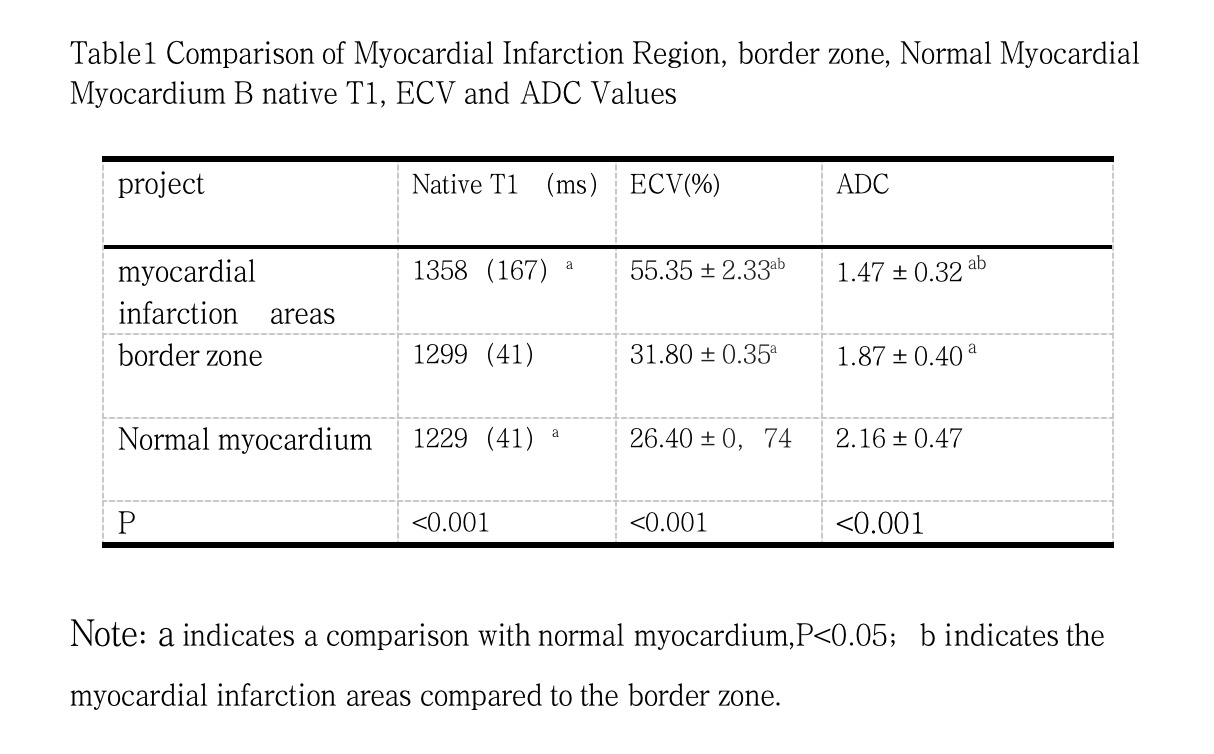
The study indicated that initial T1 values in the acute myocardial infarction (MI) areas were significantly higher (1358 ± 167 ms) compared to normal myocardium (1229 ± 41 ms) (P < 0.001). The extracellular volume fraction (ECV) among the MI, border zone, and normal myocardium differed significantly, with values of 55.35 ± 2.33%, 31.80 ± 0.35%, and 26.40 ± 0.74%, respectively (P < 0.001). Apparent diffusion coefficient (ADC) values also varied significantly across the three regions, with 1.47 ± 0.32 for the infarcted myocardium, 1.87 ± 0.40 for the border zone, and 2.16 ± 0.47 for healthy myocardium (P < 0.001).Discussion
The findings confirmed that diffusion-weighted imaging (DWI) can significantly differentiate between infarcted, border zone, and healthy myocardium based on ADC values, demonstrating the potential of DWI in accurately diagnosing myocardial infarction. The decline in ADC values in acute MI areas could be attributed to cytotoxic edema in myocardial cells, which reduces extracellular space and restricts the movement of water molecules. DWI is sensitive to these changes as it reflects the diffusion and restriction of water molecules within tissues and lesions.Conclusion
The combination of myocardial diffusion imaging, T1 mapping, and ECV quantification is highly valuable for identifying and quantifying acute myocardial infarction.Acknowledgements
noneReferences
1.Pattanayak P, Bleumke DA. Tissue characterization of the myocardium: state of the art characterization by magnetic resonance and computed tomography imaging. Radiol Clin North Am. 2015;53(2):413-423. doi:10.1016/j.rcl.2014.11.0052. Lovblad KO, Laubach HJ, Baird AE, Curtin F, Schlaug G, Edelman RR, Warach S. Clinical experience with di usion-weighted MR in patients with acute stroke. AJNR Am JNeuroradiol 1998;19(6):1061-1066.3. Deux JF, Maatouk M, Vignaud A, Luciani A, Lenczner G, Mayer J, Lim P, Dubois-Rande JL, Kobeiter H, Rahmouni A. Di usion-weighted echo planar imaging in patients withrecent myocardial infarction. Eur Radiol 2011;21(1):46-53.4. Laissy JP, Gaxotte V, Ironde-Laissy E, Klein I, Ribet A, Bendriss A, Chillon S, Schouman-Claeys E, Steg PG, Serfaty JM. Cardiac di usion-weighted MR imaging in recent,subacute, and chronic myocardial infarction: a pilot study. J Magn Reson Imaging 2013;38(6):1377-1387.5. Xiang SF, Zhang XQ, Yang SJ, Gao YY, Gao BL, Shi QL, Li S. Intravoxel Incoherent Motion Magnetic Resonance Imaging with Integrated Slice-speci c Shimming for oldmyocardial infarction: A Pilot Study. Sci Rep 2019;9(1):19766.Figures