1808
Diffusion Tensor CMR in Assessing Biventricular Myocardial Fiber Orientation: A Comparative Study with Histology in a Miniature-Swine Model1Department of Magnetic Resonance Imaging, Fuwai Hospital, Chinese Academy of Medical Sciences and Peking Union Medical College, Beijing, China, 2Clinical & Technical Support, Philips Healthcare, Beijing, China
Synopsis
Keywords: Myocardium, Heart, Diffusion tensor imaging, Cardiac magnetic resonance, Myocardium, Swine model
Motivation: Diffusion tensor cardiac magnetic resonance (DT-CMR) has shown potential for non-invasive characterization of myocardial fiber orientation.
Goal(s): The purpose of this study is to validate the accuracy of high-resolution DT-CMR in evaluating the arrangement of biventricular myocardial fibers in a miniature-swine model, using histological findings as the reference standard.
Approach: High-resolution ex-vivo DT-CMR data of one healthy miniature-swine were acquired by a 3.0 T MRI system. Helix angles (HAs) in each segment from DT-CMR was compared with that from histology.
Results: HAs evaluated by DT-CMR were closely correlated with those derived from histology (r = 0.958, P < 0.001) in 64 myocardial segments.
Impact: Using histological validation in a miniature-swine model, high-resolution DT-CMR demonstrated good performance in non-invasively evaluating the arrangement of biventricular myocardial fibers. It provides histological evidence for DT-CMR to add more diagnostic and prognostic information in human cardiovascular diseases.
Introduction
Diffusion tensor cardiac magnetic resonance (DT-CMR) is a novel tool that shows potential for non-invasive assessment of myocardial microstructures, specifically the cardiac fiber orientation. However, limited data are available on the comprehensive validation of biventricular myocardial fiber orientation assessed with DT-CMR against histology. The purpose of this study is to validate the accuracy of high-resolution DT-CMR in evaluating the arrangement of biventricular myocardial fibers in a miniature-swine model, using histological findings as the reference standard.Methods
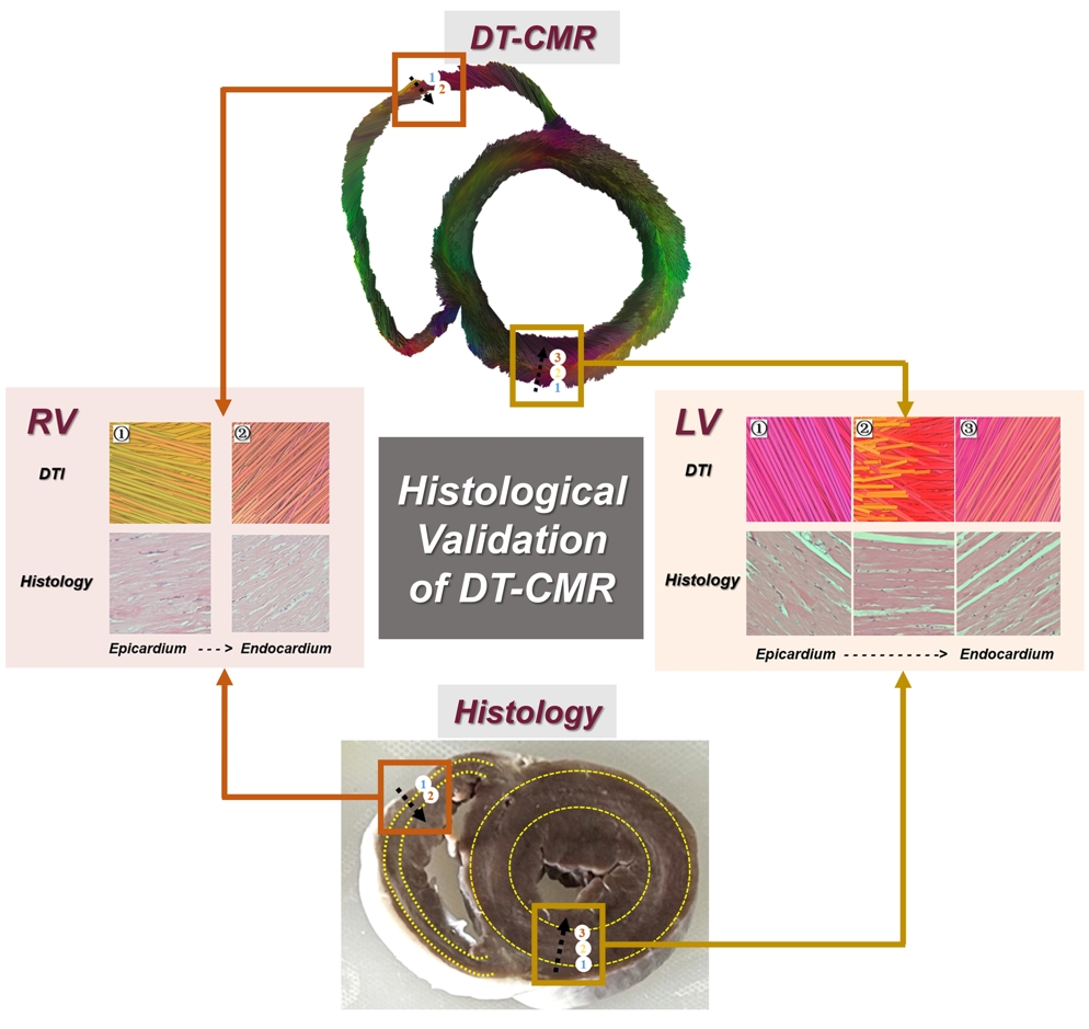
High-resolution ex-vivo DT-CMR data of one healthy miniature-swine were acquired by a 3.0 T MRI system (Ingenia 3.0, Philips Healthcare) using a second-order motion-compensated single-shot spin echo planar imaging sequence at the systolic state, which is similar with that used in a human study (DOI: 10.1148/radiol.2021203208), but with the electrocardiography trigger and respiratory navigator turned off for time saving. Each data set constituted 32 non-collinear diffusion-weighted acquisitions with b-values of 600 s/mm2. Fiber tracking was performed in three myocardial layers (subepicardial, intramyocardial, and subendocardial wall) according to the American Heart Association (AHA) 16-segment model for the left ventricle (LV) wall, and two layers (subepicardial and subendocardial wall) according to an 8-segment model for the right ventricle (RV) wall. Histology with hematoxylin and eosin (HE) staining was compared in each myocardial segment. The correlations between helix angles (HAs) assessed with DT-CMR and histology were analyzed by linear regression.Results
A total of 64 segments including 48 LV segments and 16 RV segments were head-to-head analyzed. In this normal heart, LV myocardial fibers followed a symmetric arrangement around the midmyocardium, with negative and positive HAs in the subepicardial and subendocardial wall, respectively. The subepicardial HAs in basal segments were -63.9 ± 5.5 degrees in DT-CMR and -51.9 ± 6.7 degrees in histology, while the subepicardial HAs in mid-cavity segments were -48.3 ± 8.7 degrees in DT-CMR and -40.4 ± 7.3 degrees in histology, which suggested that the subepicardial fibers may become more circumferential with higher HAs when approaching apex segments (all P<0.05). The main fiber orientation of the RV wall from subepicardial to subendocardial layers was asymmetric and presented HAs of close-to-zero to positive degrees in both DT-CMR and histology. HAs evaluated by DT-CMR were closely correlated with those assessed with histology (r = 0.958, P < 0.001) in 64 myocardial segments.Conclusions
Using histological validation in a miniature-swine model, high-resolution DT-CMR demonstrated good performance in non-invasively evaluating the arrangement of biventricular myocardial fibers. It provides histological evidence for DT-CMR to add more diagnostic and prognostic information in human cardiovascular diseases.Acknowledgements
No acknowledgement found.References
No reference found.Figures