1804
Free-breathing, Non-ECG Cardiovascular Magnetic Resonance Multitasking for the Assessment of Left Ventricular Function1Radiology, West China Hospital, Sichuan University,, Chengdu, China, 2UIH America, Inc, Houston, TX, United States
Synopsis
Keywords: Myocardium, Heart, cardiac function
Motivation: To solve the problem that cardiac magnetic resonance requires ECG gating and breath holding
Goal(s): To evaluate the feasibility of cine images acquired through cardiac magnetic resonance (CMR) Multitasking and compare with those measured through multi-breath-hold bSSFP sequence in clinical setting
Approach: Study design to test the hypothesis
Results: Left ventricular ejection fraction was not significantly different between the two sequences in both healthy volunteers and patients.
Impact: Multitasking can be used to quantify the LV functions without ECG gating or breath holding in healthy volunteers and patients. Multitasking may become a replacement of bSSFP in certain conditions.
Introduction
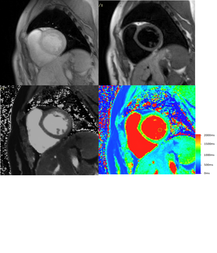
Precise evaluation of ventricular function is of great value for diagnosis, treatment, and prognosis for cardiovascular patients [1, 2]. Cardiac magnetic resonance is the gold standard for evaluating cardiac function [3]. Conventional cine sequences require electrocardiogram (ECG) gating and multiple breath-holds, which are not suitable for patients with arrhythmia or having difficulty holding their breath. The recently developed Multitasking technique, based on a low-rank tensor model, does not rely on ECG gating and can be performed while patients are freely breathing. Multidimensional images encompassing three dimensions, namely cardiac, respiration and T1 inversion recovery, can be reconstructed from a single scan (Figure 1) [4, 5]. This is in contrary to conventional imaging techniques who try to ‘freeze’ respiration and cardiac motions by breath-holding and ECG gating.Despite prior efforts in evaluating Multitasking’s T1 mapping accuracy, no attempt was made to assess its performance in ventricular function analysis. Therefore, the aim of this study was to evaluate the feasibility of cine images acquired through cardiac magnetic resonance (CMR) Multitasking, free of ECG gating and breath holding, in measuring left ventricular (LV) volume indexes and mass, compared with those measured through multi-breath-hold bSSFP sequence in clinical setting.Methods
A total of 30 healthy volunteers and 20 patients were enrolled for CMR examination. Each subject underwent bSSFP sequence with multiple breath-hold and Multitasking sequence under free breathing respectively.The Multitasking sequence was the same as in a previous publication [5]. Even readout lines were acquired along 0 degree, while the angles of odd readout lines were incremented by a golden angle of 112.15 degrees. Non-selective inversion recovery was used to create T1 contrast. The short-axis images were analyzed in CVI42 (Circle Cardiovascular Imaging, Calgary, Alberta, Canada), by a radiologist with 2 years’ experience in CMR. Paired t-test, linear regression, and Bland-Altman analysis were performed to compare five continuous LV function indices: LV ejection fraction (LVEF), LV end-diastolic mass (LVEDM), LV end-diastolic volume (LVEDV), LV end-systolic volume (LVESV), and LV stroke volume (LVSV). Image quality was assessed by image quality score and contrast-to-noise (CNR).Results
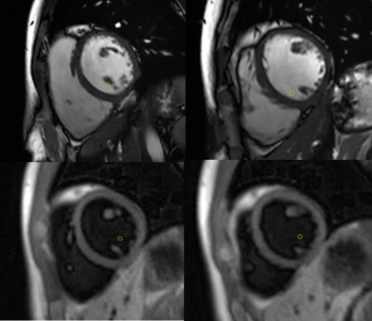
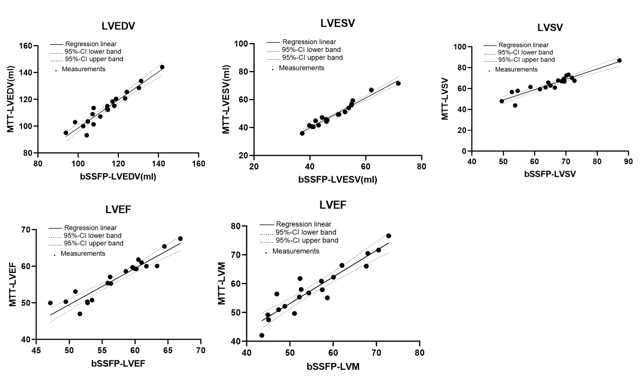
Typical images are shown in Figure 2. LV ejection fraction (EF) was not significantly different between the two sequences in both healthy volunteers (p=0.451) and patients (p=0.709). However, LV end-diastolic mass (EDM) was overestimated by Multitasking in both healthy volunteers (bSSFP: 56.2 ±9.3 g, Multitasking: 58.3 ±9.1 g, p=0.005)and patients (bSSFP: 65.6 ±13.5 g, Multitasking: 67.9 ±14.1 g, p=0.013). Linear regression analysis of data from the whole cohort showed good correlation between the two methods for all parameters (Figure 3). CNR of the myocardium and the blood pool showed no difference between Multitasking and bSSFP sequences (p=0.155). Moreover, both sequences achieved acceptable and comparable image quality score in LV function measurements.Discussion
CMR Multitasking is an innovative MRI sequence designed to efficiently acquire cardiac, respiratory and T1 recovery information without the use of ECG gating and respiration control. Its application in deriving cine images and LV function assessment has not been validated. This study showed that left ventricular function assessment on black-blood cine images using Multitasking sequence—in a state of free breathing and independent of ECG—is comparable to that on traditional white-blood cine images acquired with retrospectively cardiac gated bSSFP sequence. Further, similar blood-myocardium CNR and image quality were achieved by the cine images acquired by Multitasking sequence when compared to that from bSSFP. However, there is a small overestimation of left ventricular end-diastolic mass and underestimation of LVEDV when Multitasking cine images were used. Another finding of our study was that the cine image at end diastole and systole from Multitasking sequence can achieve satisfactory and diagnostic-level image quality. Especially, for patients with poor breath-hold, Multitasking could have better performance than bSSFP.Conclusion
The feasibility of Multitasking for quantification of the LV functions without ECG gating or breath holding was tested successfully in healthy volunteers and patients. Multitasking may become an alternative technique to bSSFP in certain conditions.Acknowledgements
No acknowledgement found.References
1. Ito, K., et al., Left ventricular dimensions and cardiovascular outcomes in systolic heart failure: the WARCEF trial. ESC Heart Fail, 2021. 8(6): p. 4997-5009.
2. Kato, M., et al., Left Ventricular End-Systolic Volume Is a Reliable Predictor of New-Onset Heart Failure with Preserved Left Ventricular Ejection Fraction. Cardiol Res Pract, 2020. 2020: p. 3106012.
3. Grothues, F. and R. Braun-Dullaeus, Serial assessment of ventricular morphology and function. Heart Fail Clin, 2009. 5(3): p. 301-14, v.
4. Christodoulou, A.G., et al., Magnetic resonance multitasking for motion-resolved quantitative cardiovascular imaging. Nat Biomed Eng, 2018. 2(4): p. 215-226.
5. Shaw, J.L., et al., Free-breathing, non-ECG, continuous myocardial T1 mapping with cardiovascular magnetic resonance multitasking. Magn Reson Med, 2019. 81(4): p. 2450-2463.
6. Piqueras-Flores, J., et al., Structural and functional alterations of the heart in Parkinson's disease. Neurol Res, 2018. 40(1): p. 53-61.
7. Kido, T., et al., Assessment of Left Ventricular Function and Mass on Free-Breathing Compressed Sensing Real-Time Cine Imaging. Circ J, 2017. 81(10): p. 1463-1468.
Figures