1803
Deep Learning based Acquisition and Reconstruction for Cardiac Magnetic Resonance Imaging in Patients with Chronic Kidney Disease1Department of radiology, Renmin Hospital Wuhan University, Wuhan, China, 2GE Healthcare, MR Research China, Beijing, China
Synopsis
Keywords: Myocardium, Cardiovascular
Motivation: Cardiac abnormalities and arrhythmias increased risk of cardiovascular mortality especially in patients with Chronic Kidney Disease (CKD).
Goal(s): To provide objective and subjective assessment in supproting the feasibility of deep learning acqusition and reconstruction cardiac magnetic resonance (CMR) imaging in patients with Chronic Kidney Disease (CKD).
Approach: Qualitative and quantitative evaluation of Sonic DL-based CMR images including breath-hold acquisition within one-beat-interval, free-breathing acquisition in comparison with traditional cine images.
Results: Sonic DL significantly accelerated acuqisition time (e.g. 11 seconds) but offered diagnosis-suffcient image quality and reliable strain values ompared to conventional sequences.
Impact: Sonic DL cine MRI showed equivalent image quality to conventional one, offered reliable strain values for diagnosis of a CKD patient lack of breath-hold ability and presentce with arrhythmias. Even it acquire a whole heart cine image in 11 seconds.
INTRODUCTION
Patients with Chronic Kidney Disease (CKD) often have myocardial abnormalities and arrhythmias [1,2]. Early identification of myocardial structural changes using cardiac magnetic resonance (CMR) and timely intervention can improve the survival rate of CKD patients [3]. Strain analysis for cine images can reflect cardiac dysfunction and future cardiovascular events via overall circumferential strain (GCS) and global radial strain (GRS) besiddes ejection fraction (LVEF) [4]. Strain is a more sensitive marker of cardiac function [2]. Furthermore, there are strong positive correlations of strain and clinical data such as estimated glomerular filtration rate and negative correlations of uric acid levels [5]. As conventional 2D breath-hold bSSFP cine sequences demands relative in-complianc patients, that is, stable breath-holding for at least 19 second image acquisition is challenging for CKD patients due to difficulties in maintaining breath-hold and involuntary arrhythmias, which may result in incomplete scans [6-8]. Partial solutions to the problem of free-breathing bSSFP include using multiple signal averages or respiratory gating but still possibly lead to poorer image quality and even increased scan time [6-8]. A recent deep learning based k-space acceleration and reconstruction method (Sonic DL) has been proposed to directly reconstruct cardiac images from sparsely sampled k-space data with high spatial-resolution cine images [8]. This study is the first report on the feasibility of Sonic DL cine in CKD patients who cannot hold their breath and have arrhythmias.METHODS
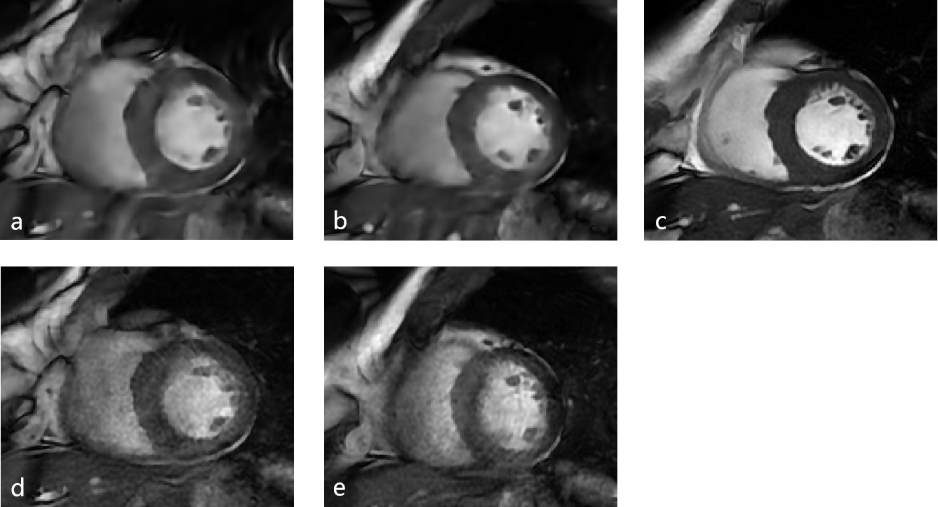
This study was approved by the hospital and prospectively recruited 35 patients who underwent CMR examinations from September 2023 to October 2023 on 3.0 T MRI scanner (Signa Architect, GE Healthcare) at our hospital. CMR examinations included Cine sequences: conventional standard cine images (CineST) (spatial resolution = 0.19×0.15×8, total breath-hold scan time 84 seconds) and Sonic DL cine images (spatial resolution = 0.16×0.16×8, breath-hold and free-breathing scan time 11 and 25 seconds) including DLR and noDLR 1RR breath-hold Cine images (Cine1RR BH-DL, Cine1RR BH) and DLR and noDLR 1RR free-breathing Cine images (Cine1RR FB-DL, Cine1RR FB),. The percentage of myocardium enhancement area (Parea) was assessed for cardiovascular disease diagnosis (Fig.1). Qualitative and quantitative evaluations of CMR images were conducted by two radiologists, subjective image quality evaluation was assessed at the workstation and strain was assessed using Cvi42 software. Statistical analysis was performed using R-project (version 4.0.4, http://www.r-project.org), and Likert scale was used to assess the subjective image quality evaluation, Kendall's coefficient of synergy tests were used to evaluation of inter-, intra-observer agreement, The paired t-test was used to evaluate the differences between measurements, the ICC test was used to evaluate the consistency of changes between measurements, and the Person test was used to evaluate the correlation of changes between measurements. p<0.05 was considered statistically significant.RESULTS
A total of 35 suspicious UMI volunteers were studied (18 males, and 17 females, mean age: 53.97 years ± 13.39). Likert scores of CineST, Cine1RR BH-DL, Cine1RR BH, Cine1RR FB-DL, Cine1RR FB is 4.50±0.50, 3.33±0.50、3.11±0.60、3.89±0.65、3.50±0.43 (Fig.1),Kendall's concordance coefficient test showed high intra-observer and inter-observer consistency (all W values > 0.8, p < 0.05). There are differences in LVEF between LVEFST and LVEF1RR BH as well as between LVEF1RR BH and LVEF1RR BH-DL (t = 2.96 and 2.57, respectively, p < 0.05), while there are no statistically significant differences in LVEF between the other groups (p > 0.05). GCSST and GCS1RR BH as well as GRSST and GRS1RR BH showed statstically different (t = -7.50 and 2.43, respectively, p < 0.05), while there are no statistically significant differences in other strain measures (p > 0.05). Each LVEF, GCS and GRS measurements of all Sonic DL cine images were good inter-consistency to LVEFST (ICC > 0.80, p < 0.001), GCSST (ICC > 0.70, p < 0.05), and GRSST (ICC > 0.70, p < 0.05) when compared (Fig.2, Fig.3) .DISCUSSION
For CKD patients, irregular heartbeat and breath-hold scanning hamper clinical diagnosis due to failure in cardiac imaging. The image quality of both Cine1RR BH DL and Cine1RR FB DL sequences was significantly improved compared to Cine1RR BH and Cine1RR FB sequences and almost equivalent to CineST.CONCLUSION
Overall, free-breathing cine images possessed superior image quality to breath-hold. We suggested Cine1RR BH DL for patients who can cooperate well with breath-holding and have no arrhythmias and Cine1RR FB DL for CKD patients who cannot cooperate with breath-holding and have arrhythmias.Acknowledgements
None.References
[1] Hayer MK, Radhakrishnan A, Price AM, et al. Defining Myocardial Abnormalities Across the Stages of Chronic Kidney Disease: A Cardiac Magnetic Resonance Imaging Study. JACC Cardiovasc Imaging 2020;13(11):1-11.
[2] Kenneth Mangion, Kirsty McDowell, Patrick B. Mark, et al. Characterizing Cardiac Involvement in Chronic Kidney Disease Using CMR - a Systematic Review. Current Cardiovascular Imaging Reports 2018;11(2):1-10.
[3] Singh AK, Antiochos P, Singh AT, et al. Multiparametric Cardiac Magnetic Resonance for Chronic Kidney Disease.Mapping the Footprints of a “Silent Killer”? JACC Cardiovasc Imaging 2020;13(11) :2368-2370.
[4] Sobh DM, Batouty NM, Tawfik AM, et al. Left Ventricular Strain Analysis by Tissue Tracking- Cardiac Magnetic Resonance for early detection of Cardiac Dysfunction in children with End-Stage Renal Disease. J Magn Reson Imaging 2021;26(5).
[5] Yi Zhang, Jin Wang, Yan Ren, et al. The additive efects of kidney dysfunction on left ventricular function and strain in type 2 diabetes mellitus patients verifed by cardiac magnetic resonance imaging. Cardiovasc Diabetol 2021;11(1):1-11.
[6] Evan J. Zucker, Christopher M. Sandino, Aya Kino, et al. Free-breathing Accelerated Cardiac MRI Using Deep Learning: Validation in Children and Young Adults. Radiology 2021; 300: 539-548.
[7] Julio A. Oscanoa, Matthew J. Middione, Cagan Alkan, et al. Deep Learning-Based Reconstruction for Cardiac MRI : A Review Bioengineering (Basel) 2023;10(3).
[8] Makoto Orii, Misato Sone, Takeshi Osaki, et al. Reliability of respiratory-gated real-time two-dimensional cine incorporating deep learning reconstruction for the assessment of ventricular function in an adult population. The International Journal of Cardiovascular Imaging (2023) 39:1001-1011.
Figures