1792
A cine-ASL magnetic resonance method detects microvascular dysfunction in a mouse model of heart failure with preserved ejection fraction1Calico Sciences, South San Francisco, CA, United States, 2Centre de Résonance Magnétique Biologique et Médicale (CRMBM), Aix-Marseille University, CNRS, Marseille, France
Synopsis
Keywords: Heart Failure, Perfusion, Heart failure, preclinical imaging, HFpEF animal model, MRI
Motivation: Addressing the challenges in heart failure with preserved ejection fraction (HFpEF) diagnosis, the study aimed to validate cine-ASL as a precise non-invasive imaging tool.
Goal(s): The study sought to explore nuanced aspects of HFpEF, enabling a better understanding of its complexities through advanced imaging techniques.
Approach: Employing advanced cardiac magnetic resonance imaging with arterial spin labeling (cine-ASL), our study investigates longitudinally myocardial perfusion in a two-hit mouse HFpEF model.
Results: Significant reductions in myocardial perfusion in a HFpEF-like mouse model revealed the technique's efficacy in capturing HFpEF-related changes.
Impact: These findings highlight early coronary microcirculation impairment in HFpEF-like myocardium, shedding light on potential diagnostic biomarkers for HFpEF and paving the way for improved early diagnosis and understanding of the HFpEF pathophysiology.
Introduction
Heart failure (HF) affects over 6 million Americans and is the second leading cause for adult hospitalization(1,2). HF can be classified based on left ventricular ejection fraction (EF) as HF with preserved EF (HFpEF) and heart failure with reduced EF (HFrEF) each constituting roughly 50% of HF cases(2). Diagnosing HFpEF remains challenging due to poorly understood pathophysiology and a lack of easily measurable diagnosis criteria(2). Patients with HFpEF commonly exhibit a range of comorbidities associated with cardiac and vascular disturbances, including but not limited to diabetes mellitus, obesity, pulmonary hypertension, coronary artery disease, chronic renal failure, and systemic inflammation(3), all of which contribute to endothelial dysfunction, cardiomyocyte hypertrophy, and cardiac fibrosis(3,4). In this study, we employed non-invasive cardiac magnetic resonance imaging (IRM) and myocardial blood flow-sensitive arterial spin labeling (cine-ASL) to assess resting myocardial perfusion in a mouse model of HFpEF. By exploring the intricate relationship between myocardial perfusion and HFpEF, our research aims to unravel essential disease mechanisms, potentially paving the way for improved diagnostic and therapeutic strategies for this complex condition.Materials and Methods
Animals: Experiments were approved by the IACUC. Eighteen diet-induced obese 25-week-old male C57BL/6J mice were obtained from The Jackson Laboratory and housed in our facility on a 12:12 light-dark cycle, controlled temperature, and free access to food and water.Two-hit HFpEF model: The model was induced by combining diet induced obesity started at 6 weeks of age and one bolus i.p. injection of hypertension inducing Desoxycorticosterone pivalate (DOCP, 75 mg/kg) at 26 weeks of age(5). Mice were imaged longitudinally starting prior to DOCP injection (baseline, 0), 7 days, 14 days and 1-month post-DOCP (fig.1).MRI Scans: Mice were anesthetized with isoflurane (1.5%) and the body temperature was maintained at 37±0.3°C inside the MRI bore using a water and air heating system. Cine-ASL mapping was performed with a preclinical 9.4T Bruker BioSpec (Ettlingen, Germany) using 4-channel cardiac array coil. T1 cine sequences in short-axis, 2 and 3-chambers view were acquired for myocardial anatomical and functions assessments. To acquire myocardial perfusion mapping at rest, cine-ASL scans were performed using a previously published sequence(6) with the following parameters: TR/TE = 6.64/1.73 ms, FA = 8°, FOV = 25 × 25 mm, matrix size = 128 × 128, slice thickness = 1 mm, temporal resolution within one cardiac cycle = 6.64 ms, 25 averaged cine blocks for both tag and control images. Throughout the ASL measurements, a two-lead electrode was used to detect ECG signals of the mouse heart. The ECG trace was monitored with a dedicated module for small animals (SA Instruments Inc.). Image acquisition was gated upon detection of the ECG’s R-wave.Data analysis: The cine-ASL mapping was reconstructed and analyzed using custom python-based analysis software (version 3.11) developed by Frank Kober. Quantitative myocardial perfusion maps were generated using cine-ASL at rest, and myocardial perfusion values were determined based on left ventricle segmentation(6). At 1-month post-DOCP, hearts were histologically examined to confirm myocardial interstitial fibrosis and assess small vessel remodeling, including vessel density and perivascular fibrosis in each group.Statistical analysis was performed with GraphPad Prism (version 10.0.1).Results and Discussion
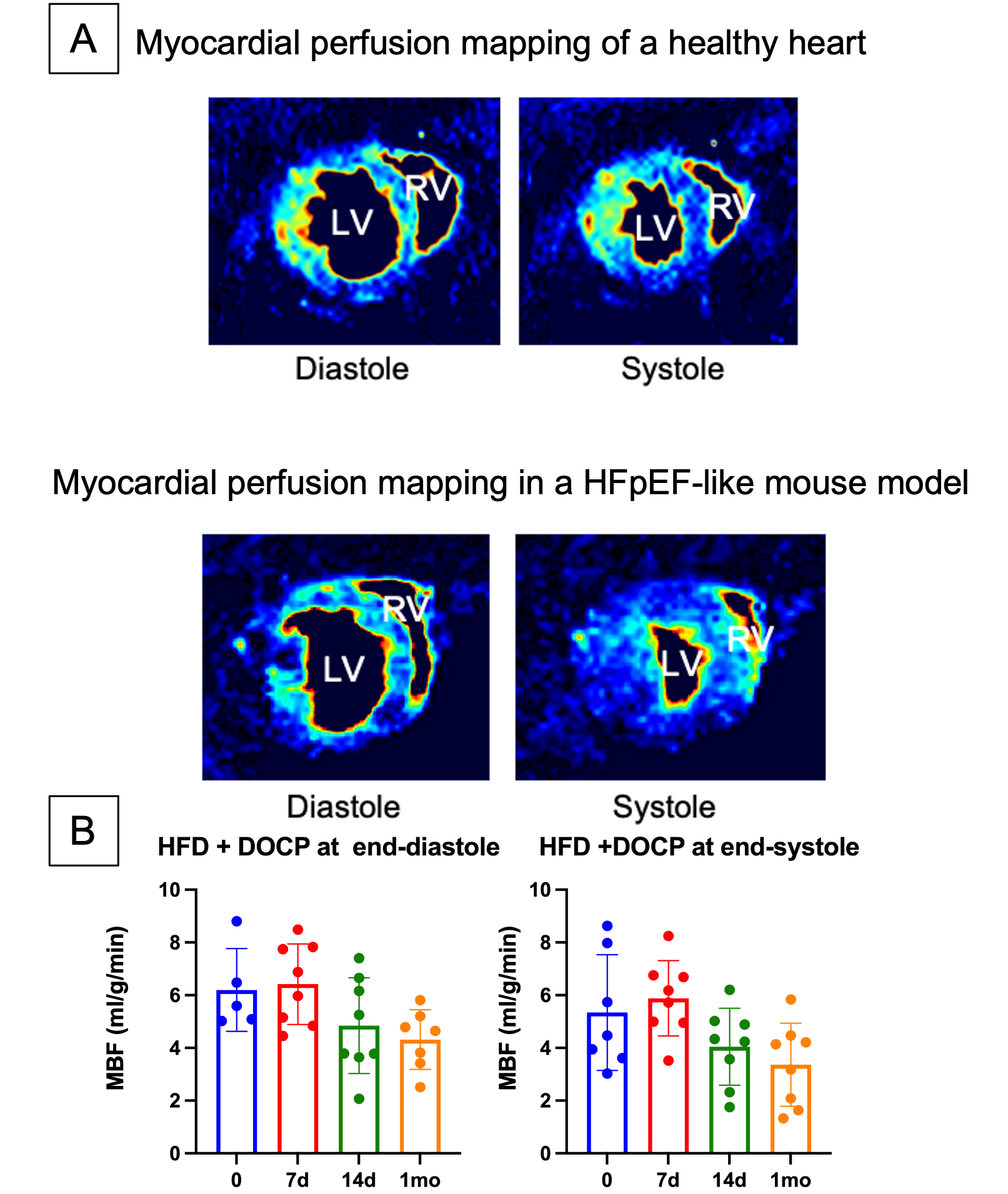
Significant differences in mid-ventricular myocardial perfusion (myocardial blood flow; MBF, fig.2A) were observed at 14 days and 1 month after DOCP injection compared to the baseline, both at end-systolic (p=0.0220, one-way ANOVA test) and end-diastolic phases of the cardiac cycle (p=0.0450, one-way ANOVA test) (Fig. 2B). Specifically, at the end-diastole, MBF decreased by 22% 14 days post-DOCP and 31% one month after DOCP injection compared to baseline. Similar decreases were observed at end-systole (24% and 37% decline respectively) indicating an early reduction in myocardial perfusion detectable at 14 days post-DOCP, which persisted one month post-DOCP injection (Fig. 2B). Further analysis of cardiac MRI data and histological measures of vascular density and fibrosis will be correlated with theses in vivo outcomes in order to confirm the impaired myocardial perfusion and characterize the importance of vascular remodeling in this HFpEF-like model.Conclusion
This study introduces a novel in vivo imaging biomarker for HFpEF using cine-ASL, enabling the assessment of myocardial vascular remodeling. Observed changes in myocardial perfusion will be complemented with histological and gene expression data to confirm the robustness of this non-invasive in vivo method and to elucidate if early remodeling of coronary microcirculation is an important contributor to HFpEF progression. These findings may improve the early diagnosis and our understanding of HFpEF pathophysiology.Acknowledgements
No acknowledgement found.References
1- Redfield, M. M. (2016). Heart failure with preserved ejection fraction. New England Journal of Medicine, 375(19), 1868-1877.
2- Redfield, M. M., & Borlaug, B. A. (2023). Heart failure with preserved ejection fraction: a review. Jama, 329(10), 827-838.
3- Nair, N. (2020). Epidemiology and pathogenesis of heart failure with preserved ejection fraction. Reviews in cardiovascular medicine, 21(4), 531-540.
4- Alem, M. M. (2019). Endothelial dysfunction in chronic heart failure: assessment, findings, significance, and potential therapeutic targets. International Journal of Molecular Sciences, 20(13), 3198.
5- Deng, Y., Xie, M., Li, Q., Xu, X., Ou, W., Zhang, Y., ... & Li, T. (2021). Targeting mitochondria-inflammation circuit by β-hydroxybutyrate mitigates HFpEF. Circulation research, 128(2), 232-245.
6- Troalen, T., Capron, T., Cozzone, P. J., Bernard, M., & Kober, F. (2013). Cine‐ASL: a steady‐pulsed arterial spin labeling method for myocardial perfusion mapping in mice. Part I. Experimental study. Magnetic Resonance in Medicine, 70(5), 1389-1398.
Figures