1789
Long-Term Outcomes Prediction in Diabetic Heart Failure with Preserved Ejection Fraction: A Cardiac Magnetic Resonance Imaging Study1Fuwai Hospital, State Key Laboratory of Cardiovascular Disease, National Center for Cardiovascular Diseases, Chinese Academy of Medical Sciences and Peking Union Medical College, Beijing, China
Synopsis
Keywords: Heart Failure, Diabetes, global longitudinal strain; heart failure with preserved ejection fraction; diabetes mellitus; prognosis
Motivation: While diabetes mellitus has a high prevalence in HFpEF and being associated with poorer outcomes, limited data are available on the cardiac MRI features of diabetic HFpEF
Goal(s): We aimed to explore imaging features including tissue characterization and myocardial deformation in diabetic HFpEF patients by MRI, and investigate its prognostic value for adverse outcomes
Approach: Feature-tracking derived strain and strain rates parameters and myocardial fibrosis were assessed by cardiac MRI in patients with diabetic HFpEF
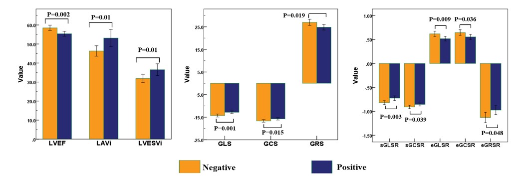
Results: Diabetic HFpEF patients were characterized by more impaired strains and myocardial fibrosis. Tissue characterization and global longitudinal strain obtained from MRI-FT provided incremental value for risk prediction
Impact: Our findings suggested that MRI-derived variables especially GLS played a crucial role in risk stratification and predicting worse prognosis in diabetic HFpEF, which could assist in identifying high-risk patients and guide therapeutic decision making.
Introduction
Cardiac MRI feature-tracking(FT) is a useful tool for predicting adverse outcomes in heart failure with preserved ejection fraction(HFpEF). However, limited data are available on the prognostic value of MRI-FT for patients with diabetic HFpEF, despite diabetes mellitus(DM) having a high prevalence in HFpEF and being associated with poorer outcomes. We aimed to evaluate the differences of MRI-derived imaging features between diabetic HFpEF subjects and those without DM and to determine whether the comprehensive MRI imaging provided independent and additional prognostic value beyond conventional clinical indices.Methods
In this retrospective study, patients with HFpEF who underwent cardiac MRI between January 2010 and December 2016 were enrolled in this study. Global circumferential strain(GCS), longitudinal strain(GLS) and radial strain(GRS) as well as peak systolic and diastolic strain rates(SRs) were derived from FT analysis and myocardial fibrosis was assessed by contrast enhanced MRI. Cox proportional regression analysis was performed to determine the association between strain analysis derived from MRI-FT with primary outcomes in diabetic HFpEF. Primary outcomes were all-cause death or heart failure hospitalization during the follow-up period.Results
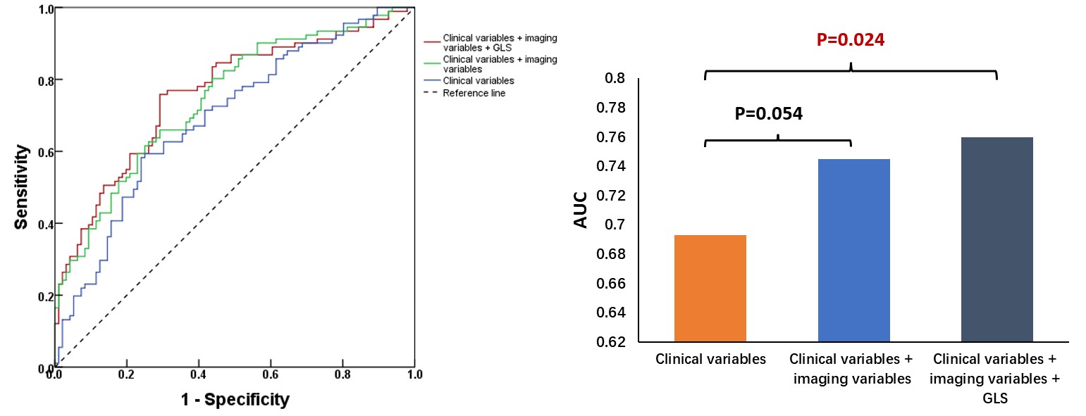
Of the 335 enrolled patients with HFpEF, 191 had DM(mean age: 58.7 years±10.8; 137 men). Diabetic HFpEF patients showed more impaired strains and myocardial fibrosis by MRI. During a median follow-up of 10.2 years(interquartile range: 8.0 to 11.3 years), 91 diabetic HFpEF and 56 non-diabetic HFpEF patients experienced primary outcomes. DM was a significant predictor of worse prognosis in HFpEF patients. In diabetic HFpEF, addition of conventional imaging variables(left ventricular ejection fraction, left atrial volume index, extent of LGE) and GLS resulted in a significant increase in the area under the receiver operating characteristic (ROC) curve (from 0.693 to 0.760, P<0.05). After adjustment for multiple clinical and imaging variables, each 1% worsening in global longitudinal strain(GLS) was associated with an 9.8% increased risk of adverse events(P=0.004), and the optimal cutoff value of the GLS for the primary end point was -13.64% from ROC analysis.Discussion
The present study provided several key findings and insights into the prognostic value of MRI-FT in diabetic cohorts with HFpEF. Compared to non-diabetic HFpEF patients, diabetic HFpEF patients exhibited more severe cardiac dysfunction and LV remodeling by MRI. The potential pathophysiological mechanisms of DM, such as oxidative stress, release of proinflammatory cytokines, mitochondrial dysfunction and so on, align to more severe clinical features and outcomes, and promote more explorations in this phenotype of HFpEF. GLS is of particular value in identifying myocardial dysfunction in many progressive myocardial diseases in their early stages, as well as in HFpEF. A more sensitive and robust marker may be needed to demonstrate risk stratification in a defined diabetic HFpEF group. Our study suggested the prognostic implications of MRI would allow better delineation of those as high risk in diabetic phenotype and suggested GLS as an accessible and supplemental parameter in patients with diabetic HFpEF.Conclusion
Diabetic HFpEF is characterized by more severely impaired strains and myocardial fibrosis, which was identified as high-risk HFpEF phenotype. In diabetic HFpEF, cardiac MRI including LGE and strain analysis provides incremental value in predicating prognosis. Particularly, MRI-FT measurement of GLS is an independent predictor of adverse outcome in diabetic HFpEF.Acknowledgements
NAReferences
1. Ritchie RH, Abel ED. Basic Mechanisms of Diabetic Heart Disease. Circ Res 2020; 126(11): 1501-25.
2. Claus P, Omar AMS, Pedrizzetti G, Sengupta PP, Nagel E. Tissue Tracking Technology for Assessing Cardiac Mechanics: Principles, Normal Values, and Clinical Applications. JACC Cardiovasc Imaging 2015; 8(12): 1444-60.
3. Romano S, Judd RM, Kim RJ, et al. Feature-Tracking Global Longitudinal Strain Predicts Death in a Multicenter Population of Patients With Ischemic and Nonischemic Dilated Cardiomyopathy Incremental to Ejection Fraction and Late Gadolinium Enhancement. JACC Cardiovasc Imaging 2018; 11(10): 1419-29.
4. He J, Sirajuddin A, Li S, et al. Heart Failure With Preserved Ejection Fraction in Hypertension Patients: A Myocardial MR Strain Study. J Magn Reson Imaging 2021; 53(2): 527-39.
Figures