1778
Higher oxygen extraction fraction in coronary artery disease is associated with lower cognition and cardiorespiratory fitness in males.1Quantitative Physiology Imaging Lab (QPI Lab), Department of Physics, Concordia University, Montreal, QC, Canada, 2Centre Epic and Research Center, Montreal Heart Institute, Montreal, QC, Canada, 3Hurvitz Brain Sciences Program, Clinical evaluative Sciences, Sunnybrook Research Institute, Toronto, ON, Canada, 4Department of Biomedical Science, Faculty of Medicine, Université de Montreal, Montreal, QC, Canada, 5Department of Radiology, Université de Sherbrooke, Sherbrooke, QC, Canada, 6Sherbrooke Connectivity Imaging Lab (SCIL), Computer Science Department, Faculty of Science, University of Sherbrooke, Sherbrooke, QC, Canada, 7Department of Psychology, Concordia University, Montreal, QC, Canada, 8Chemistry and Biochemistry, Concordia University, Montreal, QC, Canada, 9Department of Medicine, Université de Montreal, Montreal, QC, Canada, 10Research Center, Institut Universitaire de Gériatrie deMontréal, Montreal, QC, Canada, 11Physics, Concordia University, Montreal, QC, Canada, 12PERFORM Centre, Concordia University, Montreal, QC, Canada
Synopsis
Keywords: Heart Failure, Oxygenation, Quantitative Susceptibility Mapping (QSM), Venography, Cardiorespiratory fitness, Cognition
Motivation: There is a lack of knowledge about the physiological underpinnings of the effects of coronary artery disease (CAD) on human brain health.
Goal(s): Here we investigated the effect of CAD on brain oxygen extraction fraction (OEF)
Approach: Using quantitative susceptibility mapping (QSM) and its relation to cognitive performance and cardiorespiratory fitness.
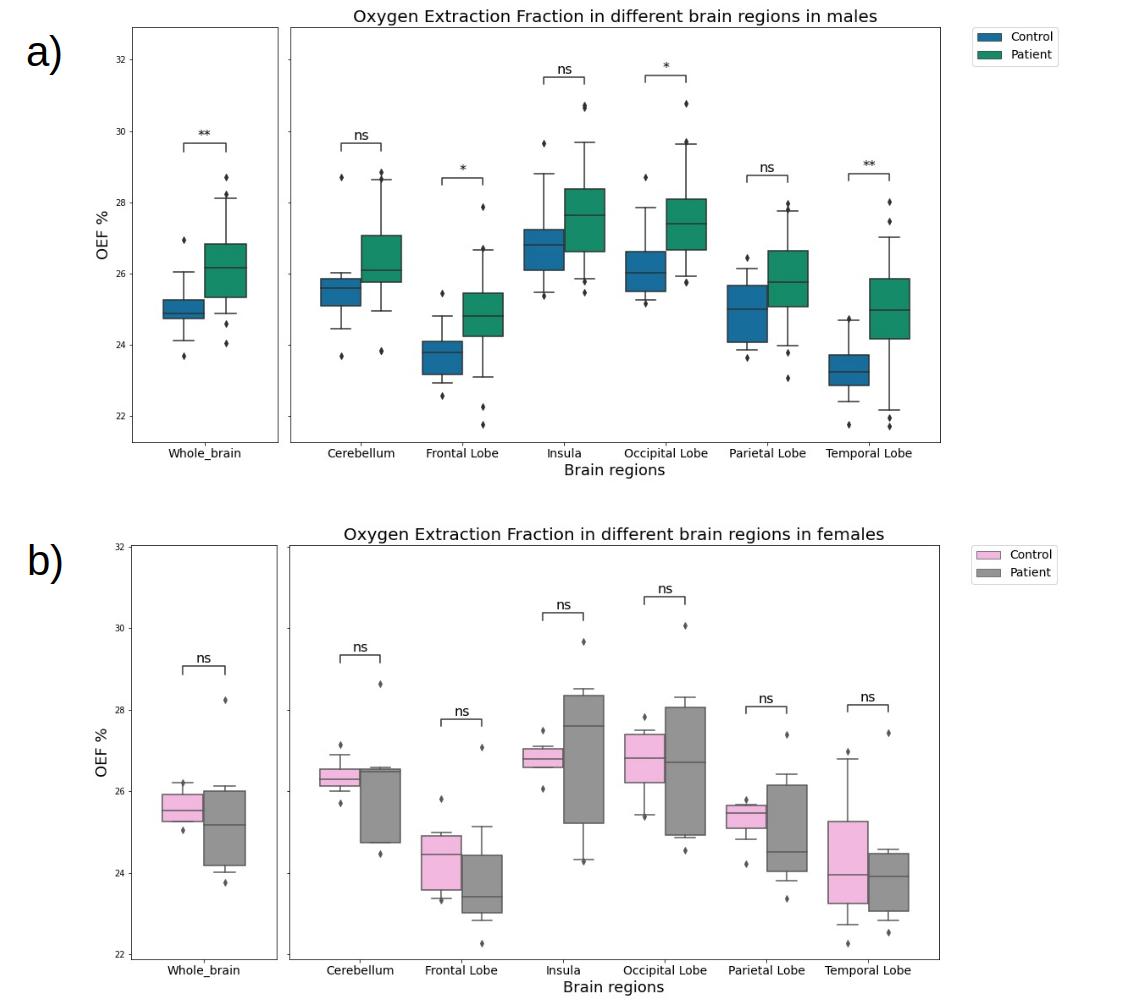
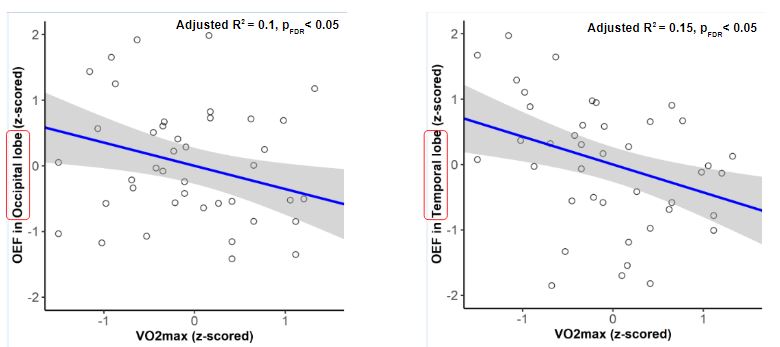
Results: The results of the sex-disaggregated analysis in a preliminary dataset show that CAD male patients have significantly higher OEF in whole gray matter, frontal, occipital, and temporal lobes. Furthermore, higher OEF in males is associated with lower cognitive performance, and higher OEF is associated with lower VO2max.
Impact: Sex differences in the effects of coronary artery disease (CAD) on brain health are unknown. Our results show that CAD increases OEF, and that higher OEF is associated with lower cognitive performance , and lower cardiorespiratory fitness in males.
Introduction
Coronary artery disease (CAD) is the most prevalent cardiovascular disease condition1 and is linked to cerebrovascular damage and cognitive decline, particularly in executive functions2. Our understanding of the nature of brain changes and how they are linked to cognition is currently limited, though there are indications that brain structure3 and vascular health1 are both impacted. Because brain function relies on consistent oxygen delivery and CAD is known to be associated with both vascular4 and metabolic dysfunctions5 outside the brain, oxygen extraction fraction (OEF) is likely to be a valuable biomarker. OEF assesses the balance between oxygen usage and delivery and OEF is affected in various diseases, including Alzheimer's disease6 and stroke7. However, the effect of CAD on OEF remains unclear, and any link with cognition is unknown. Recent progress in Quantitative Susceptibility Mapping (QSM) now allows noninvasive and accurate cerebral OEF measurements8. Additionally, some of the effects of CAD can be mitigated by rehabilitation programs that include exercise. Exercise has also been shown to improve brain health9 and cognition9, though its exact effects on the brain are still under investigation10 and the effect of cardiorespiratory fitness on OEF is unknown. Finally, important sexual dimorphisms have been observed in the presentation of CAD, where females often have a preserved ejection fraction11, in cognition12, and in the effect of exercise13. Therefore, this study will explore the effects of CAD on OEF and the relationship between OEF and cognition and cardiorespiratory fitness in sex-disaggregated data.Methods
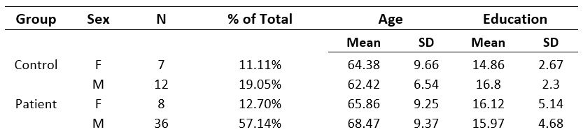
This preliminary data includes 44 CAD patients (36 males) and 19 healthy controls (HC) (12 males) over 50 years, without significant comorbidities or cognitive impairment (Table 1). Participants completed executive function assessments14,15. Executive function scores were derived as in15,16. QSM data acquisitions were performed using a 3T Siemens Skyra MRI scanner with a 32-channel coil, using a 3D multi-echo gradient echo sequence (TR/TE1/TE2/TE3/TE4/flip angle = 20 ms/6.92 ms/13.45 ms/19.28 ms/26.51 ms/9°, 0.7 x 0.7 x 1.4 mm3 voxel size). Raw unwrapped phase data were combined and wrapped using the ROMEO toolbox17. The QSM maps were reconstructed using the TGV toolbox18. QSM maps were referenced to ventricular CSF susceptibility values. Then, we used a Recursive Ridge Filtering method19 to extract draining veins7 and calculate venous OEF8. VO2max values were obtained from a maximal effort test on a bicycle ergometer as in20. Linear models were performed on sex-disaggregated data in R to assess the link between lobar OEF and executive function or VO2max. A Benjamini-Hochberg post-hoc test was employed to control for false discovery rate (FDR) error with pFDR < 0.05.Results
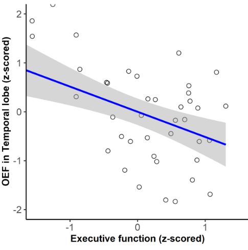
Table 1 includes demographic information of our preliminary sample. Figure 1 shows the lobar OEF of CAD and HCs in each sex. Linear models revealed that there was a statistically significant relationship between males’ OEF and executive function scores (figure 2). Figure 3 shows the significant relationship between OEF in the temporal and occipital lobes with VO2max in males. No regional OEF in females showed a significant relationship with cognition or VO2max.Discussion
Figure 1a shows that the whole brain, frontal, occipital, and temporal lobe OEF in CAD was significantly higher than HC in males which is consistent with low perfusion in CAD21. Figure 1b compares lobar OEF in females but not significantly. The negative relationship between temporal lobe OEF in males and executive function performance in Figure 2 indicates that the higher OEF observed in some participants is associated with lower cognitive performance. A combination of QSM with perfusion imaging would allow us to disentangle whether this is indicative of preserved metabolism with misery perfusion, or whether metabolism is increased. Consistent with the adverse effects of higher OEF, Figure 3 shows that lower VO2max in males is associated with higher OEF in the temporal and occipital lobes, indicating that exercise may be a promising avenue to counteract the effects of higher OEF on cognitive function. Finally, the lack of significant differences in females may reflect the preliminary nature of this analysis (i.e., a smaller sample size), however, data acquisition for this project is still ongoing.Conclusion
This study explores the sex-specific effects of CAD on OEF, and the relationships between OEF, cognition, and cardiorespiratory fitness. CAD was found to be associated with higher OEF, while higher OEF was found to be associated with poorer executive function and fitness, though only in males. More data is needed, as well as complementary information from perfusion imaging to fully disentangle these effects.Acknowledgements
We would like to acknowledge the contribution of Paule Samson, MR technologist, Julie Lalongé, research assistant and medical electrophysiology technologist, and of all the staff of the EPIC center that has contributed to this project. We also want to thank all students and research assistants who have helped in data acquisition: Roni Zaks, Robert Hovey, Stephanie Beram, Alexandre bailey, Agathe Godet and Kathia Saillant and the research participants who took part in this study.
SAT was supported by the Canadian Institutes of Health Research (CIHR: FBD 175862).
CJG was supported by the Heart and Stroke Foundation New Investigator Award and J.M. Barnett fellowship, the Michal and Renata Hornstein Chair in Cardiovascular Imaging, and the Heart and Stroke Foundation Grant-in Aid G-17-0018336.
LB was supported by Mirella and Lino Saputo Research Chair in Cardiovascular Health and the Prevention of Cognitive Decline from the Universite de Montreal at the Montreal Heart Institute.
References
- Anazodo, U. C., Shoemaker, J. K., Suskin, N., Ssali, T., & Wang, D. J. (2015). Impaired Cerebrovascular Function in Coronary Artery Disease Patients and Recovery Following Cardiac Rehabilitation. Frontiers in Aging Neuroscience.https://doi.org/10.3389/fnagi.2015.00224
- Joosten, H., van Eersel, M. E., Gansevoort, R. T., Bilo, H. J., Slaets, J. P., & Izaks, G. J. (2013). Cardiovascular risk profile and cognitive function in young, middle-aged, and elderly subjects. Stroke, 44(6), 1543–1549. https://doi.org/10.1161/STROKEAHA.111.000496
- Au, R., Massaro, J. M., Wolf, P. A., Young, M. E., Beiser, A., Seshadri, S., D'Agostino, R. B., & DeCarli, C. (2006). Association of white matter hyperintensity volume with decreased cognitive functioning: the Framingham Heart Study. Archives of neurology, 63(2), 246–250. https://doi.org/10.1001/archneur.63.2.246
- Matsuzawa, Y., & Lerman, A. (2014). Endothelial dysfunction and coronary artery disease: assessment, prognosis, and treatment. Coronary artery disease, 25(8), 713–724. https://doi.org/10.1097/MCA.0000000000000178
- Mahalle, N., Garg, M. K., Naik, S. S., & Kulkarni, M. V. (2014). Association of metabolic syndrome with severity of coronary artery disease. Indian journal of endocrinology and metabolism, 18(5), 708–714. https://doi.org/10.4103/2230-8210.139238
- Liu, Y., Dong, J., Song, Q., Zhang, N., Wang, W., Gao, B., Tian, S., Dong, C., Liang, Z., Xie, L., & Miao, Y. (2021). Correlation Between Cerebral Venous Oxygen Level and Cognitive Status in Patients With Alzheimer's Disease Using Quantitative Susceptibility Mapping. Frontiers in neuroscience, 14, 570848. https://doi.org/10.3389/fnins.2020.570848
- Fan, A. P., Khalil, A. A., Fiebach, J. B., Zaharchuk, G., Villringer, A., Villringer, K., & Gauthier, C. J. (2020). Elevated brain oxygen extraction fraction measured by MRI susceptibility relates to perfusion status in acute ischemic stroke. Journal of cerebral blood flow and metabolism: official journal of the International Society of Cerebral Blood Flow and Metabolism, 40(3), 539–551. https://doi.org/10.1177/0271678X19827944
- Fan, A. P., Bilgic, B., Gagnon, L., Witzel, T., Bhat, H., Rosen, B. R., & Adalsteinsson, E. (2014). Quantitative oxygenation venography from MRI phase. Magnetic resonance in medicine, 72(1), 149–159. https://doi.org/10.1002/mrm.24918
- Bherer, L., Erickson, K. I., & Liu-Ambrose, T. (2013). A review of the effects of physical activity and exercise on cognitive and brain functions in older adults. Journal of aging research, 2013, 657508. https://doi.org/10.1155/2013/657508
- Intzandt, B., Sanami, S., Huck, J., PREVENT-AD Research group, Villeneuve, S., Bherer, L., & Gauthier, C. J. (2023). Sex-specific relationships between obesity, physical activity, and gray and white matter volume in cognitively unimpaired older adults. GeroScience, 45(3), 1869–1888. https://doi.org/10.1007/s11357-023-00734-4
- Barton, M., & Meyer, M. R. (2020). Heart Failure With Preserved Ejection Fraction in Women: New Clues to Causes and Treatment. JACC. Basic to translational science, 5(3), 296–299. https://doi.org/10.1016/j.jacbts.2020.02.001
- Deckers, K., Schievink, S. H. J., Rodriquez, M. M. F., van Oostenbrugge, R. J., van Boxtel, M. P. J., Verhey, F. R. J., & Köhler, S. (2017). Coronary heart disease and risk for cognitive impairment or dementia: Systematic review and meta-analysis. PloS one, 12(9), e0184244. https://doi.org/10.1371/journal.pone.0184244
- Glassberg, H., & Balady, G. J. (1999). Exercise and heart disease in women: why, how, and how much?. Cardiology in review, 7(5), 301–308. https://doi.org/10.1097/00045415-199909000-00015
- Boidin, M., Handfield, N., Ribeiro, P. A. B., Desjardins-Crépeau, L., Gagnon, C., Lapierre, G., Gremeaux, V., Lalongé, J., Nigam, A., Juneau, M., Gayda, M., & Bherer, L. (2020). Obese but Fit: The Benefits of Fitness on Cognition in Obese Older Adults. The Canadian journal of cardiology, 36(11), 1747–1753. https://doi.org/10.1016/j.cjca.2020.01.005
- Besnier, F., Bérubé, B., Gagnon, C., Olmand, M., Ribeiro, P. A. B., Nigam, A., Juneau, M., Blondeau, L., White, M., Gremeaux, V., Bherer, L., & Gayda, M. (2020). Cardiorespiratory Fitness Mediates Cognitive Performance in Chronic Heart Failure Patients and Heart Transplant Recipients. International journal of environmental research and public health, 17(22), 8591. https://doi.org/10.3390/ijerph17228591
- Andrade C. Z Scores, Standard Scores, and Composite Test Scores Explained. Indian Journal of Psychological Medicine. 2021;43(6):555-557. doi:10.1177/02537176211046525
- Dymerska, B, Eckstein, K, Bachrata, B, et al. Phase unwrapping with a rapid opensource minimum spanning tree algorithm. Magn Reson Med. 2021; 85: 2294–2308. https://doi.org/10.1002/mrm.28563
- Langkammer, C., Bredies, K., Poser, B. A., Barth, M., Reishofer, G., Fan, A. P., Bilgic, B., Fazekas, F., Mainero, C., & Ropele, S. (2015). Fast quantitative susceptibility mapping using 3D EPI and total generalized variation. NeuroImage, 111, 622–630. https://doi.org/10.1016/j.neuroimage.2015.02.041
- Bazin, P. L., Plessis, V., Fan, A. P., Villringer, A., & Gauthier, C. J. (2016). Vessel segmentation from quantitative susceptibility maps for local oxygenation venography. In 2016 IEEE International Symposium on Biomedical Imaging: From Nano to Macro, ISBI 2016 - Proceedings (pp. 1135-1138). Article 7493466 (Proceedings - International Symposium on Biomedical Imaging; Vol. 2016-June). IEEE Computer Society. https://doi.org/10.1109/ISBI.2016.7493466
- Gayda, M., Gremeaux, V., Bherer, L., Juneau, M., Drigny, J., Dupuy, O., Lapierre, G., Labelle, V., Fortier, A., & Nigam, A. (2017). Cognitive function in patients with stable coronary heart disease: Related cerebrovascular and cardiovascular responses. PloS one, 12(9), e0183791. https://doi.org/10.1371/journal.pone.0183791
- Alosco, M. L., Gunstad, J., Jerskey, B. A., Xu, X., Clark, U. S., Hassenstab, J., Cote, D. M., Walsh, E. G., Labbe, D. R., Hoge, R., Cohen, R. A., & Sweet, L. H. (2013). The adverse effects of reduced cerebral perfusion on cognition and brain structure in older adults with cardiovascular disease. Brain and behavior, 3(6), 626–636. https://doi.org/10.1002/brb3.171
Figures