1774
Tissue Metabolomics Characterization of Human Valve Diseases using NMR spectroscopy1CTVS, AIIMS, NEW DELHI, DELHI, India, 2NMR, AIIMS, NEW DELHI, DELHI, India, 3Pathology, AIIMS, NEW DELHI, DELHI, India, 4Cardiology, AIIMS, NEW DELHI, DELHI, India
Synopsis
Keywords: Vessels, Heart, NMR spectroscopy
Motivation: Heart valve disease (HVD) is a multifactorial process and its pathophysiology has not been fully understood.
Goal(s): To investigate and determine the metabolite tissue using the 1H-NMR based metabolomics study to discriminate the aortic valve and mitral valve.
Approach: proton NMR spectroscopy
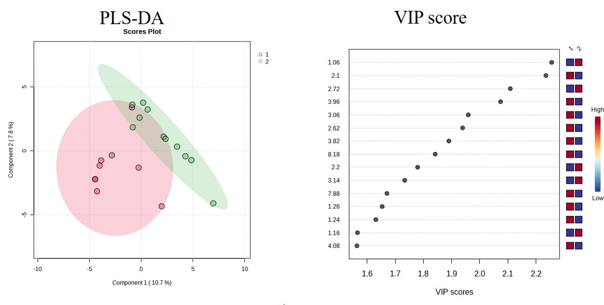
Results: The results obtained from PLS-DA and VIP score plots of metabolites in tissue showed a separation between patients with aortic valves and mitral valves.
Impact: The present study revealed discrimination of metabolic profiling of tissue of aortic valve patients from mitral valve replacement implying that 1H-NMR-based metabolomics may provide an insight into understanding the potential metabolic alteration.
Heart valve disease (HVD) is a common term that includes several etiologic entities with different pathophysiologic mechanisms that lead to anatomic disruption of the valve apparatus. Functional abnormalities due to alterations in matrix architecture and cellular components impair the function of heart valves resulting in abnormal blood flow patterns through the chambers of the heart. Degenerative and rheumatic HVD is caused by the interaction of several risk factors such as infections, age-related changes, ischemic heart disease, congenital, heart failure, stroke, genetic, inflammatory, autoimmune, and oxidative stress. Thus, to investigate the metabolic profiling of aortic, mitral, and double valve repair and replacement (aortic and mitral valve) tissue samples of patients using proton (1H) NMR spectroscopy.
Methods
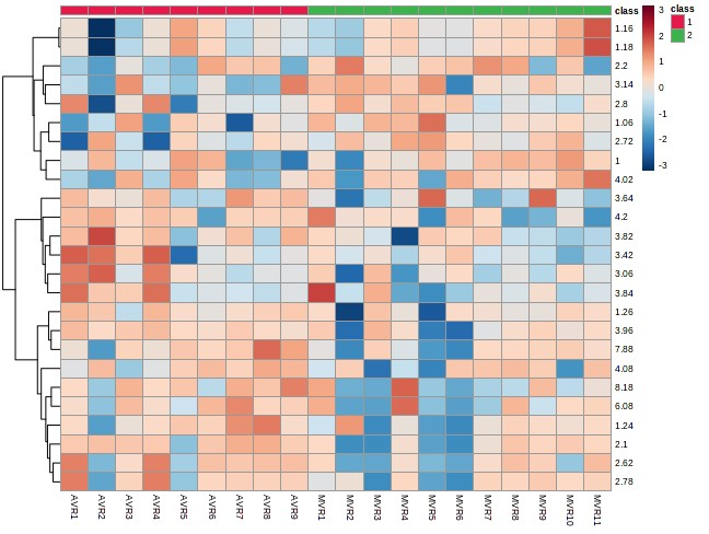
Tissue samples were collected from HVD patients with aortic valves (n =9) and mitral valve (n=11) after valve repair and replacement and stored at -80°C until NMR experiments were performed. Water-soluble metabolites were extracted from heart valve tissue using perchloric acid extraction (PCA). The frozen tissue was weighted, crushed, sonication then thoroughly homogenized in 6% PCA. The homogenate tissue was centrifuged at 10,000 rpm at 4 o C for 10 minutes. After centrifuging supernatant will be collected and then neutralized using 3M KOH to pH 7.4 and the precipitate was removed by centrifugation. The supernatant obtained will be lyophilized for 8 to 10 hours at -80oC. The resulting sample will be dissolved in 0.6mL of Deuterium oxide(D2O). TSP (Trimethyl-silyl propionic acid) was added which serves as both a chemical shift reference and concentration standard for the proton NMR studies. Proton spectra were acquired at a 700 MHz spectrometer. The spectral regions at 0.5-10 ppm were segmented into bins with equal widths of 0.04 ppm. Statistical analysis was performed using SPSS (SPSS Inc., Chicago, IL, USA). The t-test was used to analyze differences between groups. P values were two-tailed and a value < 0.05 was considered significant. The data were normalized and subjected to multivariate pattern recognition analysis using MetaboAnalyst 5.0 software. Metabolites were considered significant at VIP >1.0 for further analysis of metabolomic data analysis. Partial least square discriminant analysis (PLS-DA) and orthogonal partial least square discriminant analysis (OPLS-DA) loading plots were carried out for significant metabolite identification. Variable importance in projection (VIP) scores in PLS-DAs of metabolic profiles in the tissue of patients, VIP scores for some important metabolites with the highest contribution to the separation of the studied groups are presented. The boxes on the right refer to the relative concentrations of the appropriate metabolites in the studied groups.
Results
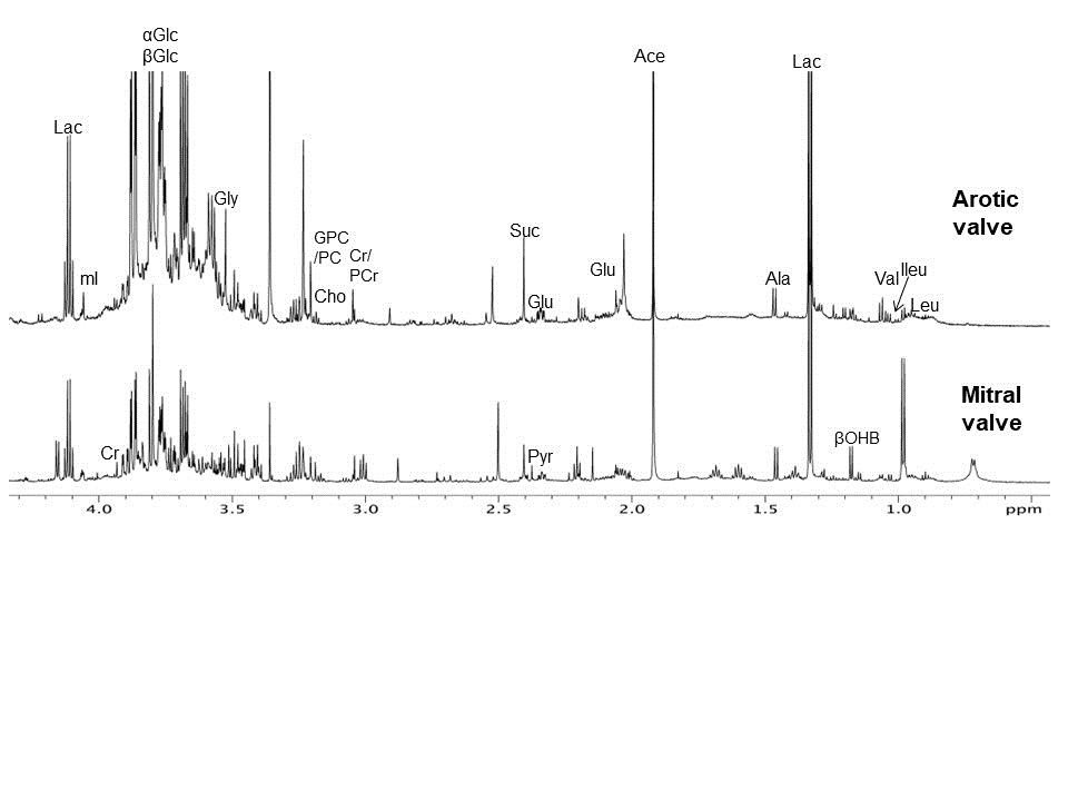
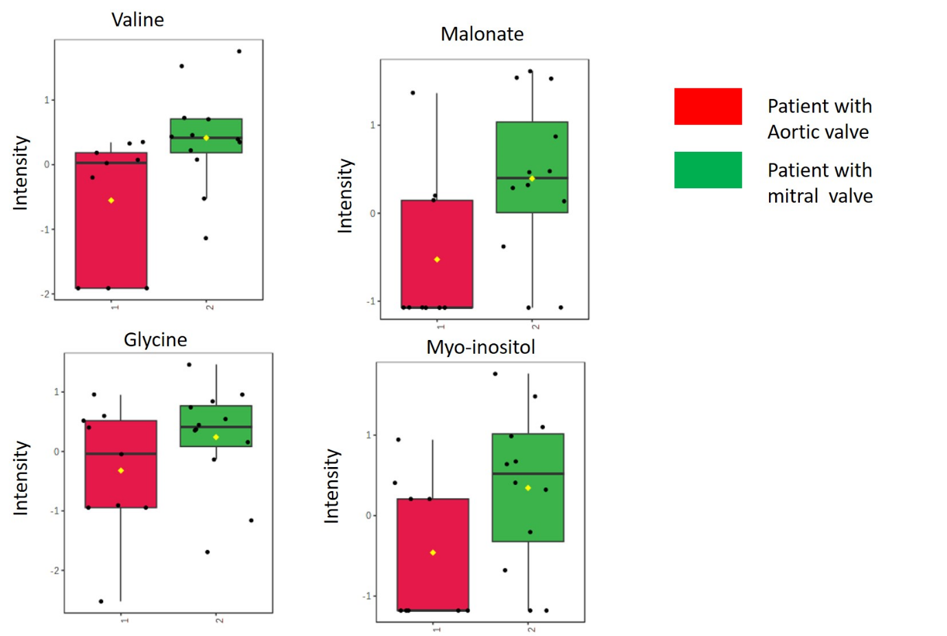
Figure 1 Representative CPMG 1H NMR spectra of tissue spectra of aortic valve and mitral valve. The tissue metabolomic profile of patients with aortic valves is characterized by decreased levels of valine malonate, glycine, and myo-inositol compared with mitral vales. The results obtained from PLS-DA and VIP score plots of metabolites in tissue showed a separation between patients with aortic valves and mitral valves shown in Figure.
Discussion
Significantly lower levels of valine malonate, glycine, and myoinositol in aortic valve as compared to mitral valve disease tissue. NMR-based metabolomics analysis enhanced the current understanding of the mechanisms involved in human valves and uncovered the therapeutic potential role of diseases. The disturbing metabolites were proposed to be the potential biomarkers that may help to predict to evaluate the efficacy of human valve diseases in the treatment. Myo-inositol is known to exert anti-diabetic, anti-oxidative, and anti-inflammatory properties. The lower level of Myo-inositol compromises the anti-inflammatory properties. In recent study, evidence has mounted in favor of the anti-inflammatory, immunomodulatory, and cytoprotective effects of the simplest amino acid L-glycine. The lower level of glycine compromises the anti-inflammatory properties. Malonate associates oxidative stress and alteration ROS system.
Conclusion
The present study revealed discrimination of metabolic profiling of tissue of aortic valve patients from mitral valve replacement implying that 1H-NMR-based metabolomics may provide an insight into understanding the potential metabolic alteration such as valine malonate, glycine, and myo-inositol metabolism associated with inflammation, oxidative stress, and tissue degradation of HVD.
Acknowledgements
Thank for Department of NMR AIIMS New DelhiReferences
1. Fu B, Wang J, Wang L, Wang Q, Guo Z, Xu M, Jiang N. Integrated proteomic and metabolomic profile analyses of cardiac valves revealed molecular mechanisms and targets in calcific aortic valve disease. Front Cardiovasc Med. 2022 Oct 13;9:944521.
2. Coffey S, Roberts-Thomson R, Brown A, Carapetis J, Chen M, Enriquez-Sarano M, et al. Global epidemiology of valvular heart disease. Nat Rev Cardiol. (2021) 18:853–64.
3. 2. Yi B, Zeng W, Lv L, Hua P. Changing epidemiology of calcific aortic valve disease: 30-year trends of incidence, prevalence, and deaths across 204 countries and territories. Aging (Albany NY). (2021) 13:12710–32.
4. Peltonen T, Ohukainen P, Ruskoaho H, Rysä J. Targeting vasoactive peptides for managing calcific aortic valve disease. Ann Med. (2017) 49:63–74.
5. Akahori H, Tsujino T, Masuyama T, Ishihara M. Mechanisms of aortic stenosis. J Cardiol. (2018) 71:215–20. 6.
Figures