1771
Multi-Slice-Aware Denoising Model for 7T MR Angiography1Bionics Research Center, Korea Institute of Science and Technology, Seoul, Korea, Republic of, 2Department of Electrical and Electronic Engineering, Yonsei University, Seoul, Korea, Republic of, 3Department of Radiology, Korea University, Seoul, Korea, Republic of
Synopsis
Keywords: Analysis/Processing, Machine Learning/Artificial Intelligence
Motivation: Noise in 7T MRA images can make it challenging to diagnose vascular diseases, and conventional denoisers tend to over-smooth in a way that blur entire image or does not preserve vascular information.
Goal(s): Our goal was to reduce noise in 7T MRA images while maintaining vascular information using deep learning method.
Approach: We devised an unsupervised denoising model using multiple slice information and cycleGAN-based neural networks.
Results: Our approach not only suppressed noise in 7T MRA images, but it also successfully preserved vessel information among the compared models.
Impact: We developed a denoising method in 7T MRA images while preserving vascular information. Clinically, our findings will help diagnose vascular-related diseases. High SNR is preserved by averaging adjacent slices, and we contribute to increase usability of 7T MRI.
Introduction
Magnetic Resonance Angiography1-2 (MRA) is a fundamental modality pivotal in diagnosing vascular-related diseases such as cerebral aneurysms, infarctions, or strokes by identifying cerebrovascular deformities or blocked blood vessels. However, noise spread throughout the 7T MRA image, due to nonoptimal sequence parameters or reconstruction errors for minimizing scan time, often hinders to recognize the small sized intracranial blood vessels3-4. Therefore, it is essential to suppress noise in the 7T MRA image to enhance the visibility of all blood vessels on it for accurate diagnosis. Although the conventional denoising methods5-6 show competitive results, they have limitations, such as loss of critical vessel information due to over-smoothing of the entire image. In particular, since the MRA image has no ground truth for the denoising task, unsupervised learning is usually required. Thus, in this work, we propose a deep learning-based approach to effectively attenuate the noise while preserving vascular information in 7T MRA images by high-Signal-to-Noise Ratio (SNR) image translation from averaging multi-slices.Method
The overall schematic diagram of the proposed denoising method is shown in Figure 1.Data acquisition
MRA images of the brain were acquired from 52 volunteers on a 7T Terra-MRI scanner (SIEMENS). Data from 50 volunteers were used for training and from the other two for validation and evaluation.
Data pre-processing
Since Averaging multiple independent images helps reduce noise power by a factor of the square root, thereby enhancing the SNR of the image. Consequently, we have devised a methodology to create high-SNR information in datasets via averaging multiple slices as shown in Figure 2. Among the MRA image slices acquired from each patient, “target slice” is defined as $$$I_t$$$ for denoising, and the neighboring slices (i.e., $$$I_{t-2}, I_{t-1}, I_{t+1},$$$ and $$$I_{t+2}$$$) adjacent to the target slice share similar image content (such as location of blood vessels and other structures of the brain). Therefore, by averaging each target slice and its adjacent slices, we can obtain higher SNR information in comparison to the original MRA image. In this experiment, we generated two datasets; one denoted as "Dataset A," which is composed of an average of five slices, including the target slice, and the other as "Dataset B," consisting of four slices, except the target slice.
Training and Data post-processing
With the two datasets above, we train a cycleGAN7-based neural networks for image translation between the paired datasets as shown in Figure 3. The deep learning model finds a mapping function that minimizes the sum of the adversarial loss and consistency loss between Dataset A and B. We can assume that both datasets have their original contents of MRA image slices, with the averaged out noise information due to the averaging effect. So, the model can only learn the image contents of the target slice from the difference between the two datasets. After network training, the denoised target slice can be obtained by post-processing function ($$$PP$$$), which is a function that extracts the noise-free target slice image from input (Dataset A) and output ($$$I_{out}$$$) generated through the networks.
$$\begin{align*}PP &= Dataset A * 5 - I_{out}*4 \\ &= I_{t-2}+I_{t-1}+I_t+I_{t+1}+I_{t+2}-(I_{t-2}+I_{t-1}+I_{t+1}+I_{t+2})\end{align*}$$
Evaluation
To test the effectiveness of the proposed approach, we qualitatively compare our method with BM3D5 and DnCNN6. For BM3D, we empirically adjusted the hyper-parameters for each slice. For DnCNN, we trained the model on a dataset from the same group of volunteers as the data used to train the proposed networks.
Results
Figure 4 shows the experimental denoising results from BM3D, DnCNN, and the proposed method, applied to the 7T MRA image. All methods are capable of producing comparable results. However, it is important to note that the results obtained using BM3D and DnCNN lose the important vascular details and tend to overly smooth the overall image, resulting in a blurriness. In contrast, our proposed method successfully suppresses noise while preserving intracranial blood vessels.Conclusion and Discussion
In this work, we propose a novel method to suppress noise in 7T MRA images while preserving vascular information using a cycleGAN-based neural networks. We construct high SNR information by averaging multi-slice images that are correlated in the target slice to ensure that the unsupervised learning preserves the original vessel information. In other words, our model uses not only the target slice but also the neighboring slice images to denoise and preserve vascular information. Through our experiments, we found that the proposed method effectively reduces the noise while preserving vessel information better than the conventional denoisers.Acknowledgements
This work is supported by KIST Institutional Programs (2V09831, 2E32341, 2E32211, 2E32271).References
1. Dumoulin, Charles L., and H. R. Hart Jr. "Magnetic resonance angiography." Radiology 161.3 (1986): 717-720.
2. Edelman, Robert R. "Basic principles of magnetic resonance angiography." Cardiovascular and interventional radiology 15 (1992): 3-13.
3. Potchen, E. J. Magnetic resonance angiography: techniques, indications and practical applications. Springer Science & Business Media, 2006.
4. Hartung, Michael P., Thomas M. Grist, and Christopher J. François. "Magnetic resonance angiography: current status and future directions." Journal of Cardiovascular Magnetic Resonance 13.1 (2011): 1-11.
5. Dabov, Kostadin, et al. "Image denoising by sparse 3-D transform-domain collaborative filtering." IEEE Transactions on image processing 16.8 (2007): 2080-2095.
6. Zhang, Kai, et al. "Beyond a gaussian denoiser: Residual learning of deep cnn for image denoising." IEEE transactions on image processing 26.7 (2017): 3142-3155.
7. Zhu, Jun-Yan, et al. "Unpaired image-to-image translation using cycle-consistent adversarial networks." Proceedings of the IEEE international conference on computer vision. 2017.
Figures
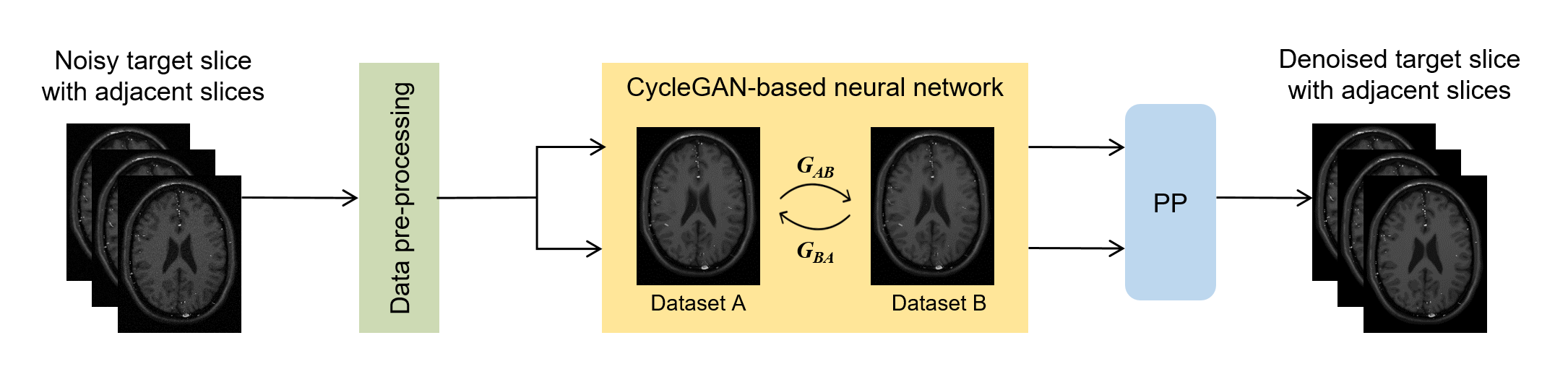
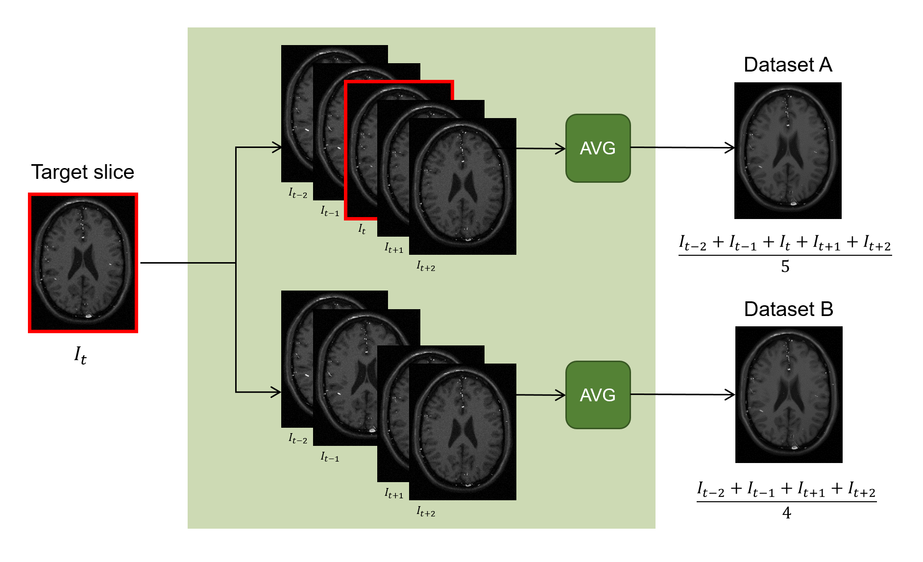
Figure 2. The procedure of data pre-processing. Training datasets can be constructed with a target slice and its adjacent slice images by averaging multiple slices. Through this procedure, two high-SNR datasets can be produced to train the cycleGAN-based neural networks.
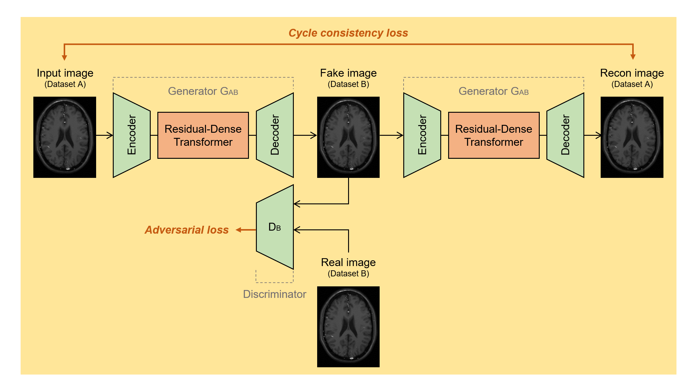
Figure 3. Architecture of the proposed cycleGAN-based neural networks.
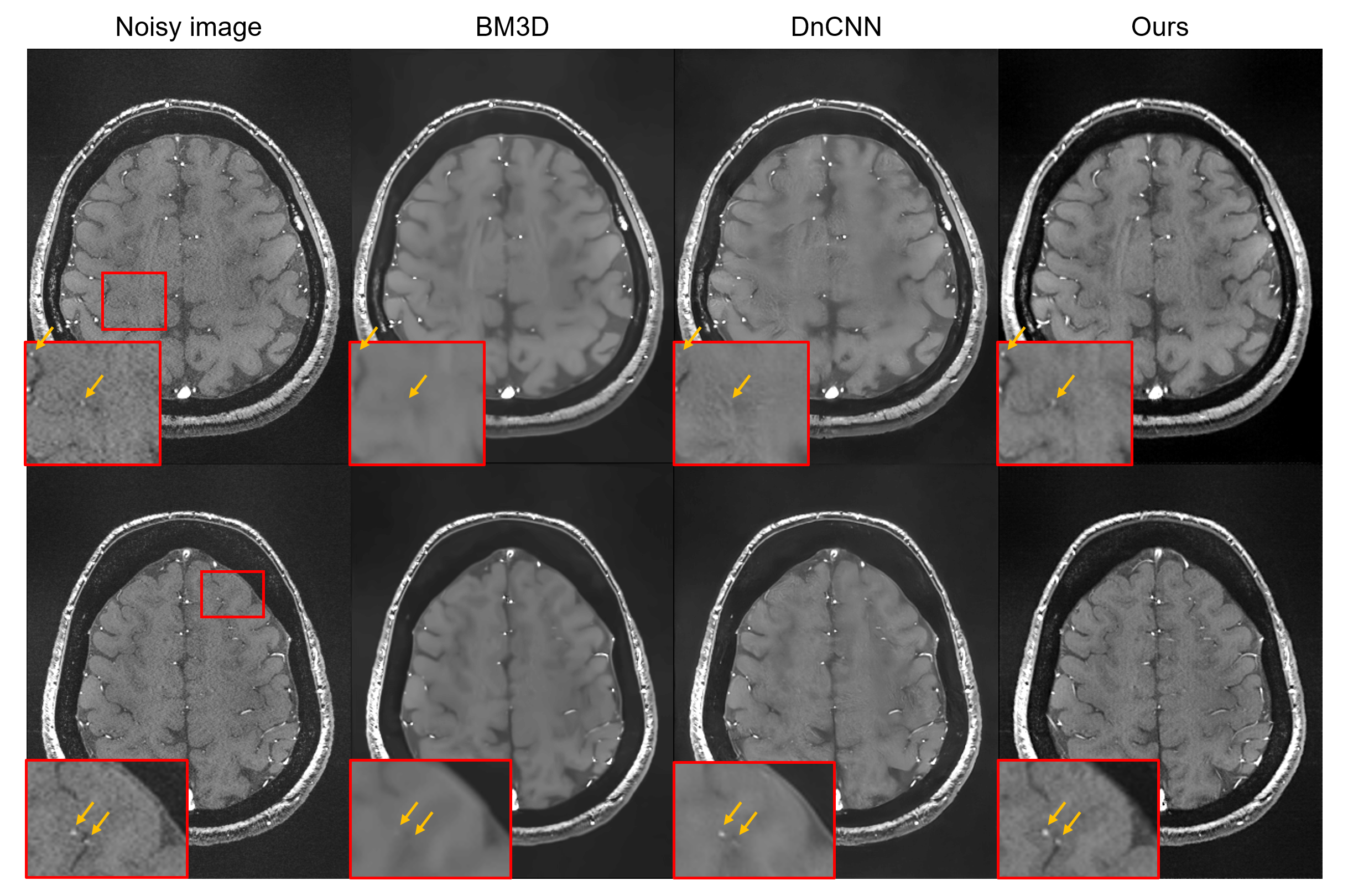
Figure 4. The experiment results of two compared methods and the proposed method. The red boxes highlight the performance of each method. The yellow arrow tips point to blood vessels.