1769
Improved lesion conspicuity on liver ADC maps using self supervised DDPMs1Boston Children's Hospital and Harvard Medical School, Boston, MA, United States
Synopsis
Keywords: AI Diffusion Models, Liver
Motivation: Abdominal DW-MRI suffers from low SNR and motion artifacts, compromising ADC reliability.
Goal(s): Improve ADC estimation in low SNR abdominal DW-MRI using a single-image acquisition per b-value.
Approach: A novel self-supervised training approach using denoising diffusion probabilistic models (ssDDPM). Tailored for multi-b-value DW-MRI images, it requires only a single gradient image per b-value for denoising, which reduces scan time.
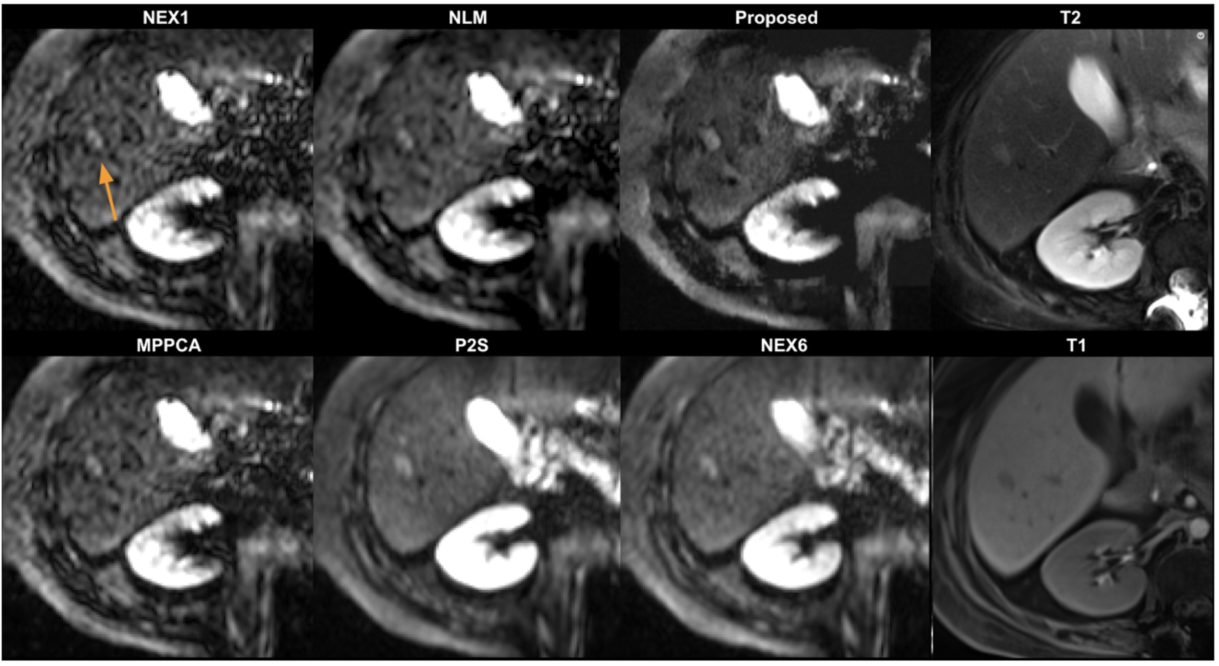
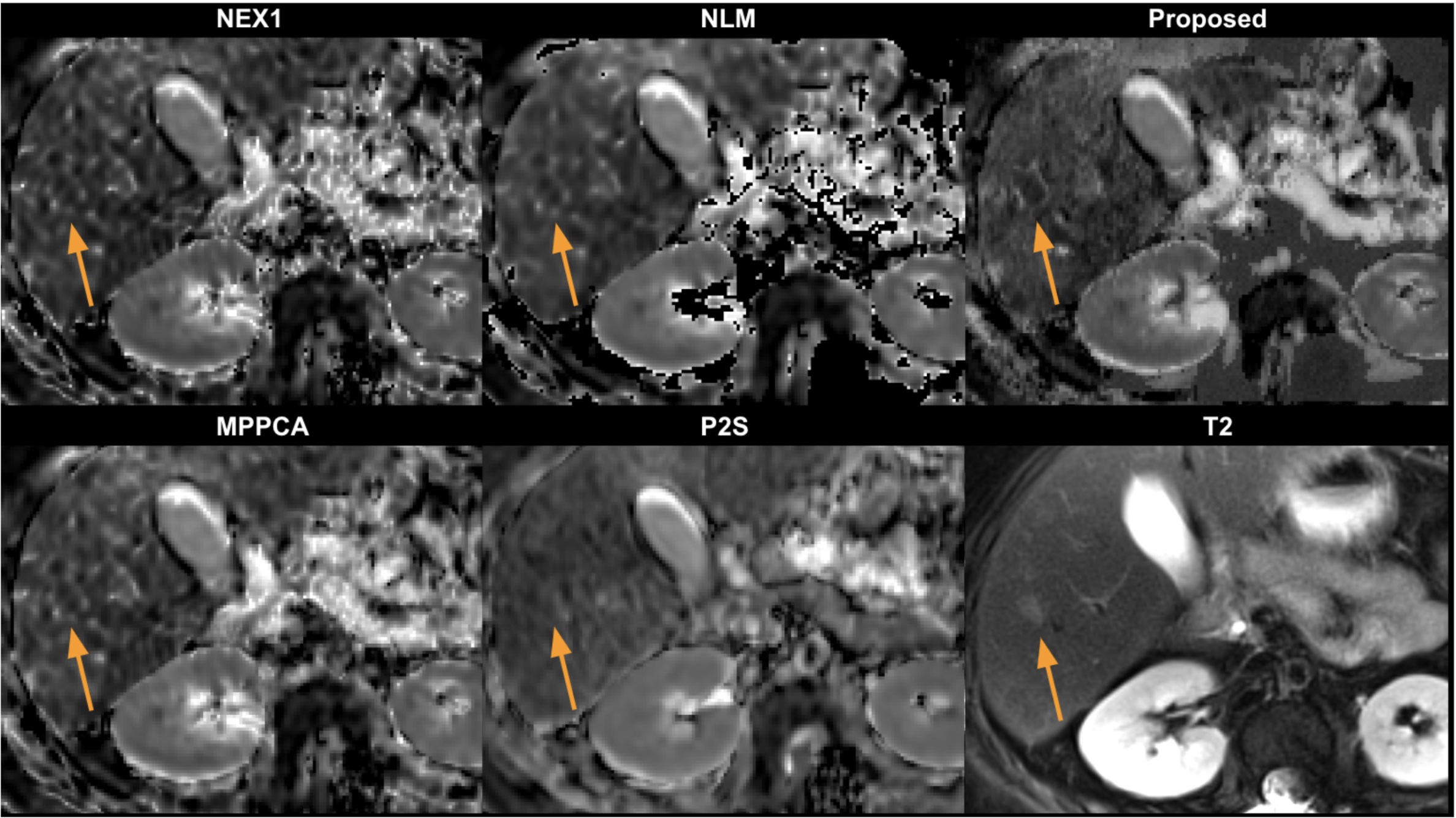
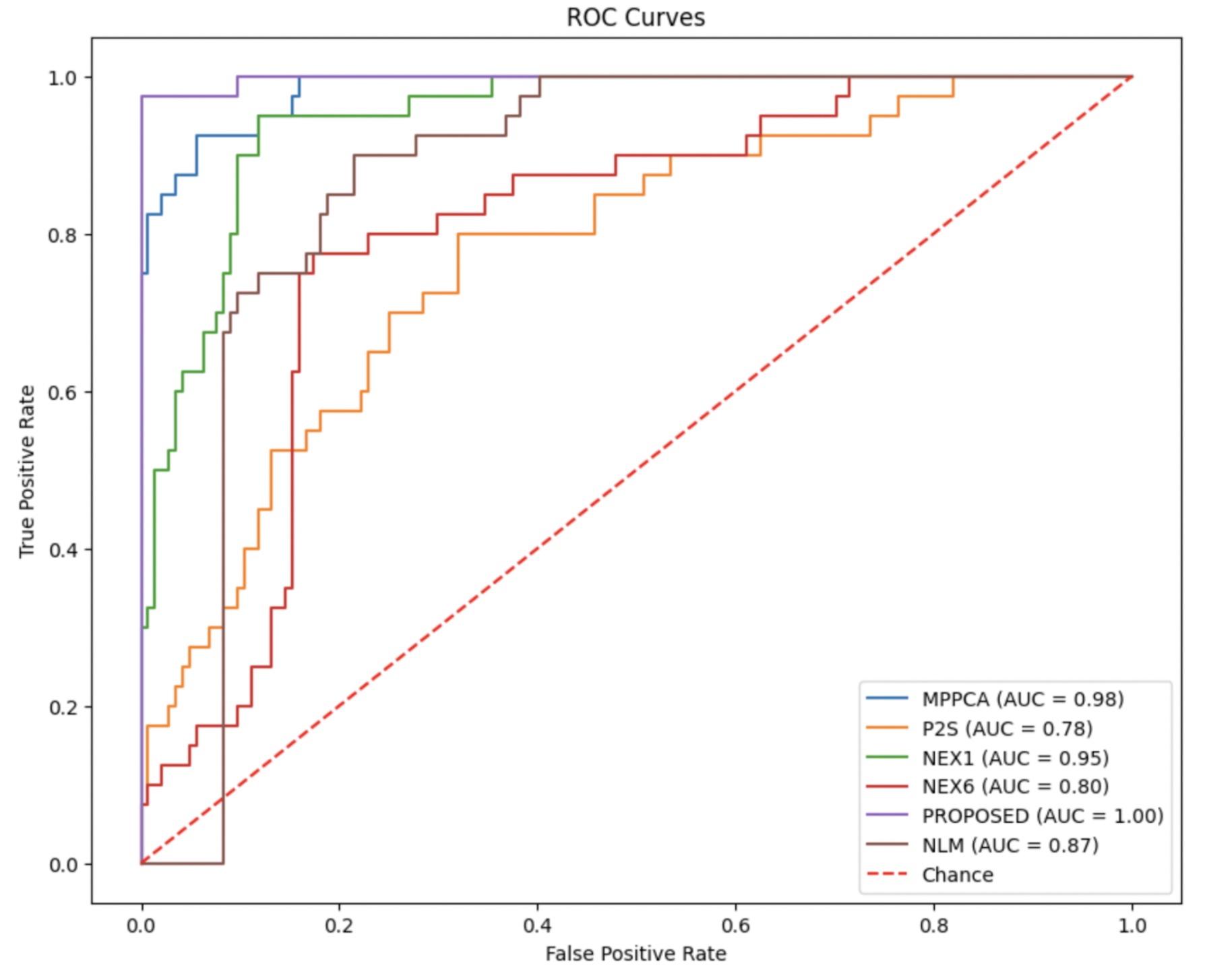
Results: ssDDPM demonstrated superior lesion conspicuity in low b-value images and quantitative ADC maps in comparison to competing methods. In lesion versus normal tissue, a logistic classifier had improved sensitivity from 0.93 to 0.98, and specificity from 0.88 to 0.97, over non-denoised NEX1 images.
Impact: ssDDPM enhances abdominal DW-MRI ADC accuracy from single acquisitions, reducing scan times and patient discomfort. This gives promise to an earlier, precise tumor detection and monitoring, impacting clinical care and healthcare efficiency.
Introduction
Diffusion-weighted MRI (DWI-MRI) provides valuable information for early tumor detection, characterization, and monitoring [1]. Apparent Diffusion Coefficient (ADC) values, which often change prior to tumor size, are obtained by fitting DWI data to an exponential decay model across different diffusion strengths or b-values. DW-MRI suffers from low SNR at high b-values, which reduces its reliability. To improve SNR, multiple acquisitions at each b-value are averaged, assuming isotropic diffusion, which prolongs scan time, adds costs, and increases patient discomfort. Moreover, in abdominal imaging, respiratory motion can blur anatomical detail or disrupt ADC estimation altogether due to misaligned voxels. This study aims to enhance quality of ADC estimates in low SNR abdominal DW-MRI, using single-image acquisition per b-value to avoid the complications of motion.Method
We propose a novel self-supervised training approach which uses a denoising diffusion probabilistic model (DDPM) [7] with a self-supervised ADC parameter estimation loss [6] in the training loop, which is designed specifically for multi-b-value inputs from DW-MRI images.In training, ADC and B0 parameters are estimated on the denoised images. An additional loss then guides the denoising network (DDPM) while minimising the ADC fitting error at the same time. The network's inputs are multi-b-value DW-MRI images. As the input images are denoised during training, they are propagated to a GPU based ADC estimation model in Pytorch to estimate S0 and ADC maps, which are in turn used in the forward equation to estimate back the original input signal. This estimated input signal is compared to actual inputs using MSE loss. Importantly, this training approach doesn't require ground truth ADC maps. One distinguishing feature of our ssDDPM approach compared to the baseline DDPM method is the initial state. In our method, this initial state is a noisy single repetition DW-MR image with low SNR. We assume that this image is akin to a high-SNR image that has had noise added to it over an unknown number of steps, denoted as 'T'. To ascertain this time step, we estimate the source SNR by measuring the SNR from existing low-SNR images [5]. During inference, we process low-SNR test data through the denoising network for 'T' steps. This results in the generation of a denoised, high-SNR image from a single repetition low-SNR image.
Data
We retrospectively collected DW-MRI data on clinically acquired DW-MRI from 105 subjects. Acquisition was based on b-values=[50,400,800] s/mm2 with 6 diffusion gradient directions (repetitions) in each. The sequence was a free-breathing single shot EPI with: TR/TE=5200/78ms; FOV= 380x310mm; in-plane resolution=1.5x1.5mm2; slice thickness = 4mm; BW = 2442 Hz/px. Additionally, each scan had a T1 post contrast VIBE and T2 weighted image available. We selected 5 subjects with hepatocellular carcinoma with small lesion sizes as our test data, on which the lesions and normal tissue were marked by a trained radiologist.Results
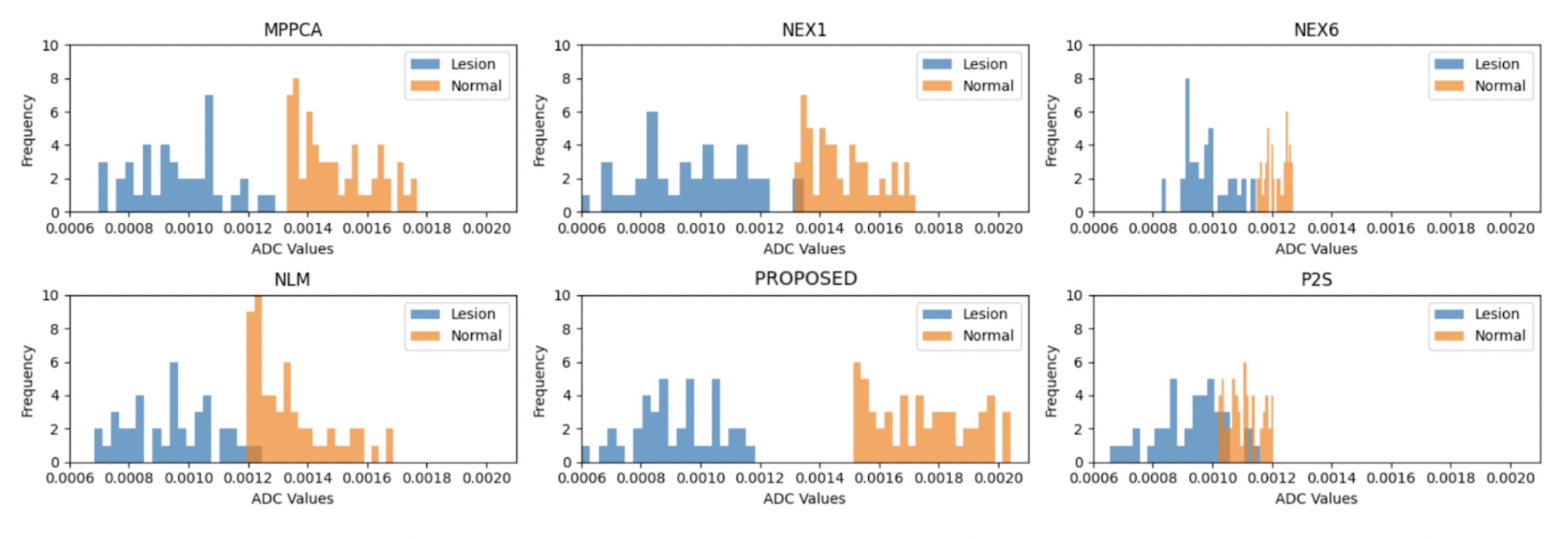
Our ssDDPM was benchmarked against well-known DW-MRI denoising methods - NLM, MPPCA and P2S [2,3,4]. While MPPCA and P2S necessitate multiple b-value repetitions, ssDDPM, similar to NLM, is effective with single-excitation (NEX1) data. ssDDPM achieved enhanced lesion conspicuity and superior ADC map delineation compared to NLM and MPPCA, and was on par with P2S, but without requiring six-fold averaged input data (NEX6). In distinguishing lesions from normal tissue, ssDDPM showed improved performance. Sensitivity and specificity in lesion classification improved to 0.98 and 0.97, respectively, up from 0.93 and 0.88 with single NEX1 acquisition.Discussion
We developed a self-supervised denoising diffusion probabilistic model (ssDDPM) for low SNR DW-MRI, needing fewer acquisitions. ssDDPM outperformed traditional denoising techniques, improving lesion detection and ADC map quality, while needing only a single NEX1 image for denoising.Acknowledgements
This work was supported partially by the National Institute of Diabetic and Digestive and Kidney Diseases (NIDDK), National Institute of Biomedical Imaging and Bioengineering (NIBIB), National Institute of Neurological Disorders and Stroke (NINDS) and National Library of Medicine (NLM) of the National Institutes of Health under award numbers R01DK125561, R21DK123569, R21EB029627, R01NS121657, R01LM013608, S10OD0250111 and by the grant number 2019056 from the United States-Israel Binational Science Foundation (BSF), and a pilot grant from National Multiple Sclerosis Society under Award Number PP-1905-34002.References
[1] Caro-Domínguez, Pablo, Abha A. Gupta, and Govind B. Chavhan. "Can diffusion-weighted imaging distinguish between benign and malignant pediatric liver tumors?." Pediatric Radiology 48 (2018): 85-93.
[2] Veraart, J.; Fieremans, E. & Novikov, D.S. Diffusion MRI noise mapping using random matrix theory Magn. Res. Med., 2016, early view, doi: 10.1002/mrm.26059
[3] Fadnavis, Shreyas, Joshua Batson, and Eleftherios Garyfallidis. "Patch2Self: Denoising Diffusion MRI with Self-Supervised Learning." Advances in Neural Information Processing Systems 33 (2020): 16293-16303.
[4] Buades, Antoni, Bartomeu Coll, and Jean-Michel Morel. "Non-local means denoising." Image Processing On Line 1 (2011): 208-212.
[5] Vasylechko, Serge, Onur Afacan, and Sila Kurugol. "Self Supervised Denoising Diffusion Probabilistic Models for Abdominal DW-MRI." In Computational Diffusion MRI, 2023. Lecture Notes in Computer Science. Cham: Springer.
[6] Vasylechko, Serge, et al. "Self‐supervised IVIM DWI parameter estimation with a physics based forward model." Magnetic resonance in medicine 87.2 (2022): 904-914.
[7] Ho, Jonathan, Ajay Jain, and Pieter Abbeel. "Denoising diffusion probabilistic models." Advances in neural information processing systems 33 (2020): 6840-6851.
Figures