1753
A Denoising Diffusion Probability Model for T1W contrast-enhanced Synthesis based on multi-parametric MRI1Department of Radiology, Minhang Branch, Fudan University Shanghai Cancer Center, Shanghai, China, 2Institute of Research and Clinical Innovations, Neusoft Medical Systems Co., Ltd, Shanghai, China, 3Department of Radiology, Children's Hospital of Fudan University, National Children's Medical Center, Shanghai, China, 4Department of Radiology, Fudan University Shanghai Cancer Center, Shanghai, China, 5Department of Oncology, Shanghai Medical College, Fudan University, Shanghai, China, 6Institute of Research and Clinical Innovations, Neusoft Medical Systems Co., Ltd, Beijing, China
Synopsis
Keywords: AI Diffusion Models, Machine Learning/Artificial Intelligence, T1CE, Synthesis
Motivation: T1W Contrast-enhanced (T1CE) images are obtained with gadolinium administration, but there might be adverse effects related to gadolinium retention.
Goal(s): To generated T1CE images from multi-parametric MRI (mp-MRI) without contrast agents.
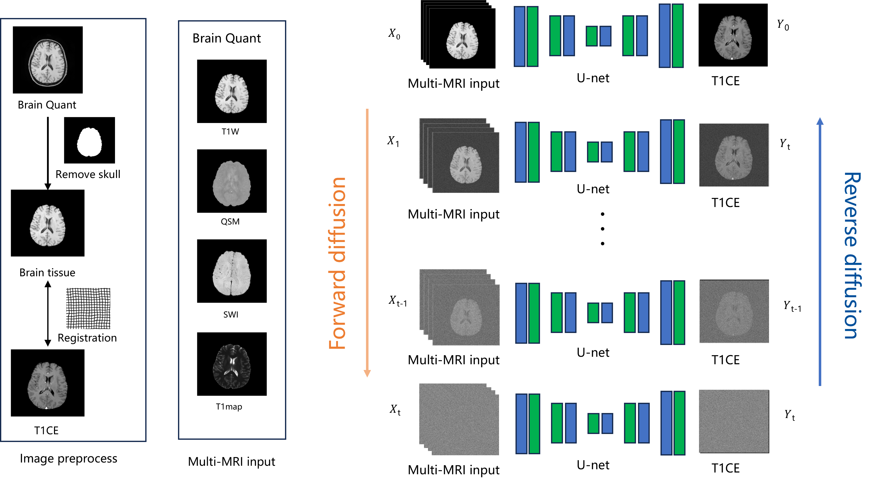
Approach: T1W, T1map, SWI and QSM were obtained from patients with brain metastasis. A novel approach for generating non-contrast enhanced images from mp-MRI was proposed based on the diffusion model, which was trained in 91 cases, evaluated and test 28 and 27 cases respectively.
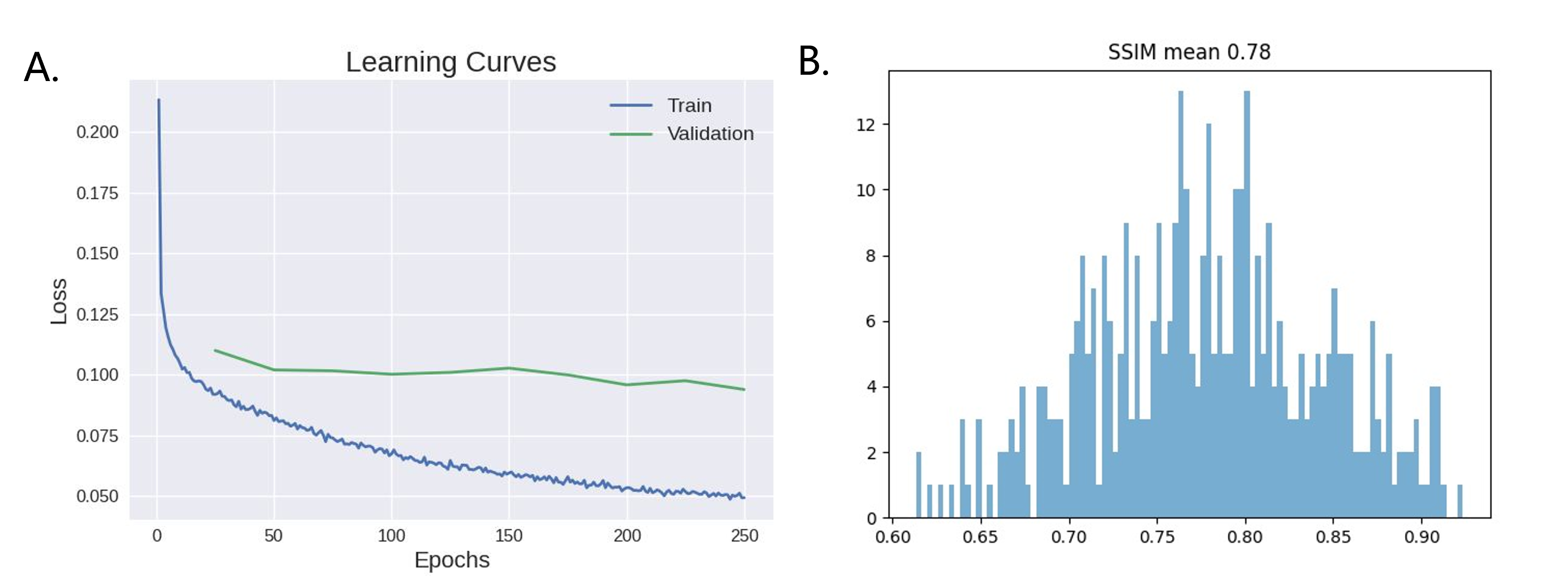
Results: The proposed model achieves the highest SSIM of 0.78, and the synthetic images are capable of revealing the details of brain tissues.
Impact: Multi-parametric MRI based DDPM provides a feasible approach for generating contrast enhanced images from non-contrast multi-parametric MRI, therefore circumventing the issue of adverse effects of gadolinium retention, which will benefit patients who have to undergo contrast enhanced MRI scans.
Introduction
Approximately 20% of cancer patients experience brain metastases (BM) that develop in a unique microenvironment and are associated with poor prognosis in advanced stages1,2. As the gold standard for clinical detection of brain metastases, MRI offers better sensitivity than enhanced CT3, 4. Gadolinium-based contrast agents (GBCA) are widely used for enhancing brain MRI images to highlight brain tumors. However, the contrast agents leading to residual gadolinium deposition not only in brain regions but also in extracranial tissues such as the liver, skin, and bone5. Therefore, non-contrast-enhanced BM detection holds significant clinical value. Artificial intelligence has recently found widespread applications in medical imaging, including image reconstruction, segmentation, and diagnostic assistance. Diffusion model represents an emerging generative approach, and many studies have shown the better performance than GAN6,7. We propose to synthesize T1W contrast-enhanced (T1CE) by mp-MRI without GBCA based on the diffusion model, and compared diffusion model with Unet of consistent network architectures.Methods
Data acquisition: A total of 160 patients with brain metastasis (BM) were split to training, validation and test cohort randomly by ratio of 6:2:2. All the subjects were scanned on a NeuMR 1.5T system (Neusoft Medical System, Shenyang, China). The imaging parameters of smart BrainQuant were as follows: FA=8°,35°,TR = 45ms,TE =10, 24,38ms,BW = 260hz/pixel,VoxelSize = 0.67*0.67*2.7mm3. A total of 3 contrast maps (T1W, PDW and Dark Fluid T2*W), 4 quantitative maps (T1 map, PD map, R2* and QSM), 2 vessel maps (SWI and true SWI) and the enhanced T1W image (T1WE) were derived in about 4 min 30 msec. Algorithm: This algorithm consists of a forward Gaussian noise process and a reverse process, where the input data is gradually perturbed over several steps by adding Gaussian noise and then learns to reverse the diffusion process to retrieve the desired noise-free data from noisy data samples. Data analysis: Image pre-processing includes skull removing and registration. The proposed image synthesis framework based on a diffusion model was evaluated by comparing it with a Unet-based network, with the metric of mean squared error (MSE) utilized. In order to compare the effects of different image inputs on the results, different combinations of T1W, T1map, SWI, and QSM from BrainQuant were used as inputs, employing the structural similarity index measure (SSIM) to evaluate image quality.Results
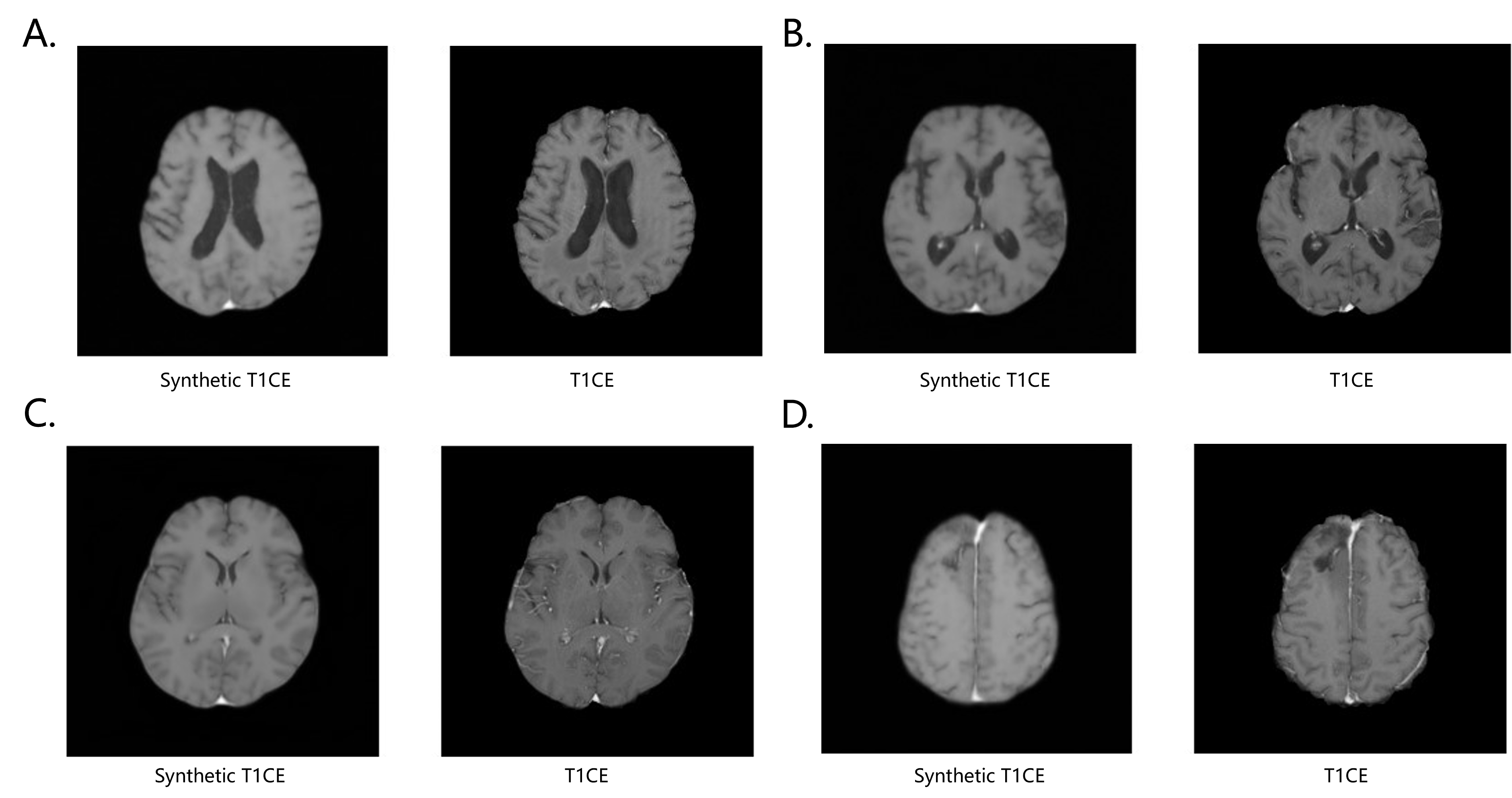
The consistency of the synthetic performance of the DDPM methods was assessed in an independent test cohort. The ablation experiment for Unet showed more down-sampling and no attention mechanism added can improve the model generation performance (Table 1), and multi-MRI DDPM achieved best SSIM value of 0.78 in the test cohort with T1W, T1map, SWI and QSM. Figure 2 shows training process and SSIM distributions in the test cohort. Qualitative results are shown in Figure 3, which provides faithful details.Discussion
In this work, we proposed a Unet-based diffusion model for T1CE synthesis from mp-MRI. The proposed DDPM framework has demonstrated the promising quality of its synthetic images, which can potentially replace contrast enhanced T1W in diagnosis, avoiding the deposition of contrast agents in patients. In terms of network design, more down-samples encoder can learn features in different resolution levels, while the decoder up-samples the features to the original input size to estimate the noise and T1CE. In contrast, the attention mechanism introduces the global receptive field, achieving inferior prediction performance due to the added noise on the image. Overall, the proposed generative method yielded the highest SSIMs with mp-MRI, indicating that the Brain Quant could provide complementary structural information.Conclusion
This work presents a DDPM-based T1CE synthesis framework that utilizes a U-net to learn a diffusion model to generate synthetic images. The proposed DDPM is able to generate T1CE images with a realistic visual appearance and high diversity. Therefore, our study provides a viable approach for obtaining contrast enhanced images from non-contrast MRI, avoiding the adverse effects of gadolinium retention.Acknowledgements
We would like to acknowledge the equal contributions of Lei Chen and Jing Zhang to this work. Both authors contributed equally to the experimental design, data analysis, and manuscript preparation.
References
1. Boire A, Brastianos PK, Garzia L, Valiente M: Brain metastasis. Nature reviews Cancer 2020; 20:4–11.
2. Achrol AS, Rennert RC, Anders C, et al.: Brain metastases. Nature reviews Disease primers 2019; 5:5.
3. G Sze, E Milano, C Johnson, L Heier: Detection of brain metastases: comparison of enhanced CT. American Journal of Neuroradiology 1990; 11:785–791.
4. Fink KR, Fink JR: Imaging of brain metastases. Surgical neurology international 2013; 4(Suppl 4):S209-19.
5. Gulani V, Calamante F, Shellock FG, Kanal E, Reeder SB, International Society for Magnetic Resonance in Medicine: Gadolinium deposition in the brain: summary of evidence and recommendations. Lancet Neurol 2017; 16:564–570.
6. Ho J, Jain A, Abbeel P: Denoising Diffusion Probabilistic Models. 2020.
7. Kazerouni A, Aghdam EK, Heidari M, et al.: Diffusion models in medical imaging: A comprehensive survey. Medical Image Analysis 2023; 88:102846.
Figures