1749
Accelerated Diffusion Tensor Imaging using A Diffusion Generative Deep Learning Model1Electrical and Computer Engineering, University of Arizona, Tucson, AZ, United States, 2Medical Imaging, University of Arizona, Tucson, AZ, United States, 3Applied Mathematics, University of Arizona, Tucson, AZ, United States, 4Biomedical Engineering, University of Arizona, Tucson, AZ, United States
Synopsis
Keywords: AI Diffusion Models, Diffusion Tensor Imaging
Motivation: High-quality diffusion tensor imaging involves fitting a large number of diffusion-encoded images to a tensor model. This challenging process requires long scans and is vulnerable to motion artifacts. There’s a need for accelerated acquisitions while preserving robust diffusion tensor estimates.
Goal(s): To develop a generative diffusion model that produces high-quality tensor metrics using a few diffusion-encoded images.
Approach: The proposed generative model was trained using 300 randomly selected subjects from the Human Connectome Project Dataset and tested on 20 subjects.
Results: Our model demonstrates the ability to generate high-quality tensor metrics for as few as 3 DWIs.
Impact: This study demonstrates that a generative diffusion model can produce high-quality tensor metrics with significant reduction in scan time, potentially eliminating image distortions and artifacts.
Introduction
Diffusion-weighted magnetic resonance imaging (dMRI) is a prominent technique, for qualitatively and quantitatively assessing microstructural characteristics of tissues. dMRI techniques such as Diffusion Tensor Imaging (DTI)1 have been utilized to study a wide range of structural and pathological processes such as neuronal connectivity2, ischemia3, tumor growth and response to therapy4. One of the major challenges of dMRI is the long data acquisition times required to obtain a large number of diffusion-weighted images (DWIs) for accurate estimation of various diffusion metrics. Recently, deep learning (DL) techniques were proposed to accelerate dMRI5,6,7,8,9. In this work, we present a novel generative diffusion DL model which produces robust and high-quality DTI metrics given an accelerated acquisition of DWIs and bypasses the need for diffusion tensor fitting.Methods
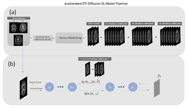
DWI datasets were acquired from the Human Connectome Project (HCP) database10. 300 randomly selected datasets were used for training/validation and 20 for testing. HCP data consists of 270 DWIs (90 each at b = 1000, 2000, 3000 s/mm2) combined with 18 b =0 images. These data were fitted to a tensor model (Fig1a), acquiring fractional anisotropy (FA), axial diffusivity (AD), and radial diffusivity (RD) maps using MRtrix.11 Our proposed method (Fig1b) is a conditional diffusion model12 built to generate DTI metrics conditioned on a small set of noisy DWIs. Let y denote the desired DTI metric and x the set of acquired DWIs combined with one b=0 image. Our overarching goal is to learn the empirical data distribution p(y|x) through a training procedure. The forward diffusion process is modeled as a Markovian process over T iterations. Gaussian noise is iteratively added to an initial data point $$${y_{0}}$$$, such that at iteration t = T, the noisy image $$${y_{T}}$$$ is well approximated by a standard Gaussian distribution. During training, given an initial data point $$${y_{0}}$$$ a noise level $$$\gamma$$$, and a noise sample $$$\epsilon \sim \mathcal{N}(0, I)$$$, a noisy image is created as $$$\widetilde{y}$$$ = $$$\sqrt{\gamma} y_0 + \sqrt{1 - \gamma} \epsilon$$$ and is used to train a model $$$f_{\theta}(x, \widetilde{y}, \gamma)$$$. The network is trained to predict the noise sample $$$\epsilon$$$ given $$$x$$$, $$$\widetilde{y}, \gamma$$$. During inference, samples from a standard Gaussian distribution are converted to samples from p(y|x) using the trained model. Our model was implemented in PyTorch and trained over 500 epochs using a linear noise schedule with 1000 timesteps, and batch size = 8. A UNet was used for denoising, which comprised three convolutional levels with 4 residual blocks each. Images were zero-padded to 192 x 192. Experiments were conducted using different number of conditioning DWIs (k=3,4, and 6 DWIs + 1 b = 0 image). For comparison, DTI metrics were obtained using Marcenko-Pastur Principle Component Analysis (MP PCA)13 and conventional DTI results for k=6 DWIs, in addition to the reference metrics which used all available data (k=270 DWIs + 18 b=0 images).Results and Discussion
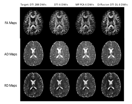
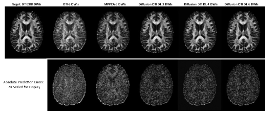
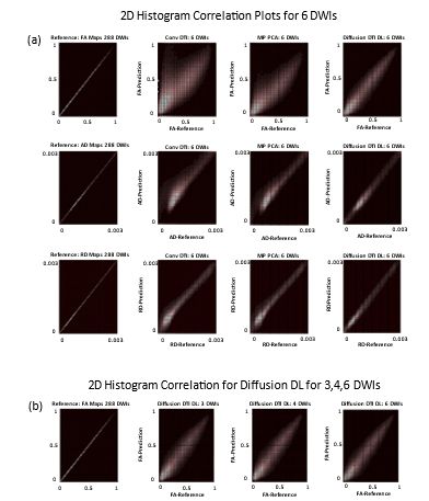
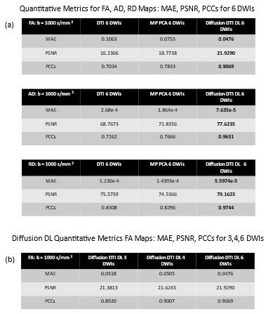
A qualitative assessment of tensor metrics FA, AD, and RD produced by the proposed method for k = 6 DWIs is shown in Fig2. All maps produced by the proposed method closely resemble the maps obtained using the full 288 image reference. Fig3 demonstrates FA maps obtained using the proposed method using k=3, 4, and 6 DWIs. It is shown that these maps can delineate structural features even using as few as 3 DWIs, which is beyond the theoretical limit of DTI modeling. Fig4 shows correlation histograms of the predicted FA, AD, and RD metrics of each method relative to the reference values over the test cohort. While the MP PCA and DTI methods show significant bias and prediction variance, the proposed DL framework predictions remain tightly distributed around the reference values. Table1 evaluates quantitative performance of our diffusion framework in calculating MAE, PSNR, and PCCs over all computational pipelines. The proposed diffusion method demonstrates superior performance over all metrics. Overall, the results indicate that our proposed model accurately predicts superior tensor metrics, even when dealing with significantly accelerated data acquisition. Additionally, it's worth noting that the proposed approach exhibits adaptability and has the potential for future expansion to encompass various MRI diffusion models. Such an extension would involve addressing the challenge of resolving crossing fibers, thereby enhancing its utility in delineating microstructural features within white matter.Conclusion
We produced an innovative generative diffusion method that possesses the capability to predict superior tensor metrics when presented with highly accelerated DWI acquisitions. Importantly, this technique bypasses the diffusion tensor fitting process. The framework opens up the exciting potential to obtain high-quality DTI metrics while drastically reducing the overall scan duration.Acknowledgements
We would like to acknowledge grant support from the Arizona Biomedical Research Commission (CTR056039), Arizona Alzheimer’s Consortium, and the Technology and Research Initiative Fund Technology and Research Initiative Fund (TRIF).References
1. Basser PJ, Mattiello J, LeBihan D. Estimation of the effective self-diffusion tensor from the NMR spin echo. J Magn Reson B 1994;103:247–254.
2. Xue, R., van Zijl, P.C., Crain, B.J., Solaiyappan, M. and Mori, S. (1999), In vivo three‐dimensional reconstruction of rat brain axonal projections by diffusion tensor imaging. Magn. Reson. Med., 42: 1123-1127. https://doi.org/10.1002/(SICI)1522-2594(199912)42:6HYPERLINK "https://doi.org/10.1002/(SICI)1522-2594(199912)42:6%3c1123::AID-MRM17%3e3.0.CO;2-H"<HYPERLINK "https://doi.org/10.1002/(SICI)1522-2594(199912)42:6%3c1123::AID-MRM17%3e3.0.CO;2-H"1123::AID-MRM17HYPERLINK "https://doi.org/10.1002/(SICI)1522-2594(199912)42:6%3c1123::AID-MRM17%3e3.0.CO;2-H">HYPERLINK "https://doi.org/10.1002/(SICI)1522-2594(199912)42:6%3c1123::AID-MRM17%3e3.0.CO;2-H"3.0.CO;2-H
3. Bockhorst KH, Narayana PA, Liu R, Ahobila-Vijjula P, Ramu J, Kamel M, Wosik J, Bockhorst T, Hahn K, Hasan KM, Perez-Polo JR. Early postnatal development of rat brain: in vivo diffusion tensor imaging. J Neurosci Res. 2008 May 15;86(7):1520-8. doi: 10.1002/jnr.21607. PMID: 18189320.
4. Lope-Piedrafita S, Garcia-Martin ML, Galons JP, Gillies RJ, Trouard TP. Longitudinal diffusion tensor imaging in a rat brain glioma model. NMR Biomed. 2008 Oct;21(8):799-808. doi: 10.1002/nbm.1256. PMID: 18470959; PMCID: PMC2857329.
5. Golkov, V., et al., 2016. q-Space deep learning: twelve-fold shorter and model-free diffusion MRI scans. IEEE Trans. Med. Imag. 35, 1344–1351.
6. Aliotta, E., Nourzadeh, H., Sanders, J., Muller, D., Ennis, D.B., 2019. Highly accelerated, model-free diffusion tensor MRI reconstruction using neural networks. Med. Phys. 46, 1581–1591.
7. Tian Q, Bilgic B, Fan Q, Liao C, Ngamsombat C, Hu Y, Witzel T, Setsompop K, Polimeni JR, Huang SY. DeepDTI: High-fidelity six-direction diffusion tensor imaging using deep learning. NeuroImage, Volume 219: 117017, 2020. https://doi.org/10.1016/j.neuroimage.2020.117017.
8. Bilgin A, Do L, Martin P, et. al., Accelerating Diffusion Tensor Imaging of the Rat Brain using Deep Learning. Proceedings of the 2021 Meeting of the ISMRM, Abstract 2444.
9. Martin, P, Altbach, M, Bilgin, A, Noise2DWI: Accelerated Diffusion Tensor Imaging with Self-Supervision and Fine Tuning. Proceedings of the 2022 Meeting of the ISMRM, Abstract 3517.
10. Van Essen, DC, et. Al. The WU-Minn Human Connectome Project: An overview. NeuroImage 80(2013):62-79.
11. Tournier JD, Smith RE, Raffelt D, Tabbara R, Dhollander T, Pietsch M, Christiaens D, Jeurissen B, Yeh C-H, and Connelly A. MRtrix3: A fast, flexible and open software framework for medical image processing and visualisation. NeuroImage, 202 (2019), pp. 116–37. 12. Saharia C, Chan W, Chang H, Lee CA, Ho J, Slimans, T, Fleet DJ, Norouzi M, Palette: Image-to-Image Diffusion Models. SIGGRAPH ’22: ACM SIGGRAPH 2022 Conference Proceedings, July 2022, Aritcle No.: 15, Pages 1-10, https://doi.org/10.1145/3528233.3530757.
13. Veraart J, Nvokov DS, Christiaens D, Ades-aron B, Sijbers J, Fieremans E, Denoising of diffusion MRI using random matrix theory. NeuroImage 142 (2016) 394-406.
Figures