1748
Generating missing M0 in ADNI ASL dataset with latent diffusion models1Laboratory of Functional MRI Technology (LOFT), Stevens Neuroimaging and Informatics Institute, University of Southern California, Los Angeles, CA, United States, 2Department of Biomedical Engineering, University of Arizona, Phoenix, AZ, United States, 3Laboratory of Neuro Imaging (LONI), Stevens Neuroimaging and Informatics Institute, University of Southern California, Los Angeles, CA, United States
Synopsis
Keywords: AI Diffusion Models, Machine Learning/Artificial Intelligence
Motivation: Alzheimer’s Disease Neuroimaging Initiative (ADNI) ASL dataset acquired on Siemens scanners missed M0 images, which prevents CBF quantification for further analysis.
Goal(s): Our goal is to generate the missing M0 for the ADNI ASL dataset using latent diffusion model (LDM).
Approach: A separate training dataset was acquired with the ADNI ASL protocol but with manually disabled background suppression to be used as the M0. A conditional LDM was trained to use acquired control images as the condition to generate M0 images.
Results: The generated M0 with the conditional LDM shows a high fidelity compared to the experimentally acquired M0.
Impact: With generated M0 images, more than 500 ADNI ASL datasets can be further analyzed for CBF to investigate AD progression.
Introduction
Alzheimer's Disease (AD) is a progressive and devastating neurological condition that affects millions of individuals. Cerebral blood flow (CBF) may provide a biomarker for early detection of AD1. Arterial spin labeling (ASL) scan can measure CBF without contrast agents or exposure to radiation. ASL has been part of the imaging protocol in the Alzheimer’s Disease Neuroimaging Initiative (ADNI) (https://adni.loni.usc.edu/). Although ADNI has collected over 1000 ASL scans, more than 500 scans acquired on Siemens scanners missed M0 images, which is necessary for CBF quantification, making it impossible for investigating CBF changes during AD progression. The diffusion model2 is a type of generative model that can generate images from random noise. Recently latent diffusion model (LDM)3 has been proposed to enable a flexible conditional generation while saving computational costs by applying diffusion models in the latent space instead of operating directly in the image space. In this study, we developed LDMs to generate M0 images, which may potentially enable CBF analysis using ADNI dataset.Methods
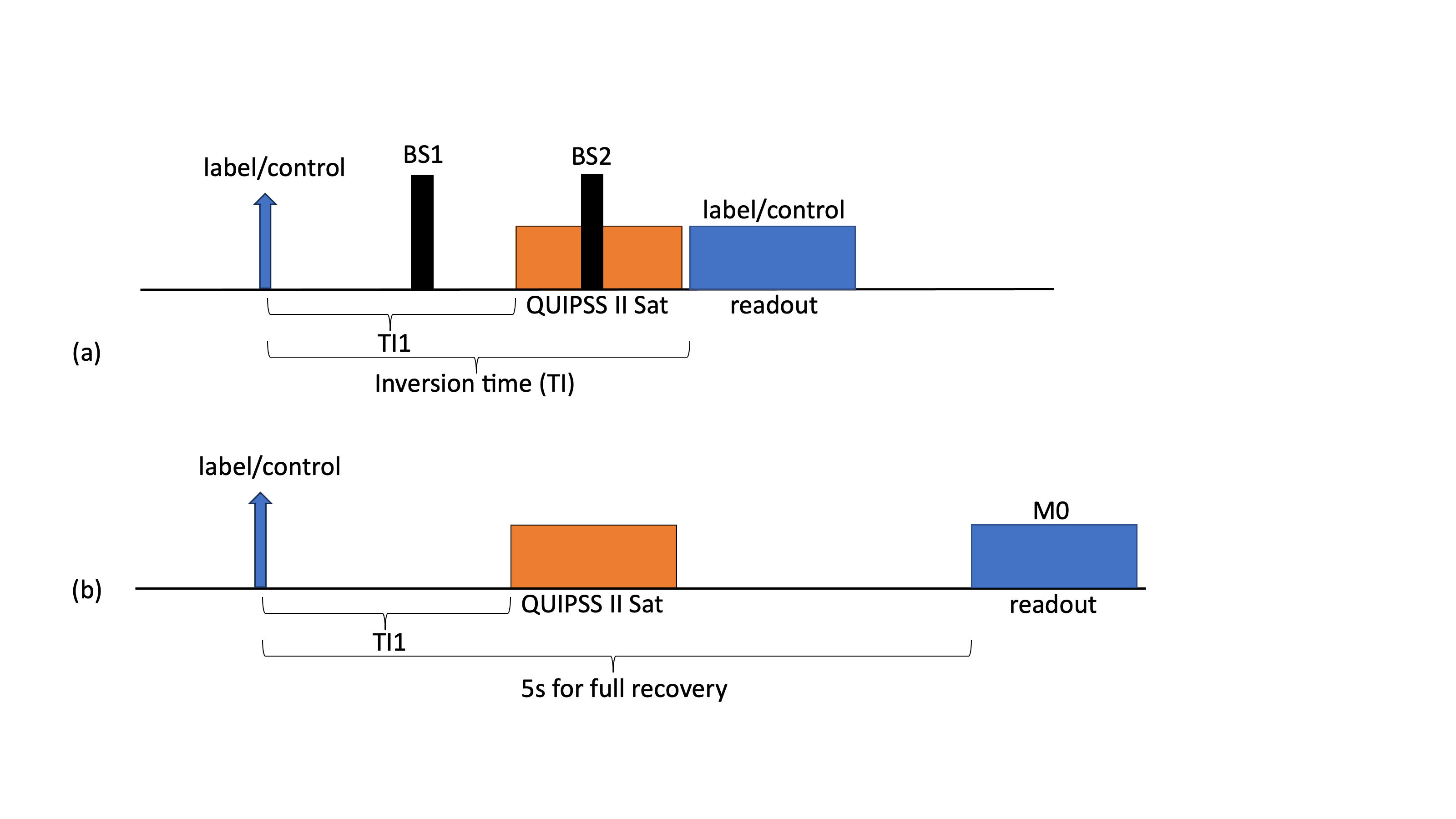
Data acquisition of the control and M0 imagesFigure 1 shows the sequence diagram of the ADNI pulsed ASL (PASL) and M0 acquisition in our experiment. Control/label images of PASL were acquired using the ADNI ASL protocol. For M0, background suppression pulses were manually disabled, and TI was extended to 5 seconds for full magnetization recovery. ASL and M0 from 36 subjects (70±11 years, 11 males) were collected using these 2 sequences on Siemens 3T Prisma scanners at two sites.
Conditional latent diffusion model
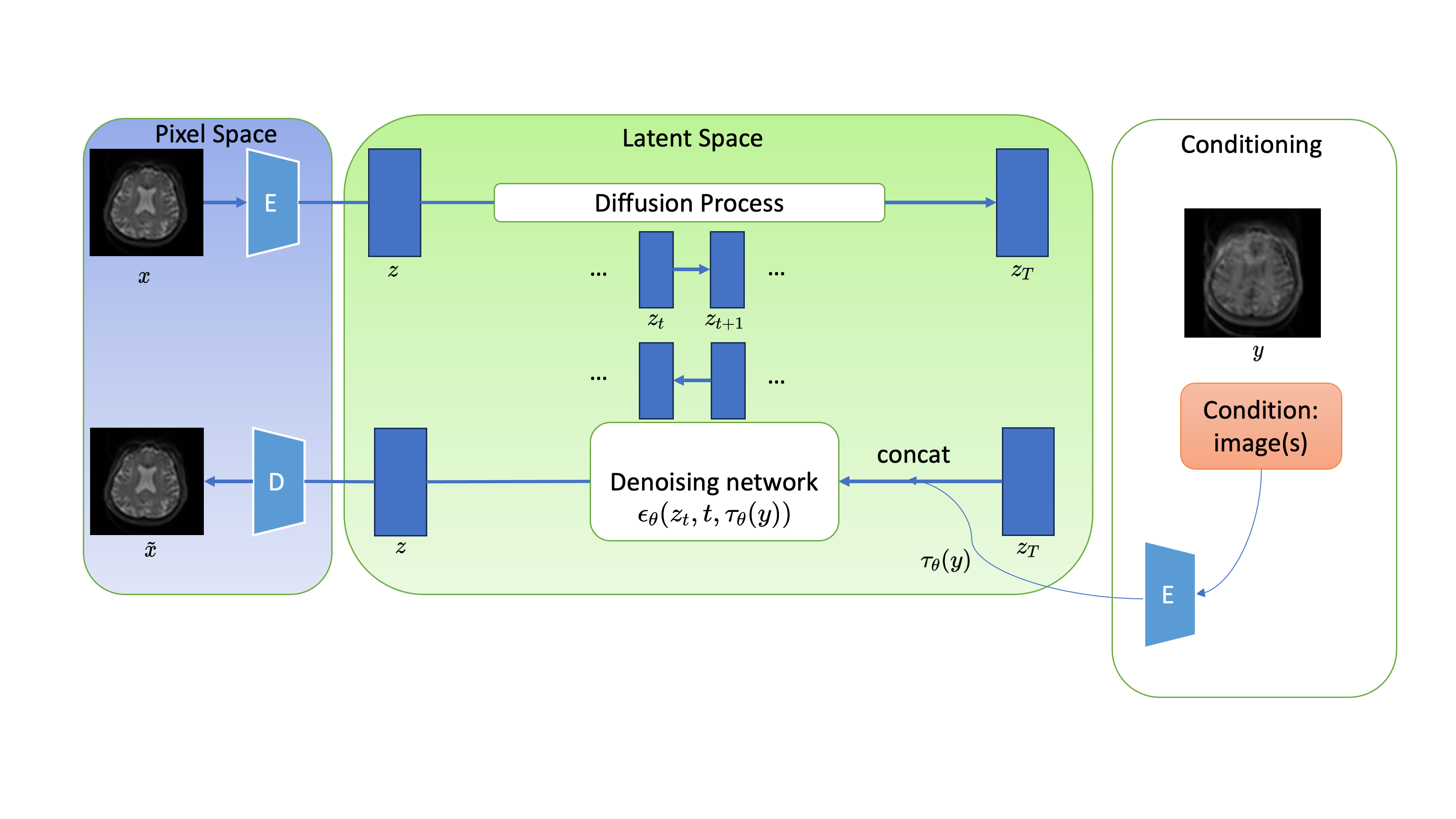
Latent diffusion model uses two steps for image generation. The images were first compressed into the latent space and the diffusion process was done in the latent space. Then the compressed image went through a decoder to generate the final image. The framework of this study is shown in Figure 2. In the forward process, M0 images went through 1000 diffusion steps, and in the reverse process, the corresponding control image was used as a condition to the network in each denoising step to generate the final M0 image.
We trained three models in total. First, a conditional LDM that generates M0 image conditioned on control images (‘M0 gen’); Second, a discriminative Resnet4 model that maps from control images to M0 images (‘direct mapping’); Third, a conditional LDM that generates CBF maps conditioned on both control and perfusion images (‘CBF gen’). The models were trained on 2D slices of 31 scans and tested on 5 scans from both sites. For generated M0 images, CBF maps were calculated with the acquired perfusion images (control - label) along with the generated M0 images.
Performance evaluation
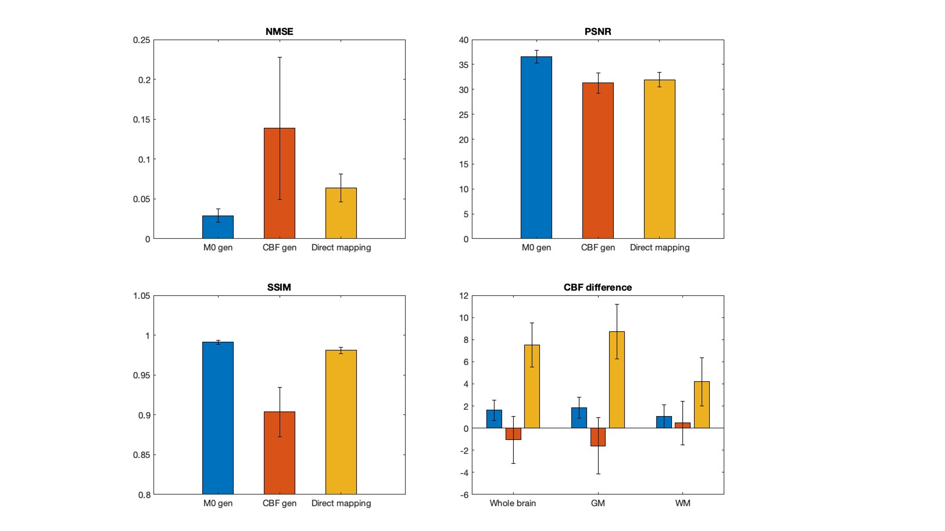
To quantitatively compare the performance, we calculated the similarity (NMSE, PSNR and SSIM) between the generated CBF maps and the ground truth. We also calculated the bias in CBF values in whole brain, GM and WM masks.
Results
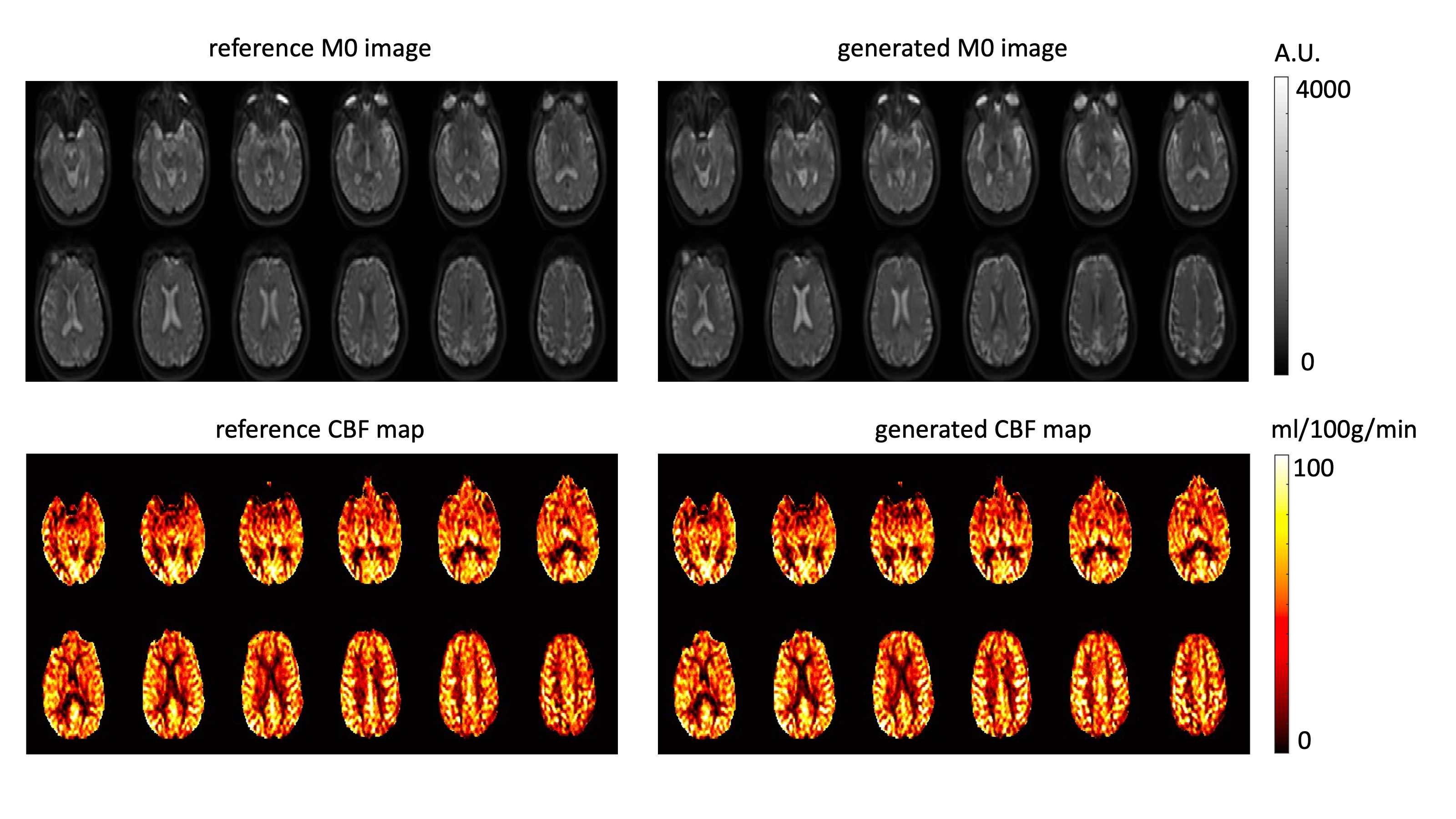
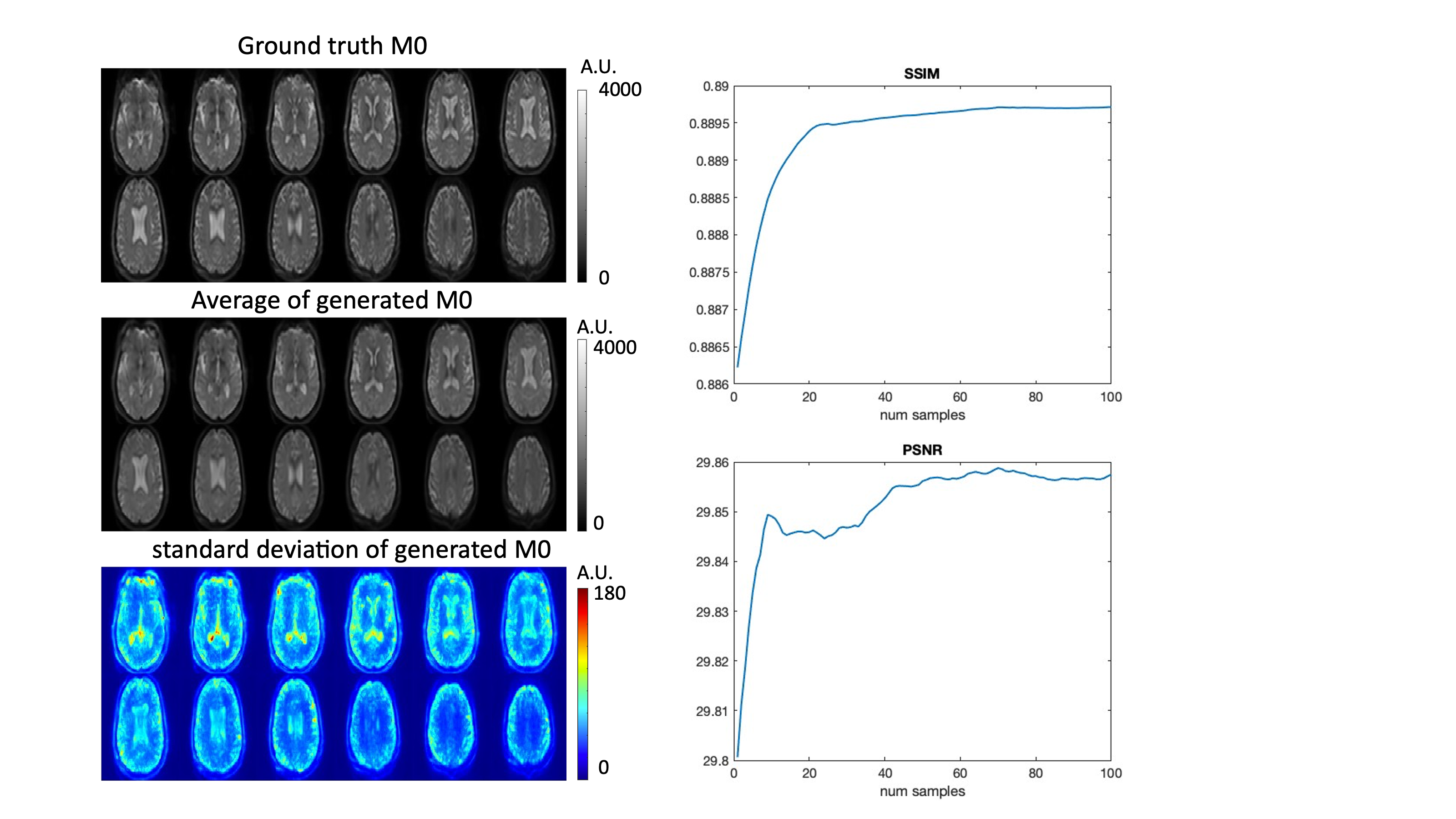
Figure 3 displays the generated M0 and CBF images with M0 gen and ground truth images. It shows that the generated M0 and CBF images by LDM has a high fidelity. Figure 4 shows the quantitative comparison of different models. It shows that the CBF maps generated from the M0 gen method has the highest similarity, with a comparably small bias as the CBF gen model. Figure 5 shows the uncertainty estimation of M0 reproduced by M0 gen. Since the generation varies for each sampling process, we generated 100 samples and calculated mean and standard deviation of these samples. The left panel shows that most variance occurs in CSF and is relatively stable elsewhere. The right panel shows that with more samples, the average generation becomes closer to the ground truth, though 20 samples already produce reasonable outcome.Discussion
In this study, we developed conditional latent diffusion models to generate M0 and subsequently quantitative CBF maps from the acquired control and/or perfusion images. Compared to the M0 gen method, CBF gen has a larger variance, likely because perfusion images have higher variability, making the model less stable. Compared to direct mapping, generative model produces a better result, probably because control images have low intensity and may include residual fat signals that may reduce the performance more in discriminative models than generative models. Future study and analysis are warranted.Conclusion
In this study, we developed conditional LDMs to generate M0 and subsequently CBF from control and/or perfusion images. This can be applied to compensate for missing M0 images in many ADNI ASL datasets, enabling quantitative CBF analyses during the progression of AD.Acknowledgements
This work was supported by US NIH grants R01-EB032169 and R01-EB028297.References
[1] H. Zhang et al., “Cerebral blood flow in mild cognitive impairment and Alzheimer’s disease: A systematic review and meta-analysis,” Ageing Research Reviews, vol. 71, p. 101450, Nov. 2021, doi: 10.1016/j.arr.2021.101450.
[2] J. Ho, A. Jain, and P. Abbeel, “Denoising Diffusion Probabilistic Models,” arXiv.org. Accessed: May 16, 2023. [Online]. Available: https://arxiv.org/abs/2006.11239v2
[3] R. Rombach, A. Blattmann, D. Lorenz, P. Esser, and B. Ommer, “High-Resolution Image Synthesis with Latent Diffusion Models.” arXiv, Apr. 13, 2022. doi: 10.48550/arXiv.2112.10752.
[4] B. Lim, S. Son, H. Kim, S. Nah, and K. M. Lee, “Enhanced Deep Residual Networks for Single Image Super-Resolution.” arXiv, Jul. 10, 2017. Accessed: Mar. 19, 2023. [Online]. Available: http://arxiv.org/abs/1707.02921
Figures