1743
Conditional Denoising Diffusion Probabilistic Model (DDPM) for Cardiac Perfusion Image Reconstruction1Stanford University, Palo Alto, CA, United States
Synopsis
Keywords: AI/ML Image Reconstruction, Machine Learning/Artificial Intelligence, perfusion
Motivation: Cardiac perfusion imposes challenges in reconstruction due to intrinsic low SNR, and large signal intensity change. Recently proposed Conditional Denoising Diffusion Probabilistic Models (DDPM) achieves exceptional performance in a broad range of inverse problems.
Goal(s): To reconstruct undersampled cardiac perfusion datasets with conditional DDPM.
Approach: We conduct the Langevin diffusion process on unacquired k-space data. Conditioning on the acquired data is explicitly embedded in the network structure, instead of utilizing Bayes rule to decouple learned unconditional DDPM prior information of perfusion images and MRI sensing model.
Results: Our experimental results validate the good performance of conditional DDPM reconstruction for R=4 accelerated perfusion imaging.
Impact: Our proposed work can help the challenging perfusion reconstruction for higher acceleration rate and benefit clinical diagnosis.
Introduction
Magnetic resonance imaging (MRI) provides high image quality yet suffers from long acquisition times. Therefore, image acquisition is accelerated by undersampling the raw data. To solve the inverse problem, many techniques have been proposed, such as parallel imaging [1], compressed sensing [2], and learning based methods [3,4]. Recently a new generative method named Denoising Diffusion Probabilistic Model (DDPM) [5] shows promising sampling quality compared to other generative methods such as GAN [6]. Invoking Bayes rule, DDPM has been combined with MRI reconstruction and shows comparable or better performance than other learning-based methods. First-pass cardiac perfusion is a challenging task due to its low SNR and possible respiratory motion. Most of the DDPM based reconstruction techniques essentially learn the distribution or the SCORE function [7] in the image domain. In this abstract we perform cardiac perfusion reconstruction based on conditional DDPM in the k-space domain [8] that estimating unacquired k-space data conditioned on the acquired k-space. In other words, the inferences of the network are directly sampled from the distribution of the unacquired k-space. Due to no alternation of acquired k-space, the data consistency is always satisfied.Methods
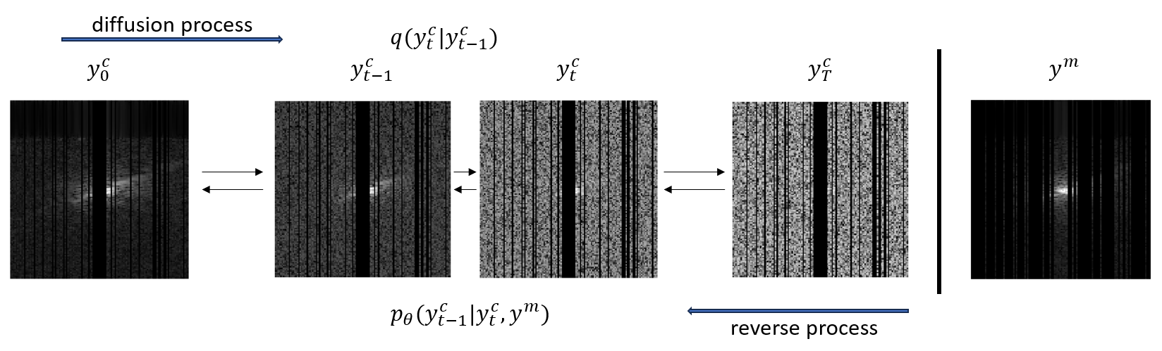
Conditional DDPM: To recover unacquired k-space, we need to estimate $$$q(\mathbf{y}^c|\mathbf{y}^m)$$$, where $$$\mathbf{y}^c$$$ is unacquired k-space, $$$\mathbf{y}^m$$$ is measured k-space. $$$\mathbf{y}^c$$$ and $$$\mathbf{y}^m$$$ sum up to be the full k-space. In conditional DDPM, $$$\mathbf{y}^c$$$ is concatenated with $$$\mathbf{y}^m$$$. As shown in Figure 1, during the diffusion process, Gaussian noise is gradually added to $$$\mathbf{y}^c$$$ over $$$T$$$ iterations with Markovian process denoted as $$$q(\mathbf{y}^c_t|\mathbf{y}^c_{t-1})$$$. We trained the network [9] with the loss function $$$\mathcal{L} = \mathbb{E}_{\mathbf{y}^c,t,\epsilon}\|\epsilon-\epsilon_\theta(\mathbf{y}^c_t,t,\mathbf{y}^m)\|$$$. In the reverse process, we generate samples from noise by iteratively calculating $$$p_{\theta}(\mathbf{y}^c_{t-1}|\mathbf{y}^c_t,\mathbf{y}^m)$$$ till $$$t=1$$$ to get target $$$\mathbf{y}^c_0$$$. Then get the full k-space with $$$\mathbf{y}^m$$$ and $$$\mathbf{y}^c_0$$$.Data: We used 21, R=2 undersampled clinically ordered patients perfusion datasets performed on a 3T Siemens Skyra scanner for training. Each dataset has three to five slices consecutively acquired within each R-R interval. We treated per frame HICU [10] reconstructions as ground truth (GT), which are then retrospectively down-sampled with an R=4 random Gaussian-density mask. Reconstruction performance was tested on other five rest perfusion datasets and one stress perfusion dataset. Due to the limited data size, we trained and inferenced per coil and combined the coil images after the inference.
Implementation: The network is trained using AdamW optimizer with learning rate of 0.0001 and exponential moving average 0.9999. Number of diffusion steps $$$T = 1000$$$. The noise is scheduled linearly from [0.0001, 0.02]. Real and imaginary parts are separated into two channels.
Results
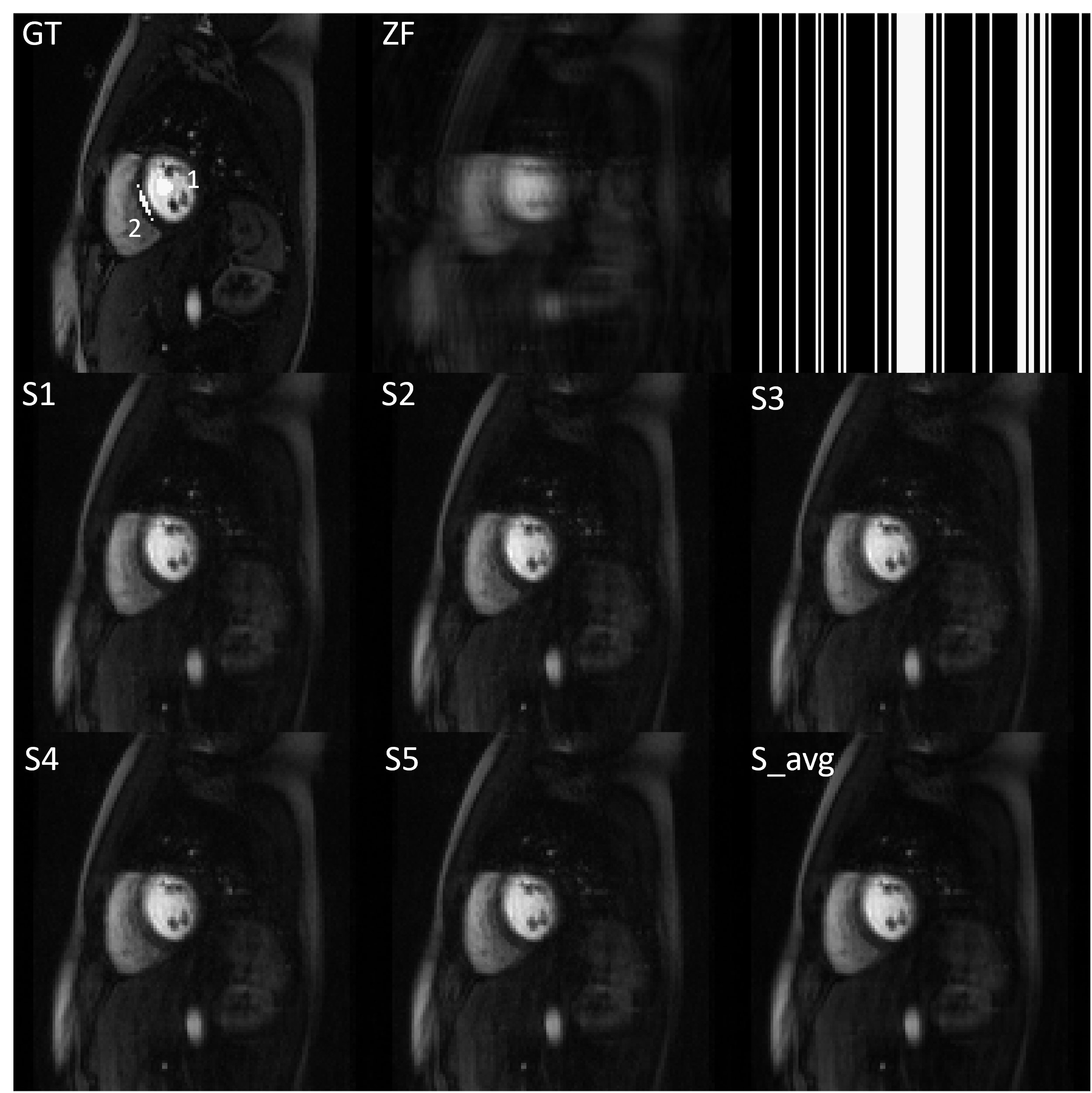
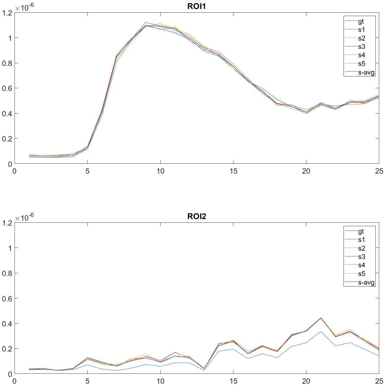
Figure 2 shows the representative image reconstruction from one clinical perfusion dataset. The inference has good consistency with the ground truth images without visual hallucinations.Figure. 3. shows the temporal fidelity of two different regions of interest (ROI) (see on Figure 2 GT image). Each sample and the average sample are of good agreement with the GT at the blood pool and myocardium.
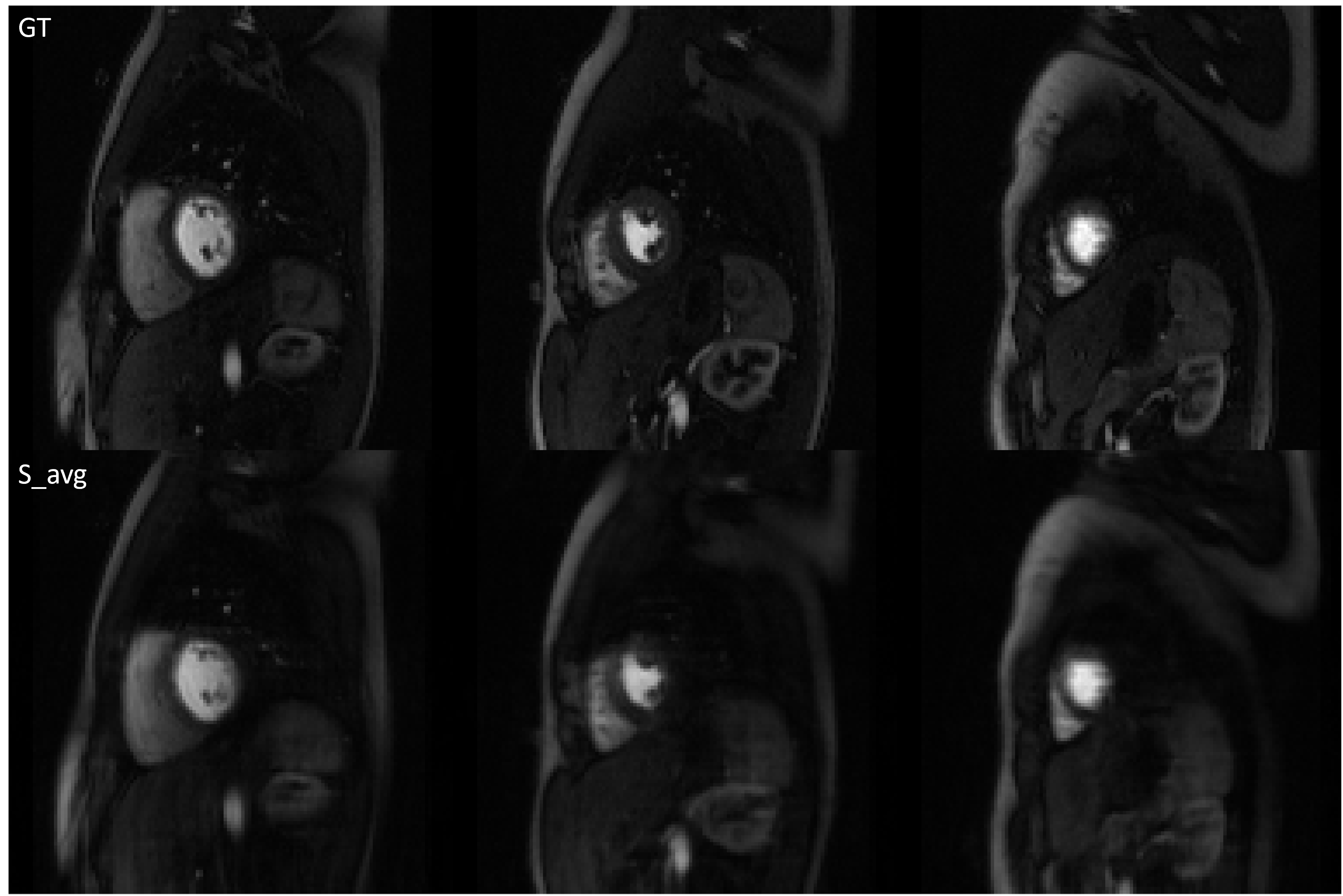
Figure 4 shows good image reconstruction quality of three slices (basal, middle, and apical) from one patient.
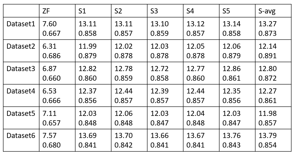
Figure 5 depicts the normalized SNR and SSIM of all six datasets. Averaged inference results have either highest nSNR or SSIM.
Conclusion and Discussion
We demonstrate conditional DDPM can be used to reconstruct rate-4 accelerated cardiac perfusion images with good image quality. While our current result can already achieve promising image quality, the current implementation is a per coil reconstruction and does not explicitly utilize parallel imaging prior information, one future direction of this work is to efficiently incorporate parallel imaging into the model. Further work will include extending the model to self-supervised or unsupervised learning, which would be appealing for cardiac perfusion imaging since there is no feasible way to acquire fully sampled data.Acknowledgements
This work is supported by NIH R01 HL131919, NIH R01 HL155962-01.References
1. Deshmane, Anagha, et al. "Parallel MR imaging." Journal of Magnetic Resonance Imaging 36.1 (2012): 55-72.
2. Lustig, Michael, et al. "Compressed sensing MRI." IEEE signal processing magazine 25.2 (2008): 72-82.
3. Hammernik, Kerstin, et al. "Learning a variational network for reconstruction of accelerated MRI data." Magnetic resonance in medicine 79.6 (2018): 3055-3071.
4. Ahmad, Rizwan, et al. "Plug-and-play methods for magnetic resonance imaging: Using denoisers for image recovery." IEEE signal processing magazine 37.1 (2020): 105-116.
5. Ho, Jonathan, Ajay Jain, and Pieter Abbeel. "Denoising diffusion probabilistic models." Advances in neural information processing systems 33 (2020): 6840-6851.
6. Mardani, Morteza, et al. "Deep generative adversarial neural networks for compressive sensing MRI." IEEE transactions on medical imaging 38.1 (2018): 167-179.
7. Song, Yang, et al. "Maximum likelihood training of score-based diffusion models." Advances in Neural Information Processing Systems 34 (2021): 1415-1428.
8. Xie, Yutong, and Quanzheng Li. "Measurement-conditioned denoising diffusion probabilistic model for under-sampled medical image reconstruction." International Conference on Medical Image Computing and Computer-Assisted Intervention. Cham: Springer Nature Switzerland, 2022.
9. Dhariwal, Prafulla, and Alexander Nichol. "Diffusion models beat gans on image synthesis." Advances in neural information processing systems 34 (2021): 8780-8794.
10. Zhao, Shen, Lee C. Potter, and Rizwan Ahmad. "High‐dimensional fast convolutional framework (HICU) for calibrationless MRI." Magnetic Resonance in Medicine 86.3 (2021): 1212-1225.
Figures