1742
Meta-Learning Enabled Score-Based Generative Model for 1.5T-Like Image Reconstruction from 0.5T MRI1Institute of Biomedical and Health Engineering, Shenzhen Institute of Advanced Technology, Chinese Academy of Sciences, Shenzhen, China, 2University of Chinese Academy of Sciences, Shenzhen, China
Synopsis
Keywords: AI Diffusion Models, Image Reconstruction, Low field
Motivation: The high-field-like image reconstruction, mainstream efforts are primarily focused on high or ultra-high fields, lacking in the reconstruction of high-field-like images from low-field ones.
Goal(s): This paper presented a model for reconstructing high-field-like MR images from low-field images with unpaired data.
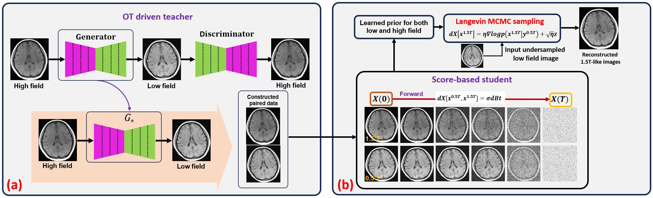
Approach: we execute a pairing using OT-driven CycleGAN, described as "teacher learning". Subsequently, we use a diffusion model to learn the joint distribution between high-field and low-field images, guiding the reconstruction from low-field to high-field.
Results: The generation experiments of T1W and T2W surpass competing experiments, and the 3-fold acceleration experiment also demonstrates the superiority of the proposed method.
Impact: The proposed method represents the first attempt in the reconstruction (acceleration and generation) of images from low-field to high-field. Its potential benefits for advancing overall healthcare standards could be significant.
Introdution
Despite high-field MRI offering exceptional SNR and contrast[1][2][3], its expensive cost and complex installation restrict its usage. Low-field MRI systems effectively alleviate these issues[4]. However, low-field MRI exhibits poorer SNR and contrast[5]. Strategies employing adversarial training have emerged in high-field-like image reconstruction[6][7][9][8]. However, the reconstruction of high-field-like images from low-field has not been explored.The work aims to overcome this limitation by reconstructing high-field-like (1.5T-like) MR images from low-field (0.5T) MRI. We have developed a meta-learning approach utilizing a teacher-student learning mechanism without paired data. Firstly, an optimal transport (OT) driven teacher is trained to approximate the degradation process from high-field to low-field MR images, thus generating pseudo-paired data. Subsequently, a score-based student is trained to solve the reconstruction inverse problem within the framework of iterative regularization. Experiments on low-field data demonstrate that the proposed method outperforms the state-of-the-art unpaired learning method in terms of reconstructed contrast, SNR and visualization of detailed structures.
Method
The degradation of 1.5T MR images to 0.5T MR images can be described by a forward model expressed as:$$x^{0.5} = f(x^{1.5}) (1)$$
Here, $$$x^{0.5}$$$ and $$$x^{1.5}$$$ are the low-field and high-field MR images, respectively, and $$$f$$$ is a degradation that maps high-field to low-field images. To reconstruct a 1.5T MR image, one needs to solve the inverse problem (1). One can be formulated as
$$T^{\beta \rightarrow \alpha} (\beta) = \alpha (2)$$
where $$$x^{1.5}\sim \alpha$$$ and $$$x^{0.5} \sim \beta$$$, and $$$\alpha$$$ and $$$\beta$$$ denotes data distribution. By doing so, the operator $$$T$$$ is able to successfully achieve $$$T^{\beta \rightarrow \alpha}(x^{0.5}) = x^{1.5}$$$. Inspired by GAN-based method, we developed a method driver by the OT-driven CyeleGAN which found a mapping from the 1.5T MR image distribution to the 0.5T MR image distribution without paired data, as following
$$T^{\alpha \rightarrow \beta} = \mathop{\arg\min}\limits_{T}{\sum_{i=1}^{N}c(T(x^{1.5}),x^{1.5}):T\alpha=\beta}, (3)$$
has a unique solution $$$T^{\alpha \rightarrow \beta} = f$$$. Sequentially, we can perform the generation and pairing process between 1.5T data and 0.5T data. The aforementioned process is referred to as "teacher".
From a probabilistic perspective, solving problem (2) can be viewed as posterior probability sampling, as follows,
$$logp(x^{1.5}, x^{0.5}|y^{0.5}) = logp(y^{0.5}|x^{0.5})+logp(x^{0.5}, x^{1.5}) (4)$$.
The prior distribution $$$logp(x^{0.5}, x^{1.5})$$$ acts as a regularization. We perturb clean 1.5T and 0.5T images with multiple scaling Gaussian noise, and then learn the joint distribution (named as joint score function) using a network. The above procedure is called as student learning. We use $$$X$$$ to denote the set $$$[x^{0.5}, x^{1.5}]$$$. We use (6) to learn the joint distribution information of $$$X$$$.
$$\phi^{\star} = \mathop{\arg\min}\limits_{\phi}\frac{1}{2L}\sum_{i=1}^{L}E_{X_{0}}E_{p(X_{i}|X_{0})}[||\varepsilon_{i}s_{\phi}(X_{i},\varepsilon_{i}) + \frac{z}{\varepsilon_{i}}||^{2}] (5)$$
Where $$$z\sim N(0,I)$$$ denotes the Gaussian distribution. $$$X_{0}$$$ represents the original data distribution. $$$s_{\phi}$$$ is the used network. $$$L$$$ is noise scale. Once we have trained a score function, we can carry out conditionally Langevin MCMC sampling based on equation (5); this is
$$X_{i+1} = X_{i} + \frac{\eta_{i}}{2}(s_{\phi^{\star}}(X^{t}, \varepsilon_{i}) - \frac{A^{H}(Ax_{i}^{0.5} - y^{0.5})}{\gamma^{2} + \varepsilon_{i}}) + \sqrt(\eta_{i})z_{i} (6)$$.
Where, $$$ \varepsilon_{i}$$$ denotes the different scales noise, $$$\gamma$$$ is an constant. $$$A$$$ represents the measurement operator from $$$x^{0.5}$$$ to $$$y^{1.5}$$$. The overall framework of the proposed method is illustrated in Fig. 1.
The IRB (institutional review board) approved in vivo experiments were performed on 0.5T OPENMARK MRI scanner (ANKE High-tech Co., Ltd, Shenzhen, China). We have compared with the mapping learning method CycleGAN to support our assertion. Moreover, we will approximate $$$p(y^{0.5}|x^{1.5})$$$ by using a Gaussian distribution. This approximation, known as Score-MRI, will enable us to demonstrate the superiority of our proposed model. Our proposed method is named as Meta-Score-MRI. We employ the primary contrast-to-noise ratio (CNR) [10] quantitative metric to assess the superiority of the proposed method.
Results
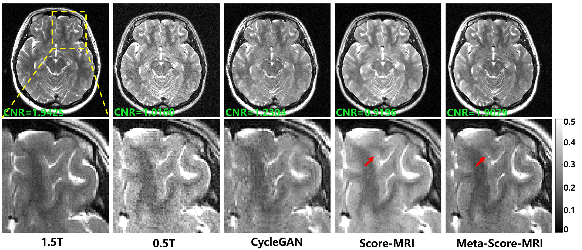
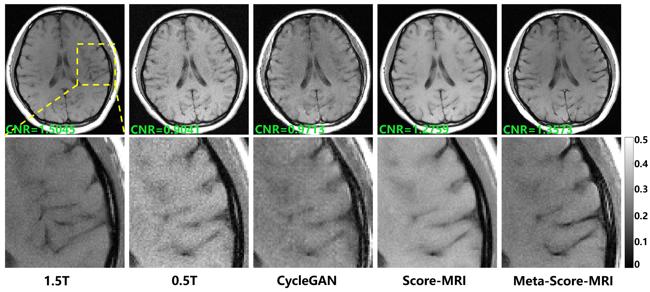
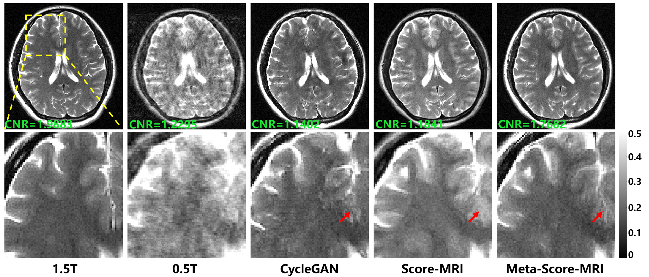
The results in reconstructing 1.5T-like T2W images are shown in Fig. (2). The proposed method, Meta-Score-MRI, reconstructed 1.5Tlike images that were visually almost indistinguishable from the real 1.5T images in terms of SNR and contrast. The results of different methods for reconstructing 1.5T T1W images are presented in Fig. (3) to verify the generalizability. Competing methods suffer from various degradations such as SNR (visual evaluation), contrast or texture loss. The proposed method shows a significant improvement in both contrast and SNR, resulting in a clear reconstruction. As shown in Fig. (4), we present the 3-fold acceleration reconstruction results. Images reconstructed by CycleGAN exhibit aliasing patterns that obscure image details. The contrast in the images produced by Score-MRI remains at the level of the 0.5T image. The Meta-Score-MRI generates the best results in terms of SNR and contrast.Conclusion
This paper presented a model for reconstructing high-field-like MR images from low-field MRI with unpaired data.Acknowledgements
Congcong Liu and Zhuo-Xu Cui contributed equally to this work. The authors would like to express their gratitude to Shenzhen Anke High-tech Co., Ltd. for providing the 0.5T and 1.5T MRI data. This work was partially supported by the National Natural Science Foundation of China (61871373, 62271474, 81830056, 61771463, U1805261, 81729003, 81901736, 12026603, 62206273 and 81971611), the National Key R&D Program of China (2023YFB3811400), the Strategic Priority Research Program of Chinese Academy of Sciences (XDB25000000 and XDC07040000), the High-level Talent Program in Pearl River Talent Plan of Guangdong Province (2019QN01Y986), the Key Laboratory for Magnetic Resonance and Multimodality Imaging of Guangdong Province (2023B1212060052), the Science and Technology Plan Program of Guangzhou (202007030002), the Key Field R&D Program of Guangdong Province (2018B030335001), the Shenzhen Science and Technology Program, Grant Award (JCYJ20210324115810030), and the Shenzhen Science and Technology Program (Grant No. KQTD20180413181834876, and KCXF20211020163408012).References
- Liu, Congcong, et al. "Accelerated submillimeter wave‐encoded magnetic resonance imaging via deep untrained neural network." Medical Physics (2023).
- Su, Shi, et al. "Accelerated 3D bSSFP using a modified wave-CAIPI technique with truncated wave gradients." IEEE Transactions on Medical Imaging 40.1 (2020): 48-58.
- Wang, Haifeng, et al. "Parameter optimization framework on wave gradients of Wave‐CAIPI imaging." Magnetic resonance in medicine 83.5 (2020): 1659-1672.
- Hori, Masaaki, et al. "Low-field magnetic resonance imaging: its history and renaissance." Investigative Radiology 56.11 (2021): 669.
- Marques, José P., Frank FJ Simonis, and Andrew G. Webb. "Low‐field MRI: An MR physics perspective." Journal of magnetic resonance imaging 49.6 (2019): 1528-1542.
- Bahrami, Khosro, et al. "Reconstruction of 7T-like images from 3T MRI." IEEE transactions on medical imaging 35.9 (2016): 2085-2097.
- Nie, Dong, et al. "Medical image synthesis with deep convolutional adversarial networks." IEEE Transactions on Biomedical Engineering 65.12 (2018): 2720-2730.
- Zhu, Jun-Yan, et al. "Unpaired image-to-image translation using cycle-consistent adversarial networks." Proceedings of the IEEE international conference on computer vision. 2017.
- Duan, Caohui, et al. "Synthesized 7T MPRAGE From 3T MPRAGE Using Generative Adversarial Network and Validation in Clinical Brain Imaging: A Feasibility Study." Journal of Magnetic Resonance Imaging (2023).
- Magnotta, Vincent A., Lee Friedman, and FIRST BIRN. "Measurement of signal-to-noise and contrast-to-noise in the fBIRN multicenter imaging study." Journal of digital imaging 19 (2006): 140-147.
Figures