1737
Abnormal connectome of dorsal raphe in methamphetamine addiction: a resting-state fMRI study1Ningbo Medical Treatment Center Lihuili Hospital, Ningbo University, Ningbo, China, 2Department of Academic Research, Ningbo Kangning Hospital, Ningbo University, Ningbo, China, Ningbo, China, 3GE Healthcare, Beijing 100176, China, Beijing, China
Synopsis
Keywords: Psychiatric Disorders, Brain Connectivity, Methamphetamine addiction, Resting-state functional connectivity, Effect connectivity, Dorsal raphe nucleus, Ventral tegmental area, Graph theory
Motivation: This study aims to investigate the role of the dorsal raphe nucleus (DRN) and the ventral tegmental area (VTA) in the mechanism of methamphetamine (MA) induced nerve injury.
Goal(s): To further explore the neural mechanism of MA addiction.
Approach: Analyze the functional connections, effect connections, and graph theory analysis between DRN and VTA with other brain regions.
Results: MA-dependent patients showed enhanced FC between the DRN and multiple brain regions in the whole brain. Topological analysis showed that multiple topological attributes of the dorsal raphe nucleus were abnormal.
Impact: Aberrations in the connecome of the dorsal raphe nucleus among individuals with methamphetamine dependence may provide novel insights into the neural mechanisms underpinning addiction to this substance
INTRODUCTION
Methamphetamine (MA) is a synthetic nervous system stimulant that is highly addictive. According to the World Drug Report 2022, an estimated 34.08 million people worldwide abused methamphetamine. The dorsal raphe nucleus (DRN), being the largest serotonergic nucleus in the brain, projects to a wide range of brain regions. DRN has been proved to be involved in reward processing and the neural mechanism of opioid addiction[1]. A 2021 study by Sepulveda M et al found that chronic methamphetamine use led to a reduction of 5-HT1A receptors in the dorsal raphe nucleus in mice[2]. However, its role in the development of human MA dependence is still largely not understood. The ventral tegmental area (VTA) plays an important role in addiction-related reward and motivational behaviors[3]. Dopaminergic neurons in the DRN were once believed to be an extension of the VTA dopaminergic neuron group, but recent studies have shown that they are actually an independent subgroup[1]. Therefore, we investigated the differences in resting-state functional connectivity (rsFC), effect connectivity(EC), and rsFC-based topological properties of VTA and DRN between MA-dependent patients and healthy controls, and the correlation between these imaging features and clinical measurements.METHODS
Patients: After receiving approval from the Ethics Committee, 46 MA addiction patients and 46 healthy volunteers were recruited as a control group, matching in terms of gender, age, and education. The psychiatric symptoms of MA-dependent patients were assessed using the Hamilton Anxiety Scale (HAMA) and the Brief Psychiatric Rating Scale (BPRS). Imaging: MRI data was acquired using a 3.0-T clinical MR image unit with an eight-channel head coil. Resting-state fMRI (RS-fMRI) data was collected for 6 minutes and 40 seconds using a T2*-weighted gradient-echo planar imaging (EPI) sequence. Data processing: DPABI software was utilized to extract time series of DRN and VTA regions of interest (ROIs) according to Automated Anatomical Labeling 3. Correlation analysis was performed between the time series of ROIs and all voxels in the entire brain. Granger causality analysis (GCA) evaluated effective connectivity between seed point time series and each voxel time series in the whole brain.The brain was divided into 246 ROIs based on Brainnetome Atlas (http://atlas.brainnetome.org), including VTA and DRN ROIs obtained from AAL3; thus resulting in a total of 248 ROIs. Pearson correlation coefficient between average time series of these two brain regions was calculated to generate a connectivity matrix sized at 248x248. Various topological attributes of rsFC networks were computed for each subject using R's brainGraph package3.RESULTS
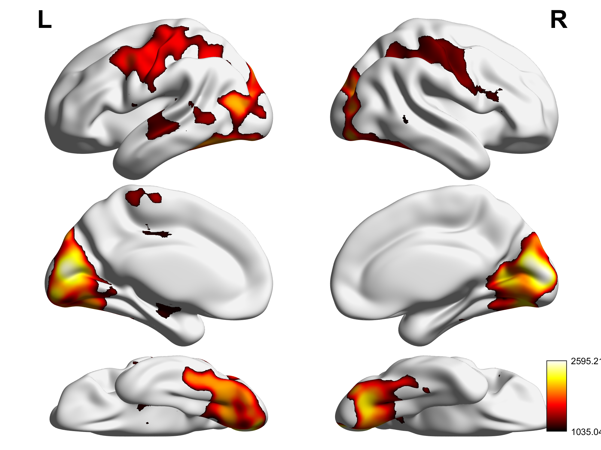
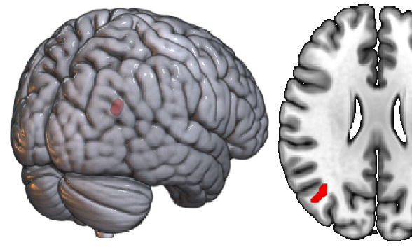
In terms of rsFC, compared with the control group, the MA group presented significantly enhanced FC between DRN and wide-spread brain regions, most of which are located in the left amygdala, visual network, somatomotor network, attention network and default mode network (DMN). VTA presented significantly enhanced FC with visual network and right hippocampus. The MA group also showed increased EC from the right Inferior Parietal Lobule to the DRN. Moreover, we dicovered significant increment of the local topological features, i.e. node efficiency(p<0.001), betweenness centrality(p<0.001), strength(p<0.001), vulnerability (p<0.001), as well as decrement of the shortest path length(p<0.001), of the DRN in the MA group. We found no evident abnormal effective connectivity and topological local features of VTA, and correlation between these imaging features of the 2 structures and clinical measurements.DISCUSSION
Previous research reported, when drug users are exposed to drug-related stimuli, there is significant activation in the visual area[3]. Consistent with these findings, our results suggest that the enhanced FC between DRN, VTA and visual network may mediate drug related cue-induced reactivity in MA-dependent patients. The enhanced FC between DRN and DMN, left amygdala may be the mechanism that causes the dysfunction of emotional control, leading to related psychiatric symptoms[5,6]. Previous studies show that interactions among the dopaminergic midbrain VTA and hippocampus, are critical for motivated memory encoding[7]. Abnormal functional connectivity between VTA and the right hippocampus may be associated with symptoms related to hypomnesia in patients. Topology analysis showed that compared with the healthy control group, the DRN was more tightly connected with other brain regions and the information transmission was more efficient in MA addiction patients. However, the higher vulnerability means that the FC between the DRN and other brain regions may be more vulnerable than other nodes when exposed to the drug.CONCLUSION
Abnormalities in the connecome of the DRN in MA-dependent patients may offer novel insights into the neural mechanisms underlying MA addiction.Acknowledgements
This study was supported by the National Natural Science Foundation of China (82071499), National Key Research and Development Program of China (2017YFC1310403), Zhejiang Basic Public Welfare Research Program Project (LGF21H090007), Zhejiang Provincial Medical and Health Science and Technology Program (2018KY708), and Ningbo Public Welfare Technology Plan Project (202002N3166).References
[1] Lin R, Liang J, Luo M. The Raphe Dopamine System: Roles in Salience Encoding, Memory Expression, and Addiction[J]. Trends in Neurosciences, 2021,44(5):366-377.
[2] Sepulveda M, Manning E E, Gogos A, et al. Long-term effects of young-adult methamphetamine on dorsal raphe serotonin systems in mice: Role of brain-derived neurotrophic factor[J]. Brain research, 2021,1762:147428.
[3] Oliva I, Wanat M J. Ventral Tegmental Area Afferents and Drug-Dependent Behaviors[J]. Front Psychiatry, 2016,7:30.
[4] Hanlon C A, Dowdle L T, Naselaris T, et al. Visual cortex activation to drug cues: A meta-analysis of functional neuroimaging papers in addiction and substance abuse literature[J]. Drug and alcohol dependence, 2014,143:206-212.
[5] Li Q, Li Z, Li W, et al. Disrupted Default Mode Network and Basal Craving in Male Heroin-Dependent Individuals: A Resting-State fMRI Study[J]. J Clin Psychiatry, 2016,77(10):e1211-e1217.
[6] Menon V. Large-scale brain networks and psychopathology: a unifying triple network model[J]. Trends Cogn Sci, 2011,15(10):483-506.
[7] Anand A, Jones S E, Lowe M, et al. Resting State Functional Connectivity of Dorsal Raphe Nucleus and Ventral Tegmental Area in Medication-Free Young Adults With Major Depression[J]. Front Psychiatry, 2018,9:765.
Figures