1736
Aberrant functional network connectivity of cerebellar network in methamphetamine-dependent patients: a resting-state fMRI study1Department of Radiology, Ningbo Medical Treatment Center Lihuili Hospital, Ningbo University, Ningbo, China, 2Department of Academic Research, Ningbo Kangning hospital, Ningbo University, Ningbo, China, 3MR Research, GE Healthcare, Beijing, China
Synopsis
Keywords: Psychiatric Disorders, fMRI (resting state), Methamphetamine, Independent component analysis, Functional network connectivity
Motivation: There are limited researches on functional network connectivity related to methamphetamine (MA), with existing studies predominantly focused on MA abstainers and lacking studies on the MA dependence.
Goal(s): This study aims to investigate the alterations in functional connectivity within and between resting-state networks (RSNs) in MA-dependence.
Approach: Using independent component analysis to acquire RSNs, calculate FCs within and between RSNs, and perform group comparison and correlation analysis.
Results: The MA group presented not only decreased rs-FC within the cerebellar network, but abnormal inter-networks rs-FC, especially between the cerebellar network and multiple cerebral networks, which is also associated with the severity of psychotic symptoms.
Impact: The cerebellar network of MA dependent individuals needs to be emphasized in future studies.
Introduction and Purpose
MA is a potent central nervous system stimulant with complex neurotoxic effects. Up to 60% abusers experience MA-related psychiatric symptoms [1]. Functional network connectivity (FNC) analysis is a data-driven and model-driven approach that allows exploration of organizational patterns of brain functional connections at a larger scale, without the need for a priori selection of regions of interest [2]. Limited efforts have been made to explore the FNC changes related to MA dependence, with existing studies predominantly focused on MA abstainers. In this study, we utilized ICA-based FNC analysis to investigate changes in FC within and between resting-state networks in MA-dependent individuals, aiming to explore the neural mechanisms of MA dependence.Materials and methods
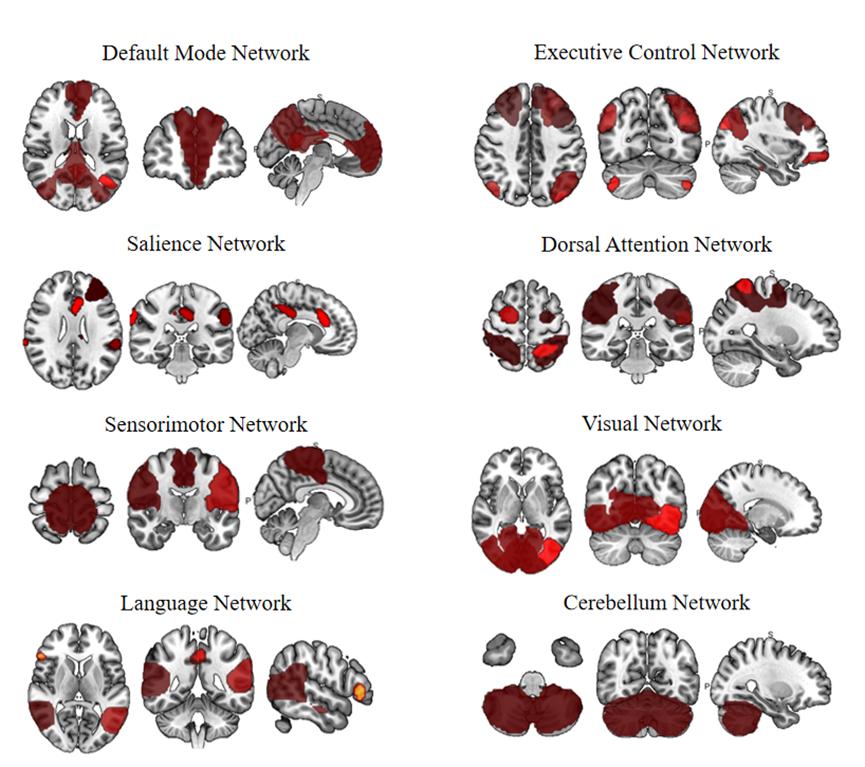
Patients: With approval from the Institutional Review Board, a total of 46 male MA-dependent individuals who were about to receive their first detoxification treatment were included in the study. The healthy control (HC) group consisted of 46 healthy male volunteers matched in age and education level to the MA group. Imaging: MRI scans were conducted using a 3.0T MRI scanner (Discovery MR750, GE) with an 8-channel phased-array head coil. Resting-state functional magnetic resonance imaging (rs-fMRI) data was acquired using a gradient recalled echo (GRE)-echo planar imaging (EPI) sequence with the following parameters: TR = 2000 ms, TE = 30 ms, matrix size = 64×64, FOV = 240mm×240mm, voxel size = 3.75×3.75×4 mm. A total of 38 axial images were acquired per time point, with a total of 200 time points collected within a scanning time of 6 minutes and 40 seconds. Data processing: The data were preprocessed using the CONN toolbox (http://www.nitrc.org/projects/conn) based on SPM12 in MATLAB v2019b. Subsequently, eight resting-state networks, i.e. default mode network (DMN), executive control network (ECN), salience network (SN), dorsal attention network (DAN), sensorimotor network (SMN), visual network (VN), language network (LN), and cerebellar network (CN), were identified on the preprocessed data ICA. After the computation of functional connectivities within and between these networks, inter-group comparisons were performed. R was used to calculate the correlation between functional connectivity and clinical measurements, such as Brief Psychiatric Rating Scale (BPRS), Hamilton Anxiety Rating Scale (HAMA) and the total duration of MA use.Results
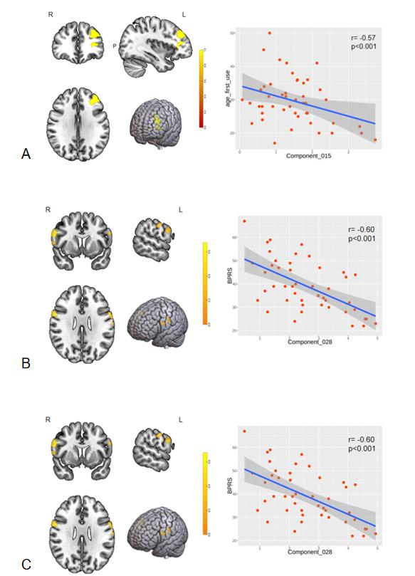
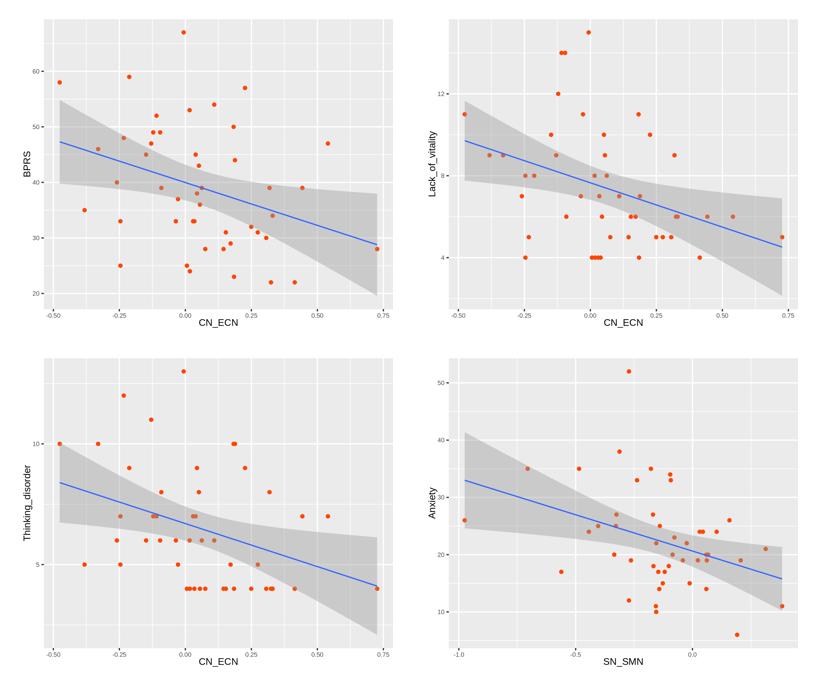
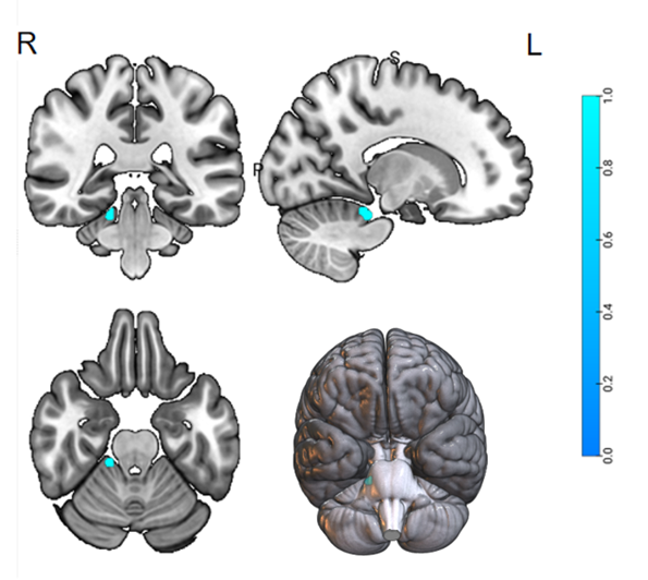
Compared with the HC group, the MA group showed decreased connectivity in the right cerebellar III-V lobules within the cerebellar network(t=-5.31, p=0.026)(Fig.2), and between the CN and 3 other networks, i.e. the SMN, DAN and VN(IC1-IC30: t=-3.41, p=0.007; IC1-IC27:t=-4.05, p=0.002; IC1-IC10:t=-2.74, p=0.028), and between the ECN, LN(IC17-IC7:t=-3.17, p=0.030), SMN, DAN and SN(IC30-IC27: t=-5.17, p<0.001; IC30-IC15: t=-2.84, p=0.028). Additionally, increased FC was detected between the CN and ECN (IC1-IC13: t=2.64, p=0.030; IC1-IC17: t=2.96, p= 0.020) (Fig.3). Significant correlations were revealed between the SN and the age of first MA use (r=-0.57, p<0.001), between the SMN and the total score of the BPRS (r=-0.60, p<0.001), and between the LN and the total duration of MA use (r=0.58, p<0.001) (Fig.4). Moreover, the FC between the CN and the ECN was negatively correlated with BPRS (r=-0.34, p=0.020), and the FC between the SN and the SMN was positively correlated with HAMA (r=-0.36, p=0.014) (Fig.5).Discussion and Conclusion
Decreased FC in the right cerebellar III-V lobules within the CN, as well as between the CN and SMN may lead to impaired motor-related functions, such as weakened plasticity of motor cortex and flexibility of movement[3]. Executive control is a highly ECN-dependent advanced cognitive activity that determines the manipulation and regulation of specific information processing for many cognitive tasks. Enhanced FC between the CN and the ECN in MA-dependent individuals may represent a compensatory change in response to long-term MA abuse, aiming to strengthen inhibitory control over habitual drug-taking behavior. The negative correlation between this FC and BPRS, indicating that this compensatory enhancement may be associated with alleviation of psychotic symptoms. The reduced functional connectivity between the ECN and LN, as well as between the CN and VN, may be related to the occurrence of MA-related hallucinations and visual symptoms in psychiatric patients. The decreased FCs between the SMN and DAN, as well as the SN, may reflect impaired attentional processing and sensory integration of incoming information in MA-dependent individuals. The weakened FC between the CN and the DAN may contribute to attention deficits in MA-dependent individuals, resulting in excessive focus on MA-related stimuli and compulsive drug-taking behavior. In summary, these abnormal functional connectivities may serve as the neurobiological basis for the occurrence of MA dependence and the emergence of related psychiatric symptoms.Acknowledgements
The study was supported by National Natural Science Foundation of China (82071499), National Key Research and Development Program of China (2017YFC1310403), Zhejiang Basic Public Welfare Research Program Project (LGF21H090007), Zhejiang Provincial Medical and Health Science and Technology Program(2018KY708), and Ningbo Public Welfare Technology Plan Project (202002N3166).References
1. Hsieh J H, Stein D J, Howells F M. The neurobiology of methamphetamine induced psychosis[J]. Front Hum Neurosci, 2014,8:537.
2. Lv H, Wang Z, Tong E, et al. Resting-State Functional MRI: Everything That Nonexperts Have Always Wanted to Know[J]. AJNR Am J Neuroradiol, 2018,39(8):1390-1399.
3. Huang X, Chen Y Y, Shen Y, et al. Methamphetamine abuse impairs motor cortical plasticity and function[J]. Mol Psychiatry, 2017,22(9):1274-1281.
Figures
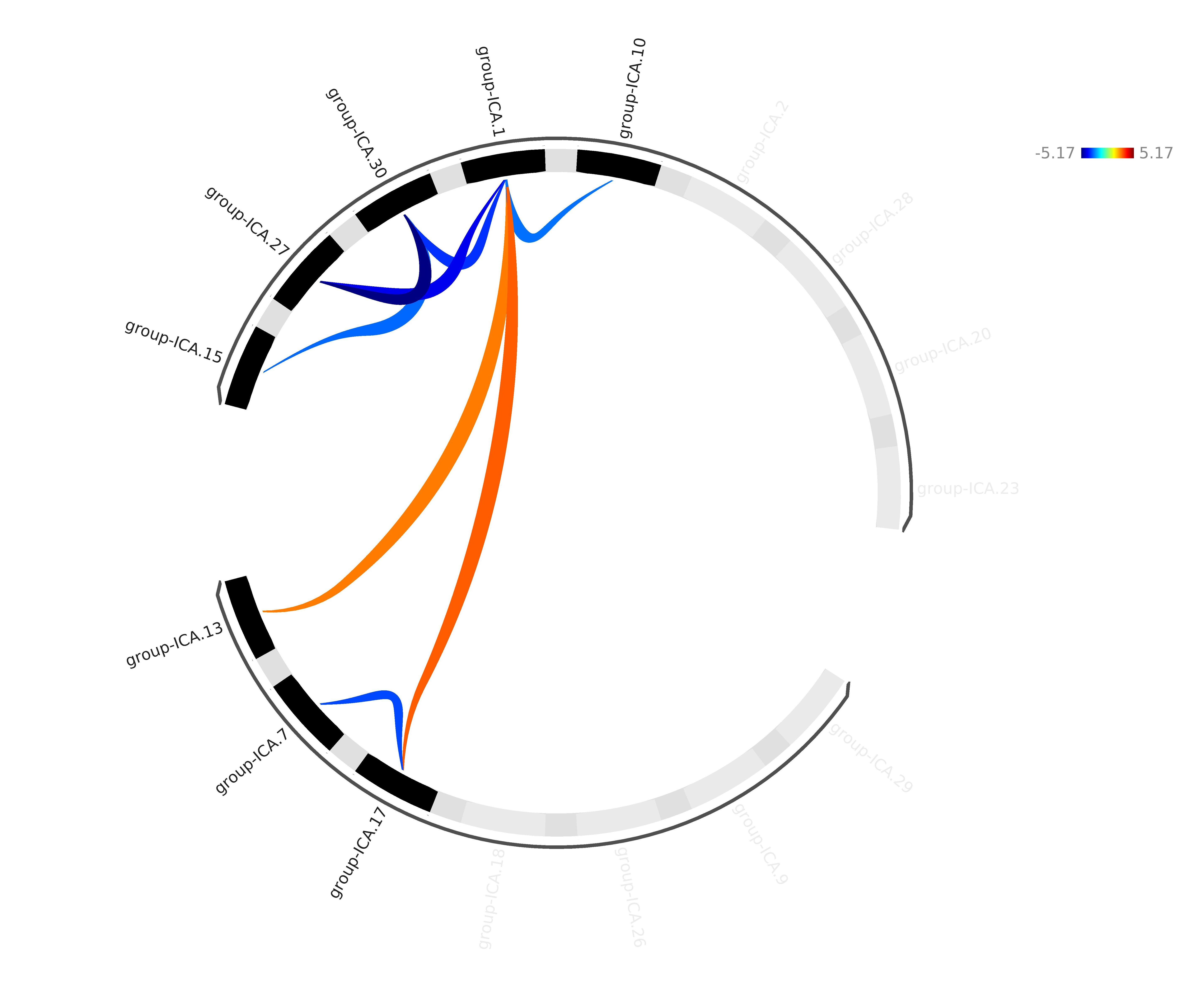
Schematic diagram of functional connectivity between RSNs with differences between the two groups
Note: Each node is coded with black color; the positive and negative connections are represented in red and blue color, respectively. The darker the lines, the more significant the difference; group-ICA7: language network; group-ICA13, group-ICA17: executive control network; group-ICA1: cerebellar network; group-ICA30: sensorimotor network; group-ICA27: dorsal attention network, group-ICA15: salience network; group-ICA10: visual network