1733
Morphological and microstructural modulation of Hypothalamic nuclei by eating disorder. A 7T MRI study1CRMBM AMU-CNRS, Marseille, France, 2SBIC, Singapore, Singapore, 3Endocrinology, AP-HM, Marseille, France, 4A-STAR, Singapore, Singapore, 5AP-HM, Marseille, France
Synopsis
Keywords: Psychiatric Disorders, Brain, Obesity, Anorexia Nervosa, quantitative T1
Motivation: The key structure for maintaining energy balance is the hypothalamus (HT), a small structure organized in multiple nuclei difficult to individualize with MR imaging.
Goal(s): To study in vivo the modulation in Obesity and Anorexia Nervosa of morphometry and microstructure of HT nuclei involved in food networks.
Approach: Characterization of T1 and volumes of HT nuclei (Neudorfer atlas) of 28 young women (13 Controls, 7 AN, 8O) using 3D-MP2RAGE 7T MRI.
Results: Differences in T1 values and volumes were observed in Obese and AN in HT nuclei involved in food networks (bilateral AN, left PVN)
Impact: Quantitative high resolution T1 MRI at 7T allows to characterize the morpho-structural modulation of HT nuclei involved in food networks in obesity and AN. These biomarkers may have a crucial role to monitor adaptation of food networks during therapeutic interventions.
Context and Objectives
The prevalence of eating disorders (ED) is constantly increasing and represents a real public health problem, with obesity now considered as an epidemic, and Anorexia Nervosa having the highest mortality rate of all psychiatric disorders. Hypothalamus (HT), a small deep brain area organized in multiple independent nuclei, appears to play a key role for maintaining energy balance. Thus, studying the characteristics HT nuclei is crucial to better understand their respective roles for behavioral changes encountered in ED. Currently, the lack of spatial resolution and poor MRI contrast prevents the precise individual characterization of HT nuclei1. UHF MRI (>=7T) has allowed a better definition of HT atlases 2. This study aimed at evaluating the sensitivity of high-resolution MRI at 7T to evidence variations in volumes and microstructures of HT nuclei associated to eating disorders.Methods
28 young women (age=26+4 years; Controls=13, Obesity=8, Anorexia Nervosa=7) were recruited by the AP-HM clinical department of Endocrinology. All subjects gave their informed consent to participate to the protocol which was approved by the Ethic Committee (CPP Est I). For all subjects, BMI, blood hormonal dosage, and eating habit questionnaires were obtained.Subjects were explored after a 12h fasting period using a 7T MR scanner (TERRA, Siemens) with a (1TX/32RX) Head coil (NOVA) using a 3D-MP2RAGE sequence (TR=5.000ms/TE=3ms/TI1=900ms/TI2=2.750ms, 256 slices, 0.6mm isotropic resolution, TA= 10min12s). Unbiased T1 maps and T1-w-UNIDEN images were obtained after B1+ inhomogeneity correction 1.
T1-weighted volumes were cropped to include basal ganglia, insula and the hypothalamus. Similar cropping was applied to the Neudorfer template/atlas (0.5mm) 2, before spatial registration (SyN procedure of ANTS 3) onto each individual cropped MRI volumes (Figure 1).
Whole brain volume (WBV) extraction was performed after segmentation using SPM12 4. Volumes and T1 values of HT nuclei were extracted using the ITKSNAP software 5 and normalized by WBV.
Non-parametric tests (Kruskal-Wallis with post hoc tests corrected for multiple comparisons (Steel-Dwass procedure) and Spearman rank correlations) were performed to evaluate between group differences and association with BMI.
Results
No significant differences in age were observed between the three groups.According to inclusion criteria, BMI were significantly different between groups (Table 1).
No significant between group differences were observed in the global HT volume, with just a trend of increase of HT volumes in the Obese group (p=0.15).
Obese patients relative to Controls showed significant increase in volumes of the bilateral fornix, as well as an increase in T1 values in the bilateral Arcuate Nuclei (p<0.05, corrected for multiple comparisons) (Figures 2-3).
AN relative to Controls showed increased T1 values in the left paraventricular nucleus (p<0.05, corrected for multiple comparisons)(Figures 2-3).
Finally for the whole cohort, BMI scores were associated to volumes of the Left Lateral Hypothalamus (rho=0.502, p=0.007) (Figure 4).
Discussion
A recent report conducted in a large cohort of subjects (n=1351) characterized by a large BMI range showed that HT volumes were associated with BMI 6. However, only volumes of the left anterior-inferior, and the right posterior parts of HT were shown to be increased in Obese patients relative to controls while the other between group comparisons did not reveal any significant differences.We demonstrate here, that in a small cohort of 28 subjects carefully selected according to age, sex, and condition (12h fasting period), high resolution quantitative T1 MRI at 7T can be sensitive enough to evidence modulation of the morpho-structural properties of specific HT nuclei by Obesity and Anorexia Nervosa.
Thus, abnormal HT regions in Obese patients included bilateral arcuate nuclei and bilateral fornix. Arcuate nuclei have been shown to be the first affected by high fat diet-induced inflammation, causing loss of the anorexigenic POMC neurons, leading to unbalancing energy homeostasis and promoting higher satiety thresholds 7,8. In addition, increased volumes of the bilateral fornix in Obese patients could be associated to inflammation and glial activation, that have been shown to be reversible by physical activity 9 or bariatric surgery 10.
Increased T1 values in the left HT PVN of Anorexia Nervosa patients could be associated to the disturbance of this central nucleus of the brain's stress systems 11.
All in all, we found a significant correlation between BMI and LHA volumes, the integrative site for signals underlying the motivation to eat 12.
These promising results open perspectives to access to robust individual data that will be used for personalized monitoring of patients with eating disorders. The Association of characteristics of HT nuclei with structural/functional connectivity as well as activations patterns related to food-cues will lead to a better understanding and monitoring of eating disorders.
Acknowledgements
This work was performed by a laboratory member of France Life Imaging network (grant ANR-11-INBS-0006) on the platform 7T-AMI, a French “Investissements d’Avenir” programme” (grant ANR-11-EQPX-0001). The CNS team is part of the Institut NeuroMarseille (A*MIDEX program). CR has received a PhD grant from the EUR Neuroschool program (ANR and A*MIDEX), and A*STAR (Singapore). This project has benefited from an A*STAR grant (Singapore).References
1. Roger C. Lasbleiz A, Guye M, Dutour A, Gaborit JP, Ranjeva JP. Role of the Human Hypothalamus in Food Intake Networks: An MRI Perspective. Front Nutr. 2022 Jan 3:8:760914.
2. Neudorfer C, Germann J, Elias GJB, Gramer R, Boutet A, Lozano AM. A high-resolution in vivo magnetic resonance imaging atlas of the human hypothalamic region. Sci Data. 2020 Sep 15;7(1):305.
3. Avants BB, Tustison NJ, Song G, Cook PA, Klein A, Gee JC. A reproducible evaluation of ANTs similarity metric performance in brain image registration. Neuroimage. 2011 Feb 1;54(3):2033-44.
4. https://www.fil.ion.ucl.ac.uk/spm/software/spm12/
5. http://www.itksnap.org/pmwiki/pmwiki.php?n=Main.HomePage
6. Brown SSG, Westwater ML, Seidlitz J, Ziauddeen H, Fletcher PC. Hypothalamic volume is associated with body mass index. Neuroimage Clin. 2023:39:103478.
7. Thaler JP, Yi CX, Schur EA, Guyenet SJ, et al. Obesity is associated with hypothalamic injury in rodents and humans. J Clin Invest. 2012 Jan;122(1):153-62.
8. Stathori et al . Alterations in Appetite-Regulating Hormones in Girls with Central Early or Precocious Puberty. Nutrients. 2023 Oct 9;15(19):4306.
9. Ma J, McGlade EC, Huber RS, Lyoo IK, Renshaw PF, Yurgelun-Todd DA. Overweight/Obesity-related microstructural alterations of the fimbria-fornix in the ABCD study: The role of aerobic physical activity. PLoS One. 2023 Jul 12;18(7):e0287682.
10. Atasoy et al. Evaluation of the white matter integrity in morbidly obese patients before and after bariatric surgery; a diffusion tensor imaging study. J Clin Ultrasound . 2023 Oct;51(8):1403-1409.
11. The selfish brain: competition for energy resources. Fehm HL, Kern W, Peters A.Prog Brain Res. 2006;153:129-40.
12. Petrovich GD. Learning and the motivation to eat: forebrain circuitry. Physiol Behav. 2011 Sep 26;104(4):582-9.
Figures
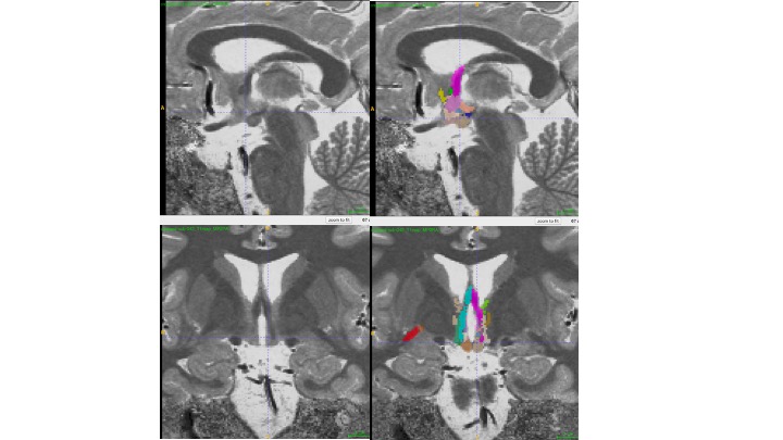
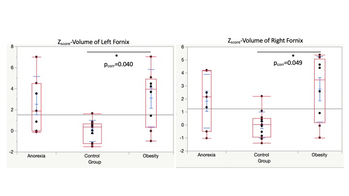
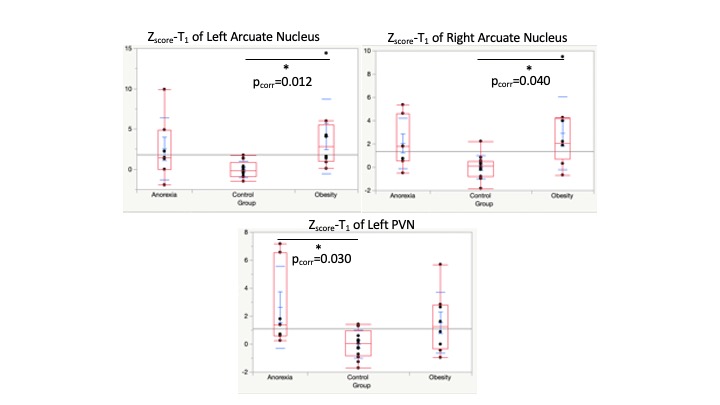
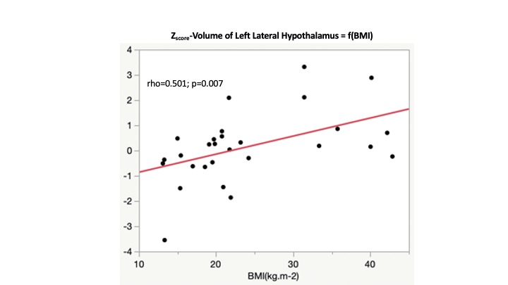
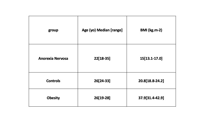
Table 1: Demographics.
Age was not different between groups (p>0.37). BMI was different between all groups (p<0.005)