1730
Graph theory-based analysis reveals neural anatomical network alterations in chronic PTSD in World Trade Center responders1Radiology and Imaging Sciences, Emory University, Atlanta, GA, United States, 2Biomedical Engineering, Georgia Institute of Technology and Emory University, Atlanta, GA, United States, 3Biomedical Engineering, Stony Brook University, Stony Brook, NY, United States, 4Family, Population, and Preventive Medicine, Stony Brook University, Stony Brook, NY, United States, 5Biomedical Engineering, Stony Brook University, Stony Brook, GA, United States, 6Stony Brook World Trade Center Wellness Program, Stony Brook University, Stony Brook, NY, United States, 7The Graduate Center and Queens College, City University of New York, New York, NY, United States, 8Environmental Medicine and Public Health, Icahn School of Medicine at Mount Sinai, New York, NY, United States, 9Environmental Health Sciences, Florida International University, Miami, FL, United States, 10Biomedical, Metabolic and Neurosciences, University of Modena, Modena, Italy, 11Psychiatry, Stony Brook University, Stony Brook, NY, United States, 12Medicine, Stony Brook University, Stony Brook, NY, United States
Synopsis
Keywords: Psychiatric Disorders, Psychiatric Disorders
Motivation: To uncover the neuropathological changes that influence the persistence and progression of symptoms in individuals with chronic Post-Traumatic Stress Disorder (PTSD), particularly among World Trade Center (WTC) responders.
Goal(s): The goal is to identify and characterize the neuroanatomical differences associated with chronic PTSD in World Trade Center responders using diffusion tensor neuroimaging and graph theory techniques.
Approach: employing graph theory to examine network alterations using brain diffusion images.
Results: The results of the study indicated significant differences in the neuroanatomical distances between white matter nodes—measured by weighted characteristic path lengths (CPL)—in World Trade Center responders with chronic PTSD compared to those without PTSD.
Impact: The impact of this study lies in advancing the understanding of PTSD as a disorder with anatomical basis. The association of increased characteristic path lengths with PTSD suggests that the disorder might involve diminished efficiency in the brain's communication networks.
Background
Multimodal imaging using network connectivity techniques can highlight neuropathology that influences Post-Traumatic Stress Disorder (PTSD) symptom maintenance and course [1,2]. The present study sought to investigate white matter neuroanatomical changes in a case-controlled PTSD group using structural diffusion data and graph theory analysis. We aimed to examine correlations of characteristic path length (CPL) with specific subtypes of chronic PTSD symptomatology, cognition, and physical functional limitations, that have been previously reported in WTC responders with chronic PTSD.Methods
World Trade Center (WTC) responders who continue to suffer chronic WTC PTSD and controls were recruited into a diffusion tensor neuroimaging protocol (N=109, 47 with PTSD, 55 WTC controls with no PTSD, and nine non-WTC-exposed controls.Diffusion data were acquired from all the volunteers on a 3T scanner (Siemens Biograph mMR). Diffusion images were acquired using single shell diffusion-weighted EPI acquisitions (TE/TR = 87.6/4680 ms, b value = 1200, 64 diffusion directions, in-plane resolution = 2 mm, slice thickness = 2 mm, matrix size = 128 x 128, multiband factor = 2).
The primary outcome in this study was weighted CPL (wCPL) as measured using diffusion tractography imaging (DTI). Connectivity matrices were generated via deterministic tractography analysis using Q-space diffeometric reconstruction with DSI-Studio [3].
Results
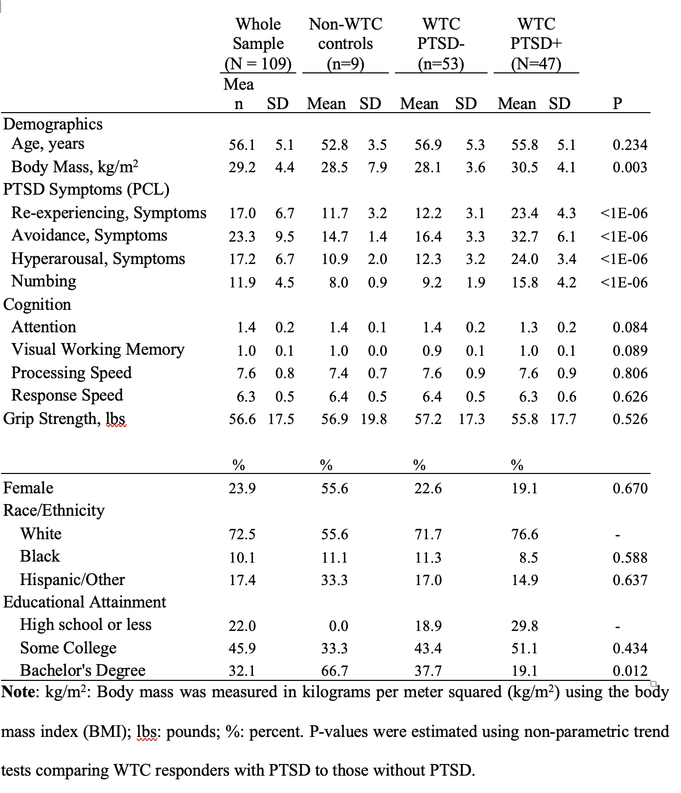
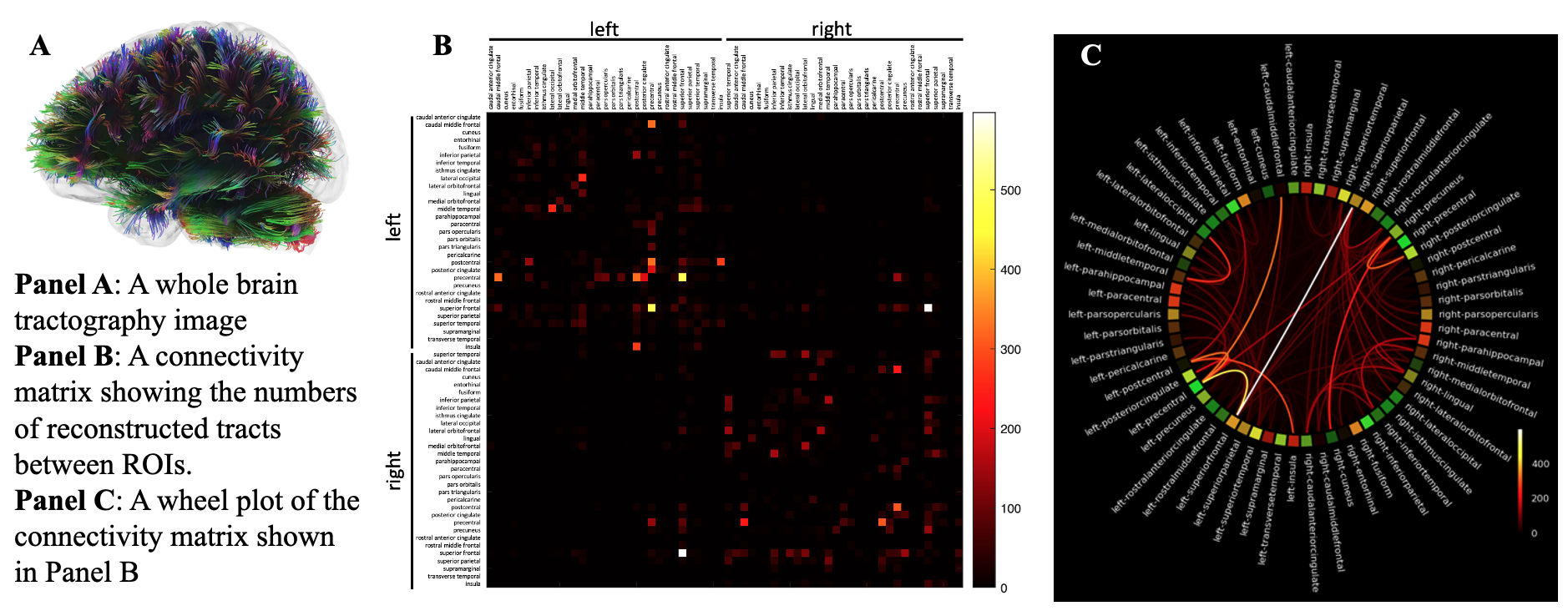
The sample was in their mid-fifties, on average, and as is consistent with the responder population the majority were Male (Figure 1).Figure 2 shows an example tractography image and the associated individual-level connectivity matrix. Together, these show the complexity and redundancy of the connected network and highlight the central interconnections between proximal and distal cortical regions.
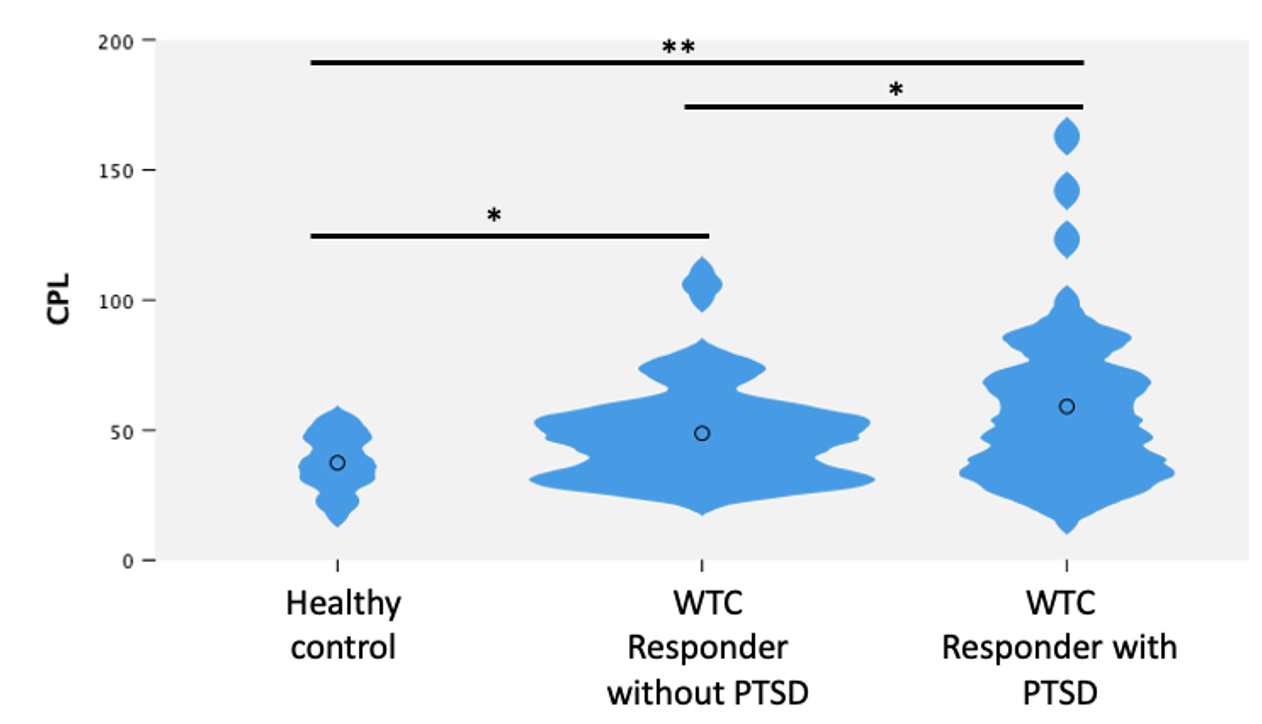
Figure 3 depicts comparisons of network analysis for healthy controls, and WTC responders with/without PTSD. One-way ANOVA found that wCPL is associated with the group (ordinal measure: healthy controls, WTC PTSD+, WTC PTSD-). Furthermore, significant differences were found in wCPL between PTSD+ and PTSD- subjects (healthy controls combined with WTC responders without PTSD, d = -0.50; p = 0.019). PTSD+ responders were found to have increased wCPL relative to unexposed controls (d = 0.76; p < 0.001), and PTSD- WTC responders (d = 0.42; p = 0.047). Finally, WTC responders without PTSD were found to have increased wCPL when compared to unexposed controls (d = 0.65; p = 0.018). However, while we did find strong associations with PTSD, we did not find associations with WTC exposure severity (p = 0.830) or duration (p = 0.601).
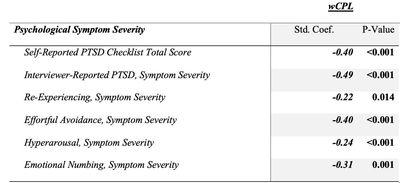
Focusing on measures of symptom severity indicated that the presence of increased PTSD symptom severity was broadly associated with decreases in wCPL in self-reported and interviewer-recorded symptom severity measures (Figure 4).
Discussions
In the largest study to date to examine the relationship between chronic PTSD and the CPL of anatomical neural connections, we found that wCPL was significantly increased in PTSD+ when compared to the PTSD- groups. Our findings are consistent with previous investigations on wCPL and PTSD status showing that wCPL was increased in pediatric patients with PTSD [1].These results expand on that study and suggest that wCPL might be increased among those experiencing any trauma. An increase in CPL for PTSD subjects supports the increasing evidence identifying neural correlates of PTSD. This study has multiple possible interpretations including that results might suggest that chronic PTSD could alter the brain’s level and communication efficiency as shorter CPL is reflective of more efficient re-entrant communication between global nodes of cortical and subcortical regions [4]. However, more research is necessary to determine whether wCPL represents a marker of increased vulnerability to PTSD or is a byproduct of other conditions.
Acknowledgements
This work was in part supported by CDC/NIOSH U01 OH011314, NIH/NIA P50 AG005138, NIH/NIA R01 AG049953. We would also like to acknowledge ongoing funding to the WTC Health and Wellness Program (CDC 200-2011-39361).
References
1. Suo, X., et al., Anatomic insights into disrupted small-world networks in pediatric posttraumatic stress disorder. Radiology, 2017. 282(3): p. 826-834.
2. Newman, M.E.J., Mixing patterns in networks. Physical Review E, 2003. 67(2): p. 026126.
3. Yeh, F.-C. and W.-Y.I. Tseng, NTU-90: a high angular resolution brain atlas constructed by q-space diffeomorphic reconstruction. Neuroimage, 2011. 58(1): p. 91-99.
4. Onoda, K. and S. Yamaguchi, Small-worldness and modularity of the resting-state functional brain network decrease with aging. Neuroscience letters, 2013. 556: p. 104-108.
Figures