1725
Investigation of resting state dynamics of cerebellar functional connectivity in schizophrenia1Institute of Neuroscience and Medicine - 4 (INM-4), Forschungszentrum Jülich, Jülich, Germany, 2Department of Psychiatry, Psychotherapy and Psychosomatics, RWTH Aachen University, Aachen, Germany, 3Center for Computational Life Sciences, RWTH Aachen University, Aachen, Germany, 4Institute of Neuroscience and Medicine 11 (INM - 11), Forschungszentrum Jülich, Jülich, Germany, 5Department of Neurology, RWTH Aachen University, Aachen, Germany, 6Department of Nuclear Medicine, RWTH Aachen University, Aachen, Germany
Synopsis
Keywords: Psychiatric Disorders, Psychiatric Disorders, Schizophrenia, Dynamic Functional Connectivity, Cerebellum
Motivation: The complex nature of schizophrenia and its unclear pathophysiology drive our research. We aim to delve into altered brain communication, especially the cerebellum, during transitions between mental states for deeper insights.
Goal(s): Our goal was to investigate cerebellar communication dynamics in schizophrenia to better understand its pathophysiology.
Approach: We used dynamic ICA analysis to study resting-state cerebello-cortical temporal dynamics.
Results: We observed implications in the dynamics of the cerebello-frontal and cerebello-occipital connections potentially indicative of higher social sensitivity and deficits in cognitive inhibition, and highlighting the role of the cerebellum as modulator between different brain circuits.
Impact: The identification of resting state temporal dynamic changes in cerebello-frontal and cerebello-occipital circuits in schizophrenia provides crucial pathophysiological insights, inspiring further research on the role of the cerebellum as a brain circuit modulator, promising advancements in treatment strategies and outcomes.
Introduction
Schizophrenia is a complex disorder characterized by an altered perception of reality in individuals. It often includes symptoms such as hallucinations, distorted beliefs, disorganized thinking, and social difficulties. Many studies have attempted to explain its underlying mechanisms, with no clear conclusions1. One widely accepted hypothesis is alterations in brain communication. Several studies, including work by Yeganeh-Doost2 and others, has highlighted the role of cerebellar communication in schizophrenia. The human mind is known to transition between different mental states even at rest3, necessitating an exploration of dynamic functional connectivity alongside the traditional static approach. This method has proven useful in identifying shifts in mindfulness and mind-wandering3, as well as abnormal transient states in schizophrenia4 and traumatic brain injury5. Therefore, this study focuses on examining cerebellar functional connectivity dynamics through dynamic ICA analysis in the context of schizophrenia.Methods
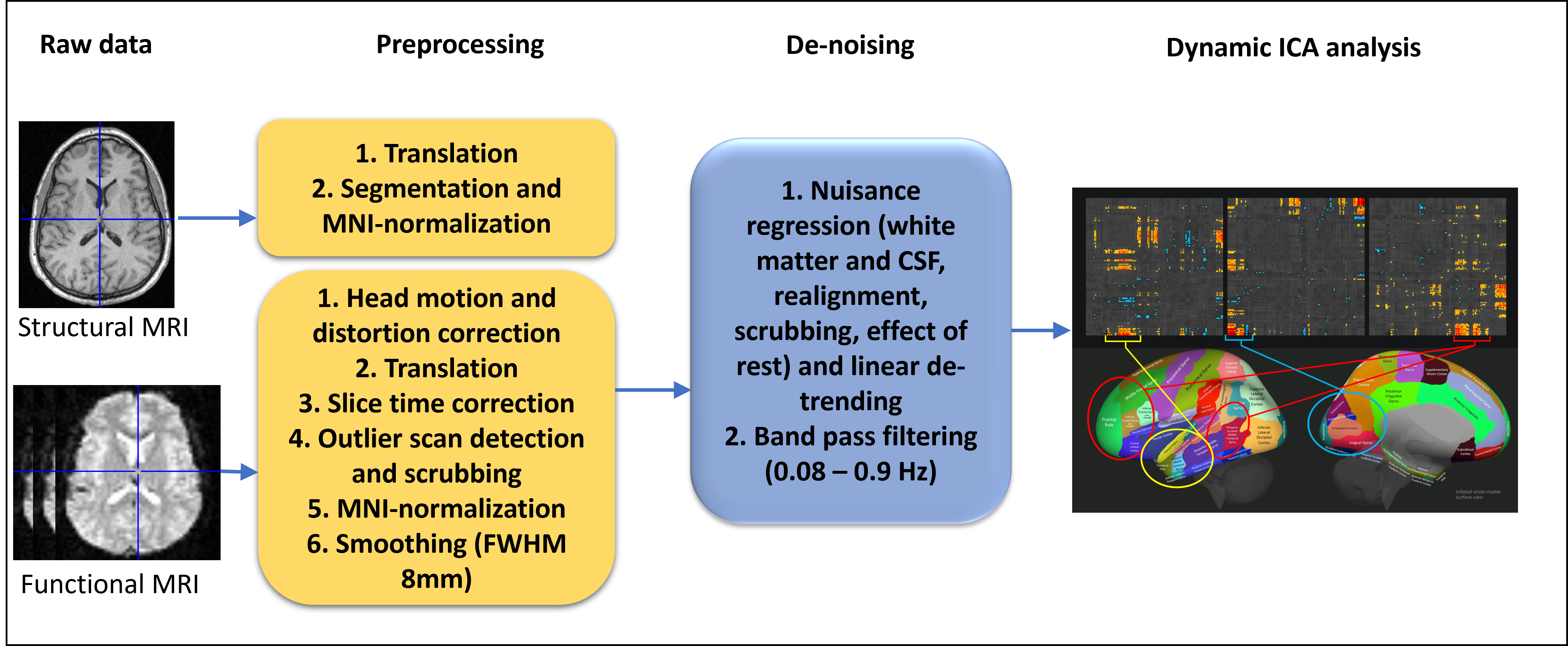
Resting state fMRI data of 28 male schizophrenia patients (age: 38 ± 10) and 41 group-level age-matched male healthy controls (age: 36 ± 11) recorded simultaneously with EEG and PET on a 3T BrainPET MR Scanner (Trio, Siemens, Germany) were used for this study. The acquisition protocol and sequence parameters can be found in another study6. The raw data were preprocessed and denoised using the steps shown in figure 1. After denoising dynamic ICA analyses were performed in the first level. All analyses were performed in CONN v2021a7.In the first-level analysis, dynamic independent component analysis (dyn-ICA) was employed to identify 20 temporal modulation factors representing changes in functional connectivity within each functional run, along with the associated circuits. The BOLD signal from 132 Harvard-Oxford atlas regions across subjects and conditions were concatenated, modelling functional connectivity using a generalized psychophysiological interaction model (gPPI) with 20 unknown psychological factors and a 30s FWHM Hanning regularization kernel. Then, a fast-ICA fixed-point algorithm was used to identify spatially independent group-level circuits from the regression coefficients of the group-level gPPI model interaction terms, and subject-specific ICA maps related to these circuits were computed for individual subjects and conditions.
In the second-level, group circuits reflecting cortico-cerebellar connectivity were identified through visual inspection, and temporal properties of these circuits such as average, frequency and variability were compared between groups using independent sample t-tests with a significance threshold of p-uncorrected<0.05.
Results
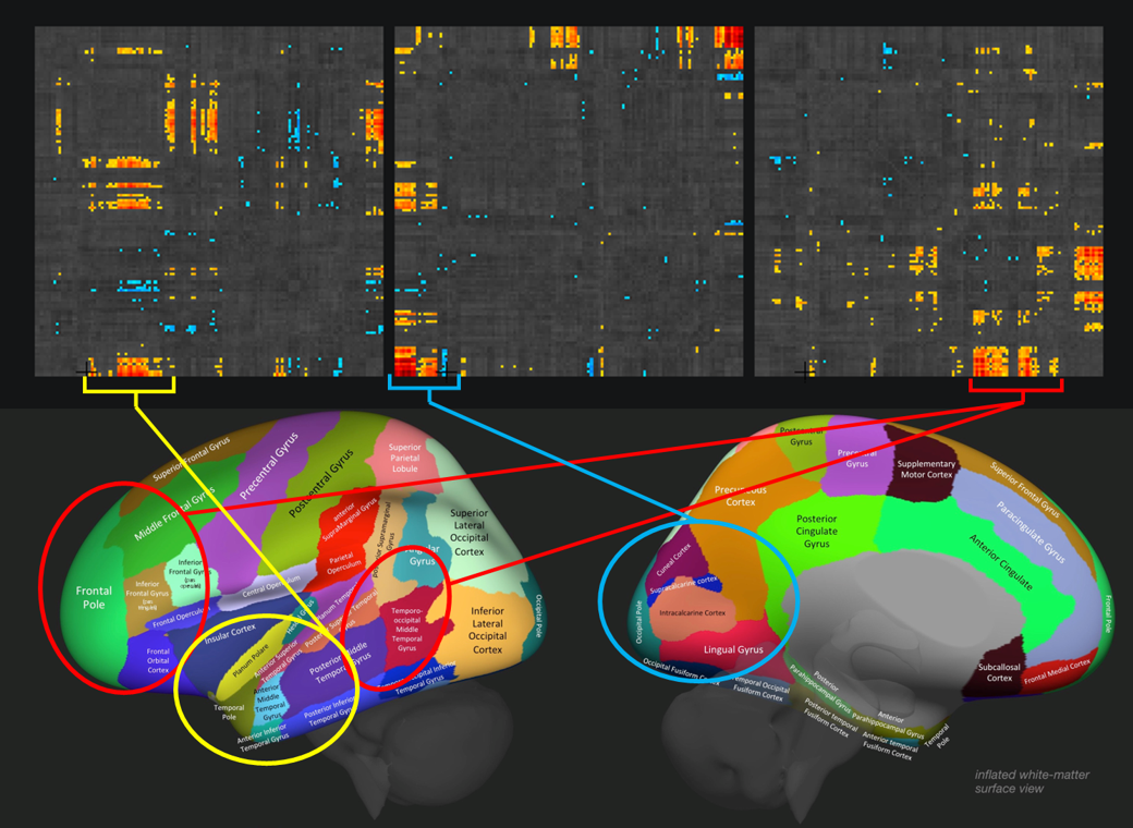
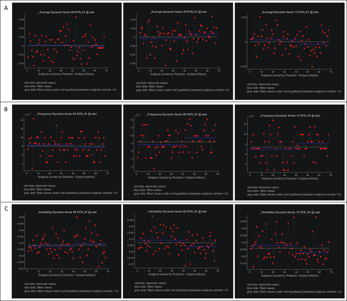
Three circuits involving cortico-cerebellar connectivity were identified, covering clusters of connections in the temporal, occipital and frontal lobes respectively (circuits 4, 5 and 13) (figure 2). Temporal averages did not differ between patients and controls significantly but showed trends of increase and decrease in the cerebello-occipital and cerebello-frontal connections respectively (figure 3A). On the other hand temporal frequency of the cerebello-occipital circuit was significantly higher (figure 3B) and the temporal variability of cerebello-occipital and cerebello-frontal circuit was significantly lower in schizophrenia patients (figure 3C). The cerebello-temporal circuit did not show any significant differences.Discussion
The cerebellum has long been associated with motor and balance functions, but recent research has revealed its involvement in higher-level processes. It's now recognized that the cerebellum plays a fundamental role in motor, default-mode (task-negative), and attentional/executive (task-positive) functions8. The occipital lobe handles visual and social processing9, while the frontal cortex is known for logical reasoning and contains important hubs of the default mode network. Analysing the temporal dynamics of their connectivity with the cerebellum offers valuable insights into the pathophysiology of schizophrenia. Increased cerebello-occipital connectivity and frequency over time may indicate heightened social sensitivity or tendencies for visual hallucinations, while reduced cerebello-frontal connectivity suggests a lack of cognitive inhibition. Both findings align with previous studies indicating decreased GABA signalling in the cerebellum in schizophrenia2. Lack of temporal variability in both of these circuits shows that the states do no change much, indicative of a control deficit in schizophrenia. Taken together, this sheds further light on the role of the cerebellum as a modulator between different brain networks, and paves way for further research in this direction.Conclusion
Schizophrenia is a complex neuropsychiatric disorder, the pathophysiological mechanisms of which are still unclear. The application of dynamic ICA to resting state functional connectivity has shown alterations in the cerebello-frontal and cerebello-occipital circuits, potentially indicating lack of cognitive inhibition, increased social sensitivity and highlighting the role of the cerebellum as a modulator between different brain circuits.Acknowledgements
*equal contribution. The authors would like to thank the data acquisition team of the entire project comprising Claudia Regio Brambilla, Linda Orth, Hasan Sbaihat, Nicolas Kaulan, Jörg Mauler, Lutz Tellmann, Karl-Josef Langen, Christoph Lerche, Silke Frensch, Susanne Schaden, Gabriel Stoffels and Andreas Matusch. This trimodal imaging project was supported by the EU FP 7 funded project TRIMAGE (Nr 602621 BMBF and Siemens 9 4 T MR PET project (Grant number 13N9121), and partial funding from the DFG (Shah DFG SH 79/2-2).References
1. Keshavan, M. S. et al. Neuroimaging in schizophrenia. Neuroimaging clinics of North America 30, 73 (2020).
2. Yeganeh-Doost, P., Gruber, O., Falkai, P. & Schmitt, A. The role of the cerebellum in schizophrenia: from cognition to molecular pathways. Clinics (Sao Paulo, Brazil) 66 Suppl 1, 71–7 (2011).
3. Mooneyham, B. W. et al. States of mind: Characterizing the neural bases of focus and mind-wandering through dynamic functional connectivity. Journal of Cognitive Neuroscience 29, 495–506 (2017).
4. Damaraju, E. et al. Dynamic functional connectivity analysis reveals transient states of dysconnectivity in schizophrenia. NeuroImage: Clinical 5, 298–308 (2014).
5. Vergara, V. M., Mayer, A. R., Kiehl, K. A. & Calhoun, V. D. Dynamic functional network connectivity discriminates mild traumatic brain injury through machine learning. NeuroImage: Clinical 19, 30–37 (2018).
6. Rajkumar, R. et al. Excitatory–inhibitory balance within EEG microstates and resting-state fMRI networks: assessed via simultaneous trimodal PET–MR–EEG imaging. Translational Psychiatry 11, 1–15 (2021).
7. Whitfield-Gabrieli, S. & Nieto-Castanon, A. Conn: A Functional Connectivity Toolbox for Correlated and Anticorrelated Brain Networks. Brain Connectivity 2, 125–141 (2012).
8. Guell, X. & Schmahmann, J. Cerebellar Functional Anatomy: a Didactic Summary Based on Human fMRI Evidence. Cerebellum 19, 1–5 (2020).
9. Ramkiran, S. et al. How brain networks tic: Predicting tic severity through rs‐fMRI dynamics in Tourette syndrome. Hum Brain Mapp 44, 4225–4238 (2023).
Figures